Essential role of the keratinocyte-specific endonuclease DNase1L2 in the removal of nuclear DNA from hair and nails - PubMed (original) (raw)
. 2011 Jun;131(6):1208-15.
doi: 10.1038/jid.2011.13. Epub 2011 Feb 10.
Sandra Szabo, Jennifer Scherz, Karin Jaeger, Heidemarie Rossiter, Maria Buchberger, Minoo Ghannadan, Marcela Hermann, Hans-Christian Theussl, Desmond J Tobin, Erwin F Wagner, Erwin Tschachler, Leopold Eckhart
Affiliations
- PMID: 21307874
- PMCID: PMC3185332
- DOI: 10.1038/jid.2011.13
Essential role of the keratinocyte-specific endonuclease DNase1L2 in the removal of nuclear DNA from hair and nails
Heinz Fischer et al. J Invest Dermatol. 2011 Jun.
Abstract
Degradation of nuclear DNA is a hallmark of programmed cell death. Epidermal keratinocytes die in the course of cornification to function as the dead building blocks of the cornified layer of the epidermis, nails, and hair. Here, we investigated the mechanism and physiological function of DNA degradation during cornification in vivo. Targeted deletion of the keratinocyte-specific endonuclease DNase1-like 2 (DNase1L2) in the mouse resulted in the aberrant retention of DNA in hair and nails, as well as in epithelia of the tongue and the esophagus. In contrast to our previous studies in human keratinocytes, ablation of DNase1L2 did not compromise the cornified layer of the epidermis. Quantitative PCRs showed that the amount of nuclear DNA was dramatically increased in both hair and nails, and that mitochondrial DNA was increased in the nails of DNase1L2-deficient mice. The presence of nuclear DNA disturbed the normal arrangement of structural proteins in hair corneocytes and caused a significant decrease in the resistance of hair to mechanical stress. These data identify DNase1L2 as an essential and specific regulator of programmed cell death in skin appendages, and demonstrate that the breakdown of nuclear DNA is crucial for establishing the full mechanical stability of hair.
Figures
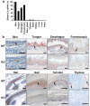
Figure 1. Expression of DNase1L2 is associated with cornification
(a) Quantification of DNase1L2 mRNA expression in mouse organs using real-time PCR. Note that the skin sample included both interfollicular epidermis and hair follicles. The results of one of two experiments with similar results are shown. (b) Immunohistochemical detection of DNase1L2 in the mouse. Tissues of DNase1L2-deficient mice were used as negative controls. In some panels, arrows point to sites of DNase1L2 expression. Scale bars: 40 μm.
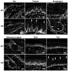
Figure 2. Deletion of DNase1L2 leads to retention of DNA in distinct pathways of terminal differentiation of keratinocytes
Thin sections of ear, sole, sebaceous glands, tongue, esophagus and the tail of DNase1L2 knockout (KO) and wild-type (WT) mice were labeled with the DNA-binding dye Hoechst 33258 (white). Arrows point to nuclear remnants within the corneocytes. Dotted lines mark the outer border of the cornified layer in all tissues except the sebaceous glands where the border of the gland is indicated. Scale bars: 40 μm.
Figure 3. DNA is aberrantly retained in the hair and nails of DNase1L2-deficient mice
(a) Hoechst 33258 labeling (white) of nails from DNase1L2 knockout (KO) and wild-type (WT) mice. The outer border of the nail is indicated by a dotted line. (b, c) Nuclear and mitochondrial DNA extracted from nails was quantified by quantitative real-time PCR. Data represent means ± s.e.m. (n = 3; asterisk, P < 0.05). (d) Hair from the back of DNase1L2 wild-type and DNase1L2-deficient mice was permeabilized and labeled with Hoechst 33258 (white). Fluorescent and bright field images of the same areas are shown. (e) Hoechst 33258 labeling of various hair types of DNase1L2-null mice. Note that the hair types differ in the number of cell rows in the medulla. (f) Quantification of nuclear DNA from hair by real-time PCR. Data represent means ± s.e.m. (n = 5; double asterisk, P < 0.01). (g) Quantification of mitochondrial DNA from hair by real-time PCR. Data represent means ± s.e.m. (n = 5; n.s., not significant). DNA quantifications were repeated in an independent experiment with similar results. KO, DNase1L2 knockout; WT, wild-tpye. Scale bars: (a) 40 μm; (d) 100 μm.
Figure 4. DNA disturbs the structural maturation of hair corneocytes
(a) Cross sections of hair follicles were double-stained for DNA using Hoechst 33258 (blue) and keratin 31 (K31, red), a marker specific for the hair cortex, using a specific antiserum. Arrows point to cavities in the keratin 31 scaffold of DNase1L2 knockout mice that are filled with DNA. The outer border of the hair cortex is indicated by a dotted line. (b) Transmission electron microscopy of hair cross sections. Arrows point to nuclear remnants that interdigitate structural proteins in the hair cortex. (c) Scanning electron microscopy of hairs. Arrows point to indentions on the surface. c, cortex, m, medulla. Scale bars: (a) 10 μm; (b) 0.2 μm and 1 μm (inset); (c) 20 μm.
Figure 5. Retention of DNA increases the fragility of hair from DNase1L2-deficient mice
(a) Schematic depiction of the mechanical stress test. Shaved hairs from wild-type and DNase1L2-deficient mice were exposed to mechanical stress in a bead mill containing ceramic beads. The resulting fragments were spread on microscope slides, photographed and measured using an automatic software algorithm. 3 slides were analyzed per bead mill treatment. (b) Untreated zigzag hairs of wild-type and mutant mice were photographed and their length was determined manually (n = 5; ns, not significant). (c, d) After exposure to mechanical stress the mean length of hair fragments (c) and the percentage of hair fragments longer than 1 mm was determined (d). Data represent means ± s.e.m. (n = 7 (WT) and 8 (KO); double asterisk, P < 0.01; asterisk, P < 0.05). Similar results were obtained in a second independent bead mill experiment using hair samples from n = 5 mice per genotype.
Similar articles
- Inactivation of DNase1L2 and DNase2 in keratinocytes suppresses DNA degradation during epidermal cornification and results in constitutive parakeratosis.
Fischer H, Buchberger M, Napirei M, Tschachler E, Eckhart L. Fischer H, et al. Sci Rep. 2017 Jul 25;7(1):6433. doi: 10.1038/s41598-017-06652-8. Sci Rep. 2017. PMID: 28743926 Free PMC article. - Terminal differentiation of nail matrix keratinocytes involves up-regulation of DNase1L2 but is independent of caspase-14 expression.
Jäger K, Fischer H, Tschachler E, Eckhart L. Jäger K, et al. Differentiation. 2007 Dec;75(10):939-46. doi: 10.1111/j.1432-0436.2007.00183.x. Epub 2007 May 9. Differentiation. 2007. PMID: 17490414 - DNase1L2 degrades nuclear DNA during corneocyte formation.
Fischer H, Eckhart L, Mildner M, Jaeger K, Buchberger M, Ghannadan M, Tschachler E. Fischer H, et al. J Invest Dermatol. 2007 Jan;127(1):24-30. doi: 10.1038/sj.jid.5700503. Epub 2006 Aug 10. J Invest Dermatol. 2007. PMID: 16902420 - Cell death by cornification.
Eckhart L, Lippens S, Tschachler E, Declercq W. Eckhart L, et al. Biochim Biophys Acta. 2013 Dec;1833(12):3471-3480. doi: 10.1016/j.bbamcr.2013.06.010. Epub 2013 Jun 20. Biochim Biophys Acta. 2013. PMID: 23792051 Review. - Invited review: formation of keratins in the bovine claw: roles of hormones, minerals, and vitamins in functional claw integrity.
Tomlinson DJ, Mülling CH, Fakler TM. Tomlinson DJ, et al. J Dairy Sci. 2004 Apr;87(4):797-809. doi: 10.3168/jds.S0022-0302(04)73223-3. J Dairy Sci. 2004. PMID: 15259213 Review.
Cited by
- A Mixture of Tocopherol Acetate and L-Menthol Synergistically Promotes Hair Growth in C57BL/6 Mice.
Ahn S, Lee JY, Choi SM, Shin Y, Park S. Ahn S, et al. Pharmaceutics. 2020 Dec 18;12(12):1234. doi: 10.3390/pharmaceutics12121234. Pharmaceutics. 2020. PMID: 33353178 Free PMC article. - The Role of Nucleases and Nucleic Acid Editing Enzymes in the Regulation of Self-Nucleic Acid Sensing.
Santa P, Garreau A, Serpas L, Ferriere A, Blanco P, Soni C, Sisirak V. Santa P, et al. Front Immunol. 2021 Feb 26;12:629922. doi: 10.3389/fimmu.2021.629922. eCollection 2021. Front Immunol. 2021. PMID: 33717156 Free PMC article. Review. - Pan‑cancer analysis of the deoxyribonuclease gene family.
Bai Q, He X, Hu T. Bai Q, et al. Mol Clin Oncol. 2023 Feb 2;18(3):19. doi: 10.3892/mco.2023.2615. eCollection 2023 Mar. Mol Clin Oncol. 2023. PMID: 36798465 Free PMC article. - Secreted mammalian DNases protect against systemic bacterial infection by digesting biofilms.
Lacey KA, Serpas L, Makita S, Wang Y, Rashidfarrokhi A, Soni C, Gonzalez S, Moreira A, Torres VJ, Reizis B. Lacey KA, et al. J Exp Med. 2023 Jun 5;220(6):e20221086. doi: 10.1084/jem.20221086. Epub 2023 Mar 16. J Exp Med. 2023. PMID: 36928522 Free PMC article. - Transglutaminase Activity Is Conserved in Stratified Epithelia and Skin Appendages of Mammals and Birds.
Sachslehner AP, Surbek M, Golabi B, Geiselhofer M, Jäger K, Hess C, Kuchler U, Gruber R, Eckhart L. Sachslehner AP, et al. Int J Mol Sci. 2023 Jan 22;24(3):2193. doi: 10.3390/ijms24032193. Int J Mol Sci. 2023. PMID: 36768511 Free PMC article.
References
- Arin MJ. The molecular basis of human keratin disorders. Hum Genet. 2009;125:355–73. - PubMed
- Blum H, Beier H, Gross HJ. Improved silver staining of plant proteins, RNA and DNA in polyacrylamide gels. Electrophoresis. 1987;8:93–99.
- Candi E, Schmidt R, Melino G. The cornified envelope: a model of cell death in the skin. Nat Rev Mol Cell Biol. 2005;6:328–40. - PubMed
- Fischer H, Eckhart L, Mildner M, et al. DNase1L2 degrades nuclear DNA during corneocyte formation. J Invest Dermatol. 2007;127:24–30. - PubMed
Publication types
MeSH terms
Substances
LinkOut - more resources
Full Text Sources
Other Literature Sources
Molecular Biology Databases


