The antiproliferative action of progesterone in uterine epithelium is mediated by Hand2 - PubMed (original) (raw)
The antiproliferative action of progesterone in uterine epithelium is mediated by Hand2
Quanxi Li et al. Science. 2011.
Abstract
During pregnancy, progesterone inhibits the growth-promoting actions of estrogen in the uterus. However, the mechanism for this is not clear. The attenuation of estrogen-mediated proliferation of the uterine epithelium by progesterone is a prerequisite for successful implantation. Our study reveals that progesterone-induced expression of the basic helix-loop-helix transcription factor Hand2 in the uterine stroma suppresses the production of several fibroblast growth factors (FGFs) that act as paracrine mediators of mitogenic effects of estrogen on the epithelium. In mouse uteri lacking Hand2, continued induction of these FGFs in the stroma maintains epithelial proliferation and stimulates estrogen-induced pathways, resulting in impaired implantation. Thus, Hand2 is a critical regulator of the uterine stromal-epithelial communication that directs proper steroid regulation conducive for the establishment of pregnancy.
Figures
Figure 1
P-regulated expression of Hand2 in the uterus is critical for implantation. (A) IHC of Hand2 protein in the uterine sections of ovariectomized WT mice treated with vehicle (a) or P (b) and PR-null mice treated with P (c). Panel d represents sections treated with non-immune IgG. (B) Uterine sections obtained from mice on days 2 to 4 of pregnancy were subjected to IHC using Hand2 antibody. Magnification 20X. (C) H and E staining of uterine sections obtained from Hand2f/f (a, b) and Hand2d/d (c, d) mice on day 5 (n=6) of pregnancy. b and d represent magnified images of a and c, respectively. Solid and dotted arrows point to embryo and luminal epithelium. L and S represent luminal epithelium and stroma, respectively.
Figure 2
Enhanced ERα activity and proliferation in the luminal epithelium of Hand2d/d uteri. (A) Real-time PCR was performed to monitor the expression of Muc1 and Ltf in the uteri of day 4 pregnant Hand2f/f and Hand2d/d mice, *P<0.001. (B) IHC of Ki67 in Hand2f/f (a) and Hand2d/d (b) uteri on day 4 of pregnancy, 20X. Panel c shows uterine sections from Hand2d/d mice treated with non-immune IgG, 40X. (C) IHC of Ki67 in the uterine sections of ovariectomized Hand2f/f and Hand2d/d mice treated with E for one day (a and b), P for three days (c and d) or two days of P treatment followed by P and E (e and f).
Figure 3
Enhanced FGFR signaling in the luminal epithelium of Hand2d/d uteri. (A) Relative level of expression of Fgf family of growth factors in the uterine stroma of Hand2f/f and Hand2d/d mice on day 4 of pregnancy, *P<0.001. The levels of p-FRS2 (B) and p-ERK1/2 (C) were examined in the uterine sections (n=3) of both genotypes by IHC. a & b represent uterine sections obtained from day 4 pregnant mice. c & d indicate uterine sections from ovariectomized mice treated with two days of P followed by P and E.
Figure 4
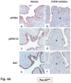
The inhibitor PD173074 (A) or PD184352 (B) was administered to one uterine horn of Hand2_d/d_ mice on day 3 of pregnancy (n=5). The other horn served as vehicle-treated control. Uterine horns were collected on day 4 morning and sections were subjected to IHC to detect p-FRS2, p-ERK1/2, and Ki67 (C) IHC of pERα and Muc-1 in uterine sections of Hand2_d/d_ mice treated with PD173074 or PD184352.
Comment in
- Cell biology. A hand to support the implantation window.
Hewitt SC, Korach KS. Hewitt SC, et al. Science. 2011 Feb 18;331(6019):863-4. doi: 10.1126/science.1202372. Science. 2011. PMID: 21330520 Free PMC article.
Similar articles
- Cell biology. A hand to support the implantation window.
Hewitt SC, Korach KS. Hewitt SC, et al. Science. 2011 Feb 18;331(6019):863-4. doi: 10.1126/science.1202372. Science. 2011. PMID: 21330520 Free PMC article. - Analysis of heart and neural crest derivatives-expressed protein 2 (HAND2)-progesterone interactions in peri-implantation endometrium†.
Šućurović S, Nikolić T, Brosens JJ, Mulac-Jeričević B. Šućurović S, et al. Biol Reprod. 2020 Apr 24;102(5):1111-1121. doi: 10.1093/biolre/ioaa013. Biol Reprod. 2020. PMID: 31982918 - Uterine Epithelial Estrogen Receptor-α Controls Decidualization via a Paracrine Mechanism.
Pawar S, Laws MJ, Bagchi IC, Bagchi MK. Pawar S, et al. Mol Endocrinol. 2015 Sep;29(9):1362-74. doi: 10.1210/me.2015-1142. Epub 2015 Aug 4. Mol Endocrinol. 2015. PMID: 26241389 Free PMC article. - Minireview: Steroid-regulated paracrine mechanisms controlling implantation.
Pawar S, Hantak AM, Bagchi IC, Bagchi MK. Pawar S, et al. Mol Endocrinol. 2014 Sep;28(9):1408-22. doi: 10.1210/me.2014-1074. Epub 2014 Jul 22. Mol Endocrinol. 2014. PMID: 25051170 Free PMC article. Review.
Cited by
- Dietary supplementation with N-acetyl-L-cysteine ameliorates hyperactivated ERK signaling in the endometrium that is linked to poor pregnancy outcomes following ovarian stimulation in pigs.
Cheng L, Shi Z, Yue Y, Wang Y, Qin Y, Zhao W, Hu Y, Li Q, Guo M, An L, Wang S, Tian J. Cheng L, et al. J Anim Sci Biotechnol. 2024 Nov 6;15(1):148. doi: 10.1186/s40104-024-01109-1. J Anim Sci Biotechnol. 2024. PMID: 39501409 Free PMC article. - Progesterone in frozen embryo transfer cycles: assays, circulating concentrations, metabolites, and molecular action.
Mandelbaum R, Stanczyk FZ. Mandelbaum R, et al. F S Rep. 2024 May 23;5(3):237-247. doi: 10.1016/j.xfre.2024.05.007. eCollection 2024 Sep. F S Rep. 2024. PMID: 39381665 Free PMC article. - Dysregulated miR-124-3p in endometrial epithelial cells reduces endometrial receptivity by altering polarity and adhesion.
Zhou W, Van Sinderen M, Rainczuk K, Menkhorst E, Sorby K, Osianlis T, Pangestu M, Santos L, Rombauts L, Rosello-Diez A, Dimitriadis E. Zhou W, et al. Proc Natl Acad Sci U S A. 2024 Oct 8;121(41):e2401071121. doi: 10.1073/pnas.2401071121. Epub 2024 Oct 4. Proc Natl Acad Sci U S A. 2024. PMID: 39365817 - DCAF2 is essential for the development of uterine epithelia and mouse fertility.
Yang M, Wang K, Zhang L, Zhang H, Zhang C. Yang M, et al. Front Cell Dev Biol. 2024 Sep 19;12:1474660. doi: 10.3389/fcell.2024.1474660. eCollection 2024. Front Cell Dev Biol. 2024. PMID: 39364135 Free PMC article. - Unraveling the Dynamics of Estrogen and Progesterone Signaling in the Endometrium: An Overview.
Dias Da Silva I, Wuidar V, Zielonka M, Pequeux C. Dias Da Silva I, et al. Cells. 2024 Jul 23;13(15):1236. doi: 10.3390/cells13151236. Cells. 2024. PMID: 39120268 Free PMC article. Review.
References
- Finn CA, Martin L. J Reprod Fert. 1974;39:195. - PubMed
- Carson DD, et al. Dev Biol. 2000;223:217. - PubMed
- Martin L, Das RM. J Endocrinol. 1973;57:549. - PubMed
- Bagchi IC, et al. Front Biosci. 2003;8:s852. - PubMed
Publication types
MeSH terms
Substances
LinkOut - more resources
Full Text Sources
Other Literature Sources
Molecular Biology Databases


