SCF(FBW7) regulates cellular apoptosis by targeting MCL1 for ubiquitylation and destruction - PubMed (original) (raw)
. 2011 Mar 3;471(7336):104-9.
doi: 10.1038/nature09732.
Shavali Shaik, Ichiro Onoyama, Daming Gao, Alan Tseng, Richard S Maser, Bo Zhai, Lixin Wan, Alejandro Gutierrez, Alan W Lau, Yonghong Xiao, Amanda L Christie, Jon Aster, Jeffrey Settleman, Steven P Gygi, Andrew L Kung, Thomas Look, Keiichi I Nakayama, Ronald A DePinho, Wenyi Wei
Affiliations
- PMID: 21368833
- PMCID: PMC3076007
- DOI: 10.1038/nature09732
SCF(FBW7) regulates cellular apoptosis by targeting MCL1 for ubiquitylation and destruction
Hiroyuki Inuzuka et al. Nature. 2011.
Abstract
The effective use of targeted therapy is highly dependent on the identification of responder patient populations. Loss of FBW7, which encodes a tumour-suppressor protein, is frequently found in various types of human cancer, including breast cancer, colon cancer and T-cell acute lymphoblastic leukaemia (T-ALL). In line with these genomic data, engineered deletion of Fbw7 in mouse T cells results in T-ALL, validating FBW7 as a T-ALL tumour suppressor. Determining the precise molecular mechanisms by which FBW7 exerts antitumour activity is an area of intensive investigation. These mechanisms are thought to relate in part to FBW7-mediated destruction of key proteins relevant to cancer, including Jun, Myc, cyclin E and notch 1 (ref. 9), all of which have oncoprotein activity and are overexpressed in various human cancers, including leukaemia. In addition to accelerating cell growth, overexpression of Jun, Myc or notch 1 can also induce programmed cell death. Thus, considerable uncertainty surrounds how FBW7-deficient cells evade cell death in the setting of upregulated Jun, Myc and/or notch 1. Here we show that the E3 ubiquitin ligase SCF(FBW7) (a SKP1-cullin-1-F-box complex that contains FBW7 as the F-box protein) governs cellular apoptosis by targeting MCL1, a pro-survival BCL2 family member, for ubiquitylation and destruction in a manner that depends on phosphorylation by glycogen synthase kinase 3. Human T-ALL cell lines showed a close relationship between FBW7 loss and MCL1 overexpression. Correspondingly, T-ALL cell lines with defective FBW7 are particularly sensitive to the multi-kinase inhibitor sorafenib but resistant to the BCL2 antagonist ABT-737. On the genetic level, FBW7 reconstitution or MCL1 depletion restores sensitivity to ABT-737, establishing MCL1 as a therapeutically relevant bypass survival mechanism that enables FBW7-deficient cells to evade apoptosis. Therefore, our work provides insight into the molecular mechanism of direct tumour suppression by FBW7 and has implications for the targeted treatment of patients with FBW7-deficient T-ALL.
Figures
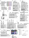
Figure 1. Mcl-1 stability is controlled by Fbw7
a, Sequence alignment of Mcl-1 with the c-Jun, c-Myc and Cyclin E Fbw7 phosphodegrons. The putative Fbw7 phosphodegron sequence present in Mcl-1 is conserved across different species. b–c, Immunoblot analysis of HeLa cells transfected with the indicated siRNA oligonucleotides. d, Immunoblot analysis of thymus cells derived from control mice or Fbw7 conditional knockout (Lck-Cre/_Fbw7_fl/fl) mice. Mcl-1 band intensity was normalized to Hsp90, then normalized to the control lane. Data was shown as mean ± SEM from three independent experiments. e, Immunoblot analysis of wild-type (WT) or _Fbw7_−/− DLD1 cells after synchronization with nocodazole and release at the indicated time points. f, Immunoblot analysis of the indicated human T-ALL cell lines. g, DND41 and Loucy cells, which contain wild-type Fbw7, were infected with the indicated lentiviral shRNA constructs and selected with 1 µg/ml puromycin to eliminate the non-infected cells. Cell lysates were collected for immunoblot analysis with the indicated antibodies. h, T-ALL cell lines with deficient Fbw7 were infected with Fbw7-expressing retrovirus construct (with empty vector as a negative control), and selected with 1 µg/ml puromycin to eliminate the non-infected cells. Cell lysates were collected for immunoblot analysis with the indicated antibodies. i, Immunoblot analysis of the indicated primary human T-ALL clinical samples. j. Immunoblot analysis of the indicated murine T-ALL cell lines derived from the Terc−/−Atm−/−p53−/− (TKO) mice. k–m. In vivo effects of Mcl-1 depletion in Fbw7-deficient T-ALL cells. (k) An in vivo model of Fbw7-deficient T-ALL was created by orthotopic engraftment of CMLT1-luciferase cells in NOD-SCID-IL2Rγnull (NSG) mice (left; CMLT1-shGFP, right; CMLT1-shMcl-1). (l) immunoblot analysis of the engineered CMLT1 cell lines. (m) Mice were injected with 1×10⁷ cells (n=7/group) via the lateral tail vein. Tumor burden was determined by quantification of total body luminescence, and are expressed as photons/second/standardized region of interest (ph/s/ROI). Data was represented as mean ± SEM with statistical significance determined by Student’s t-test.
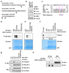
Figure 2. Phosphorylation of Mcl-1 by GSK3 triggers its interaction with Fbw7
a, In vivo Mcl-1 phosphorylation sites detected by mass spectrum analysis. b, Immunoblot analysis of HeLa cells transfected with the indicated siRNA oligonucleotides. c, Illustration of the various Mcl-1 mutants generated for this study. d–e, GSK3 phosphorylates Mcl-1 in vitro at multiple sites. Purified GSK3 protein (from New England Biolabs) was incubated with 5 µg of the indicated GST-Mcl-1 proteins in the presence of γ-32P-ATP. The kinase reaction products were resolved by SDS-PAGE and phosphorylation was detected by autoradiography. f, Phosphorylation of Mcl-1 at multiple sites by GSK3 triggers its interaction with Fbw7 in vitro. Autoradiograms showing recovery of 35S-labeled Fbw7 protein bound to the indicated GST-Mcl-1 fusion proteins (GST protein as a negative control) incubated with GSK3 prior to the pull-down assays. IN, input (5% as indicated). g, Immunoblot (IB) analysis of whole cell lysates (WCL) and immunoprecipitates (IP) derived from 293T cells transfected with HA-Fbw7 together with the indicated Myc-Mcl-1 constructs. Thirty hours post-transfection, cells were pretreated with 10 µM MG132 for 10 hours to block the proteasome pathway before harvesting. h, Immunoblot (IB) analysis of whole cell lysates (WCL) and immunoprecipitates (IP) derived from 293T cells transfected with HA-Fbw7. Thirty hours post-transfection, cells were pretreated with 20 µM MG132 for 8 hours to block the proteasome pathway before harvesting. Where indicated, 25 µM of the GSK3β inhibitor VIII (with DMSO as a negative control) was added for 8 hours before harvesting.
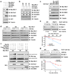
Figure 3. Fbw7 promotes Mcl-1 ubiquitination and destruction in a GSK3 phosphorylation-dependent manner
a–c, GSK3 phosphorylation-dependent degradation of Mcl-1 by Fbw7. Immunoblot analysis of 293T cells transfected with the indicated Myc-Mcl-1 and HA-Fbw7 plasmids in the presence or absence of HA-GSK3. A plasmid encoding GFP was used as a negative control for transfection efficiency. Where indicated, the proteasome inhibitor MG132 was added. d, 293T cells were transfected with the indicated Myc-Mcl-1 constructs together with the HA-Fbw7 and HA-GSK3 plasmids. Twenty hours post-transfection, cells were split into 60 mm dishes, and after another 20 hours, treated with 20 µg/ml cycloheximide (CHX). At the indicated time points, whole cell lysates were prepared and immunoblots were probed with the indicated antibodies. e, Wild-type (WT) or _Fbw7_−/− DLD1 cells were treated with 20 µg/ml cycloheximide (CHX). At the indicated time points, whole cell lysates were prepared and immunoblots were probed with the indicated antibodies. Mcl-1 band intensity was normalized to tubulin, then normalized to the t=0 controls. f, Immunoblot analysis (IB) of whole cell lysates (WCL) and His-tag pull-down of HeLa cells transfected with the indicated plasmids. Twenty hours post-transfection, cells were treated with the proteasome inhibitor MG132 overnight before harvesting. His-tag pull-down was performed in the presence of 8 M urea to eliminate any possible contamination from Mcl-1-associated proteins. g, Immunoblot analysis of wild-type (WT) or _Fbw7_−/− DLD1 cells treated with 10 µM adriamycin (ADR) for the indicated durations of time. Mcl-1 band intensity was normalized to tubulin, then normalized to the t=0 controls.
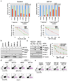
Figure 4. Elevated Mcl-1 expression protects Fbw7-deficient T-ALL cell lines from ABT-737-induced apoptosis
a, Cell viability assays showing that Fbw7-deficient T-ALL cell lines were more sensitive to sorafenib, but resistant to ABT-737 treatment. T-ALL cells were cultured in 10% FBS-containing medium with the indicated concentrations of sorafenib or ABT-737 for 48 hours before performing the cell viability assays. Data was shown as mean ± SD for three independent experiments. b, Immunoblot analysis of the indicated human T-ALL cell lines with or without ABT-737 (0.8 µM) treatment. c, Specific depletion of endogenous Mcl-1 expression restored ABT-737 sensitivity in the indicated Fbw7-deficient T-ALL cell lines. Various T-ALL cells were cultured in 10% FBS-containing medium with the indicated concentrations of ABT-737 for 48 hours before performing the cell viability assays, or with or without ABT-737 (0.8 µM) treatment for 24 hours before collecting whole cell lysates for immunoblot analysis with the indicated antibodies. For cell viability assays, data was shown as mean ± SD for three independent experiments. d, 7-Amino-Actinomycin D (7-AAD)/Annexin V double-staining FACS analysis to detect the percentage of ABT-737-induced apoptosis in the indicated Fbw7-deficient T-ALL cell lines where the endogenous Mcl-1 was depleted by lentiviral shRNA treatment (lentiviral shGFP was used as a negative control). Various T-ALL cells were cultured in 10% FBS-containing medium with or without ABT-737 (0.8 µM) treatment for 48 hours before the FACS analysis. Numbers indicate the percentage of apoptotic cells. e, 7-AAD/Annexin V double-staining FACS analysis to demonstrate that sorafenib treatment restores ABT-737 sensitivity to Fbw7-deficient HPB-ALL cells. HPB-ALL cells were cultured in 10% FBS-containing medium with the indicated concentrations of sorafenib and/or ABT-737 for 48 hours before the FACS analysis. Numbers indicate the percentage of apoptotic cells.
Similar articles
- Regulation of GATA-binding protein 2 levels via ubiquitin-dependent degradation by Fbw7: involvement of cyclin B-cyclin-dependent kinase 1-mediated phosphorylation of THR176 in GATA-binding protein 2.
Nakajima T, Kitagawa K, Ohhata T, Sakai S, Uchida C, Shibata K, Minegishi N, Yumimoto K, Nakayama KI, Masumoto K, Katou F, Niida H, Kitagawa M. Nakajima T, et al. J Biol Chem. 2015 Apr 17;290(16):10368-81. doi: 10.1074/jbc.M114.613018. Epub 2015 Feb 10. J Biol Chem. 2015. PMID: 25670854 Free PMC article. - Mcl-1 ubiquitination and destruction.
Inuzuka H, Fukushima H, Shaik S, Liu P, Lau AW, Wei W. Inuzuka H, et al. Oncotarget. 2011 Mar;2(3):239-44. doi: 10.18632/oncotarget.242. Oncotarget. 2011. PMID: 21608150 Free PMC article. Review. - The two faces of FBW7 in cancer drug resistance.
Wang Z, Fukushima H, Gao D, Inuzuka H, Wan L, Lau AW, Liu P, Wei W. Wang Z, et al. Bioessays. 2011 Nov;33(11):851-9. doi: 10.1002/bies.201100101. Epub 2011 Aug 30. Bioessays. 2011. PMID: 22006825 Free PMC article. Review. - Sensitivity to antitubulin chemotherapeutics is regulated by MCL1 and FBW7.
Wertz IE, Kusam S, Lam C, Okamoto T, Sandoval W, Anderson DJ, Helgason E, Ernst JA, Eby M, Liu J, Belmont LD, Kaminker JS, O'Rourke KM, Pujara K, Kohli PB, Johnson AR, Chiu ML, Lill JR, Jackson PK, Fairbrother WJ, Seshagiri S, Ludlam MJ, Leong KG, Dueber EC, Maecker H, Huang DC, Dixit VM. Wertz IE, et al. Nature. 2011 Mar 3;471(7336):110-4. doi: 10.1038/nature09779. Nature. 2011. PMID: 21368834 - The Fbw7 tumor suppressor regulates glycogen synthase kinase 3 phosphorylation-dependent c-Myc protein degradation.
Welcker M, Orian A, Jin J, Grim JE, Harper JW, Eisenman RN, Clurman BE. Welcker M, et al. Proc Natl Acad Sci U S A. 2004 Jun 15;101(24):9085-90. doi: 10.1073/pnas.0402770101. Epub 2004 May 18. Proc Natl Acad Sci U S A. 2004. PMID: 15150404 Free PMC article.
Cited by
- A novel small-form NEDD4 regulates cell invasiveness and apoptosis to promote tumor metastasis.
Liao CJ, Chi HC, Tsai CY, Chen CD, Wu SM, Tseng YH, Lin YH, Chung IH, Chen CY, Lin SL, Huang SF, Huang YH, Lin KH. Liao CJ, et al. Oncotarget. 2015 Apr 20;6(11):9341-54. doi: 10.18632/oncotarget.3322. Oncotarget. 2015. PMID: 25823820 Free PMC article. - EGF-mediated induction of Mcl-1 at the switch to lactation is essential for alveolar cell survival.
Fu NY, Rios AC, Pal B, Soetanto R, Lun AT, Liu K, Beck T, Best SA, Vaillant F, Bouillet P, Strasser A, Preiss T, Smyth GK, Lindeman GJ, Visvader JE. Fu NY, et al. Nat Cell Biol. 2015 Apr;17(4):365-75. doi: 10.1038/ncb3117. Epub 2015 Mar 2. Nat Cell Biol. 2015. PMID: 25730472 - FBXW7 Reduces the Cancer Stem Cell-Like Properties of Hepatocellular Carcinoma by Regulating the Ubiquitination and Degradation of ACTL6A.
Wang X, Li Y, Li Y, Liu P, Liu S, Pan Y. Wang X, et al. Stem Cells Int. 2022 Sep 14;2022:3242482. doi: 10.1155/2022/3242482. eCollection 2022. Stem Cells Int. 2022. PMID: 36159747 Free PMC article. - Clonal architecture in mesothelioma is prognostic and shapes the tumour microenvironment.
Zhang M, Luo JL, Sun Q, Harber J, Dawson AG, Nakas A, Busacca S, Sharkey AJ, Waller D, Sheaff MT, Richards C, Wells-Jordan P, Gaba A, Poile C, Baitei EY, Bzura A, Dzialo J, Jama M, Le Quesne J, Bajaj A, Martinson L, Shaw JA, Pritchard C, Kamata T, Kuse N, Brannan L, De Philip Zhang P, Yang H, Griffiths G, Wilson G, Swanton C, Dudbridge F, Hollox EJ, Fennell DA. Zhang M, et al. Nat Commun. 2021 Mar 19;12(1):1751. doi: 10.1038/s41467-021-21798-w. Nat Commun. 2021. PMID: 33741915 Free PMC article. - Regulation of GATA-binding protein 2 levels via ubiquitin-dependent degradation by Fbw7: involvement of cyclin B-cyclin-dependent kinase 1-mediated phosphorylation of THR176 in GATA-binding protein 2.
Nakajima T, Kitagawa K, Ohhata T, Sakai S, Uchida C, Shibata K, Minegishi N, Yumimoto K, Nakayama KI, Masumoto K, Katou F, Niida H, Kitagawa M. Nakajima T, et al. J Biol Chem. 2015 Apr 17;290(16):10368-81. doi: 10.1074/jbc.M114.613018. Epub 2015 Feb 10. J Biol Chem. 2015. PMID: 25670854 Free PMC article.
References
- Wood LD, et al. The genomic landscapes of human breast and colorectal cancers. Science. 2007;318:1108–1113. - PubMed
Publication types
MeSH terms
Substances
Grants and funding
- R01 GM089763/GM/NIGMS NIH HHS/United States
- R01 GM089763-01/GM/NIGMS NIH HHS/United States
- R01 GM089763-02/GM/NIGMS NIH HHS/United States
- GM089763/GM/NIGMS NIH HHS/United States
LinkOut - more resources
Full Text Sources
Other Literature Sources
Molecular Biology Databases
Miscellaneous


