Variable reliability of endoscopic findings with white-light and narrow-band imaging for patients with suspected eosinophilic esophagitis - PubMed (original) (raw)
Variable reliability of endoscopic findings with white-light and narrow-band imaging for patients with suspected eosinophilic esophagitis
Anne F Peery et al. Clin Gastroenterol Hepatol. 2011 Jun.
Abstract
Background & aims: Endoscopic findings have been used to support a diagnosis of eosinophilic esophagitis (EoE) and to assess response to therapy, but their reliability is unknown. The aim of the study was to assess inter- and intraobserver reliability of endoscopic findings with white-light endoscopy and to assess changes in interobserver reliability when narrow band imaging (NBI) was added to white light.
Methods: We collected data from 35 academic and 42 community adult gastroenterologists using 2 self-administered, online assessments of endoscopic images in patients with suspected EoE. First, gastroenterologists evaluated 35 single white light images. Next, they examined 35 paired images of the initial white light image and its NBI counterpart. To assess intraobserver reliability, a second survey to re-examine the single white light images was performed ≥2 weeks later. Agreement was determined by calculating κ values for multiple observers.
Results: Among all gastroenterologists, interobserver agreement was fair to good when white light was used to identify rings (κ = 0.56) and furrows (κ = 0.48). Interobserver agreement was poor for identification of plaques (κ = 0.29) and for images with no findings (κ = 0.34). Levels of agreement did not change in an analysis stratified by practice setting or patient volume. Agreement did not improve when NBI images were added to white light images. Levels of intraobserver agreement varied greatly and in some cases were not greater than those expected by chance.
Conclusions: Using white light endoscopy and NBI to analyze EoE, gastroenterologists identified rings and furrows with fair to good reliability, but did not reliably identify plaques or normal images. Intraobserver agreement varied. Endoscopic findings might not be reliable for supporting a diagnosis of EoE or for making treatment decisions.
Copyright © 2011 AGA Institute. Published by Elsevier Inc. All rights reserved.
Conflict of interest statement
Disclosures: No conflicts of interest exist for any author
Figures
Figure 1
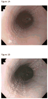
(A) Endoscopic image in white light showing linear furrows, white plaques, and subtle rings. (B) The corresponding narrowing band image of the same endoscopic findings.
Figure 2
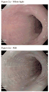
(A) Example of images for which there was good inter-observer agreement, with 88% of respondents identifying furrows, 9% no findings, 3% rings, and 1% plaques (endoscopic images in white light and NBI, respectively). (B) Example of images for which there was poor inter-observer agreement with 10% of respondents indentifying rings, 27% furrows, 43% plaques and 43% no findings (endoscopic images in white light and NBI, respectively).
Figure 3
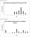
Histograms displaying the ranges of kappas for intra-observer reliability of EoE findings in white light for rings, furrows, plaques, and no findings.
Figure 3
Histograms displaying the ranges of kappas for intra-observer reliability of EoE findings in white light for rings, furrows, plaques, and no findings.
Similar articles
- Evaluating the endoscopic reference score for eosinophilic esophagitis: moderate to substantial intra- and interobserver reliability.
van Rhijn BD, Warners MJ, Curvers WL, van Lent AU, Bekkali NL, Takkenberg RB, Kloek JJ, Bergman JJ, Fockens P, Bredenoord AJ. van Rhijn BD, et al. Endoscopy. 2014 Dec;46(12):1049-55. doi: 10.1055/s-0034-1377781. Epub 2014 Sep 10. Endoscopy. 2014. PMID: 25208033 - Narrow band imaging improves observer reliability in evaluation of upper aerodigestive tract lesions.
Zwakenberg MA, Dikkers FG, Wedman J, Halmos GB, van der Laan BF, Plaat BE. Zwakenberg MA, et al. Laryngoscope. 2016 Oct;126(10):2276-81. doi: 10.1002/lary.26008. Epub 2016 Apr 14. Laryngoscope. 2016. PMID: 27074877 - Endoscopic assessment of the oesophageal features of eosinophilic oesophagitis: validation of a novel classification and grading system.
Hirano I, Moy N, Heckman MG, Thomas CS, Gonsalves N, Achem SR. Hirano I, et al. Gut. 2013 Apr;62(4):489-95. doi: 10.1136/gutjnl-2011-301817. Epub 2012 May 22. Gut. 2013. PMID: 22619364 - Clinical features of eosinophilic esophagitis.
Miehlke S. Miehlke S. Dig Dis. 2014;32(1-2):61-7. doi: 10.1159/000357011. Epub 2014 Feb 28. Dig Dis. 2014. PMID: 24603382 Review. - The role of endoscopy in eosinophilic esophagitis: from diagnosis to therapy.
Lucendo AJ, Arias Á, Molina-Infante J, Arias-González L. Lucendo AJ, et al. Expert Rev Gastroenterol Hepatol. 2017 Dec;11(12):1135-1149. doi: 10.1080/17474124.2017.1367664. Epub 2017 Aug 17. Expert Rev Gastroenterol Hepatol. 2017. PMID: 28803528 Review.
Cited by
- Endoscopic Diagnosis of Eosinophilic Esophagitis: Basics and Recent Advances.
Abe Y, Sasaki Y, Yagi M, Mizumoto N, Onozato Y, Umehara M, Ueno Y. Abe Y, et al. Diagnostics (Basel). 2022 Dec 16;12(12):3202. doi: 10.3390/diagnostics12123202. Diagnostics (Basel). 2022. PMID: 36553209 Free PMC article. Review. - Mucosal color changes on narrow-band imaging in esophageal eosinophilic infiltration.
Suda T, Shirota Y, Hodo Y, Sato K, Wakabayashi T. Suda T, et al. Medicine (Baltimore). 2022 Sep 23;101(38):e29891. doi: 10.1097/MD.0000000000029891. Medicine (Baltimore). 2022. PMID: 36197201 Free PMC article. - Linked color imaging improves the diagnostic accuracy of eosinophilic esophagitis.
Abe Y, Sasaki Y, Yagi M, Mizumoto N, Onozato Y, Kon T, Shoji M, Sakuta K, Sakai T, Umehara M, Ito M, Nakamura S, Tsuchida H, Ueno Y. Abe Y, et al. DEN Open. 2022 Jul 25;3(1):e146. doi: 10.1002/deo2.146. eCollection 2023 Apr. DEN Open. 2022. PMID: 35898847 Free PMC article. - Beige mucosa observable under narrow-band imaging indicates the active sites of eosinophilic esophagitis.
Ayaki M, Manabe N, Tomida A, Tada N, Matsunaga T, Murota M, Fujita M, Katsumata R, Kobara H, Masaki T, Haruma K. Ayaki M, et al. J Gastroenterol Hepatol. 2022 May;37(5):891-897. doi: 10.1111/jgh.15808. Epub 2022 Mar 9. J Gastroenterol Hepatol. 2022. PMID: 35229352 Free PMC article. - The Yield of Endoscopy and Histology in the Evaluation of Esophageal Dysphagia: Two Referral Centers' Experiences.
Mari A, Abu Baker F, Said Ahmad H, Omari A, Jawabreh Y, Abboud R, Shahin A, Shibli F, Sbeit W, Khoury T. Mari A, et al. Medicina (Kaunas). 2021 Dec 7;57(12):1336. doi: 10.3390/medicina57121336. Medicina (Kaunas). 2021. PMID: 34946281 Free PMC article.
References
- Furuta GT, Liacouras CA, Collins MH, et al. Eosinophilic esophagitis in children and adults: a systematic review and consensus recommendations for diagnosis and treatment. Gastroenterology. 2007 Oct;133(4):1342–1363. - PubMed
- Noel RJ, Putnam PE, Rothenberg ME. Eosinophilic esophagitis. N Engl J Med. 2004 Aug 26;351(9):940–941. - PubMed
- Straumann A, Simon HU. Eosinophilic esophagitis: escalating epidemiology? J Allergy Clin Immunol. 2005 Feb;115(2):418–419. - PubMed
Publication types
MeSH terms
Grants and funding
- T32 DK 07634/DK/NIDDK NIH HHS/United States
- KL2 TR001109/TR/NCATS NIH HHS/United States
- KL2 RR025746-04/RR/NCRR NIH HHS/United States
- T32 DK007634-22/DK/NIDDK NIH HHS/United States
- T32 DK007634/DK/NIDDK NIH HHS/United States
- KL2 RR025746/RR/NCRR NIH HHS/United States
- UL1RR025747/RR/NCRR NIH HHS/United States
- UL1 RR025747/RR/NCRR NIH HHS/United States
- KL2RR025746/RR/NCRR NIH HHS/United States
- UL1 RR025747-04/RR/NCRR NIH HHS/United States
LinkOut - more resources
Full Text Sources
Other Literature Sources
Medical


