Structure of the HECT:ubiquitin complex and its role in ubiquitin chain elongation - PubMed (original) (raw)
Structure of the HECT:ubiquitin complex and its role in ubiquitin chain elongation
Elena Maspero et al. EMBO Rep. 2011 Apr.
Abstract
Several mechanisms have been proposed for the synthesis of substrate-linked ubiquitin chains. HECT ligases directly catalyse protein ubiquitination and have been found to non-covalently interact with ubiquitin. We report crystal structures of the Nedd4 HECT domain, alone and in complex with ubiquitin, which show a new binding mode involving two surfaces on ubiquitin and both subdomains of the HECT N-lobe. The structures suggest a model for HECT-to-substrate ubiquitin transfer, in which the growing chain on the substrate is kept close to the catalytic cysteine to promote processivity. Mutational analysis highlights differences between the processes of substrate polyubiquitination and self-ubiquitination.
Conflict of interest statement
The authors declare that they have no conflict of interest.
Figures
Figure 1
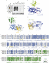
Structure of the HECTNedd4 domain in apo form and in complex with ubiquitin. (A) GST pull-down assay with the HECT domains of various Nedd4 family HECT E3 ligases. GST-fusion proteins were incubated for 2 h at 4°C in YY buffer with synthetic Lys 63-polyubiquitin chains and analysed by IB as indicated. Coomassie staining shows comparable loading of GST proteins. Similar results were obtained with linear and Lys 48-polyubiquitin chains (not shown). (B) Overall structure of HECTNedd4 (N-lobe, blue; C-lobe, green). The red dotted line indicates the boundary between the large and small subdomains of the N-lobe. (C) Overall structure of HECTNedd4 in complex with ubiquitin (yellow). The HECT structure is represented in the same orientation as in B; N-lobe, light blue; C-lobe, dark green. (D) Superposition on the large subdomain of the N-lobe of HECTNedd4 and HECTNedd4:ubiquitin. In the complex (light blue), the β5–β6 hairpin of the small subdomain of the N-lobe is closer to the large subdomain, with respect to the isolated HECT (dark blue). (E) Sequence alignment of the HECTNedd4 domain with other crystallized HECT domains. Secondary structure elements are depicted. Dotted line indicates that the residues were not visible in the electron density maps. Yellow circles indicate residues in contact with ubiquitin in the structure of HECTNedd4:ubiquitin (according to PISA; Krissinel & Henrick, 2007). Numbering refers to Nedd4 sequence. GST, glutathione _S_-transferase; IB, immunoblotting; Ub, ubiquitin.
Figure 2
HECTNedd4:ubiquitin interaction and mutant validation. (A) Close-up view of HECTNedd4 N-lobe:ubiquitin interaction. (B) GST pull-down assay with the indicated Nedd4 constructs and Lys 63-linked polyubiquitin chains was performed as described in Fig 1A. (C) Fluorescence-polarization assay with the indicated Nedd4 constructs and monomeric ubiquitin was performed. The HECTNedd4:ubiquitin interaction displays a moderate affinity with a _K_D of 11 μM, F707A mutant displays a thirty times lower affinity. Details are described in the supplementary Methods online and similar results for the Y605A mutant obtained by SPR assay are in supplementary Fig S1 online. IB, immmunoblotting; GST, glutathione _S_-transferase; SPR, surface plasmon resonance; Ub, ubiquitin; WT, wild type.
Figure 3
Disruption of HECTNedd4:ubiquitin interaction impairs substrate polyubiquitination. (A) Mutations do not affect E2 binding. Left panel: GST pull-down assay with the indicated HECT mutants and the E2 enzyme Ube2D3. IB was performed as indicated. Coomassie staining shows comparable loading of GST proteins. Right panel: the HECTNedd4:Ube2D3 interaction displays a modest affinity, that is not perturbed by the F707A and Y605A mutations. SPR assay was performed as described in the supplementary methods online. (B) Mutations do not affect the kinetics of the E2-to-HECT transthiolation process. The transfer of ubiquitin was monitored by quenching the reaction at different time points, with the addition of Laemmli buffer with or without the reducing agent (100 mM DTT). Arrow indicates thioesther HECT∼ubiquitin (−DTT) or monoubiquitinated HECT (+DTT) running at the same position. DTT-resistant higher molecular bands represent self-ubiquitinated HECT. Similar results were obtained with Y605A mutant. (C) Mutations impair substrate polyubiquitination. Upper panel: GST-γENaC ubiquitination kinetics with WT HECT and F707A mutant (ubiquitin (pellet)). Middle panel: Coomassie staining showing comparable loading of GST proteins. Lower panel: kinetics of free ubiquitin chain formation (ubiquitin (supernatant)) during the reaction. IB was performed as indicated. (D) Self-ubiquitination kinetics with WT HECT and Y605A and F707A mutants. IB was performed as indicated. Coomassie staining shows comparable loading of HECT proteins. Similar results were obtained with full-length Nedd4 mutants. DTT, dithiothreitol; ENaC, epithelial Na+ channel; GST, glutathione _S_-transferase; IB, immunoblotting; SPR, surface plasmon resonance; Ub, ubiquitin; WT, wild type.
Figure 4
Ubiquitin binding to the HECTNedd4 domain is compatible with HECTNedd4-like:E2∼ubiquitin complex and does not dictate chain specificity. (A) Substrate ubiquitination assay with the indicated ubiquitin KR mutants was performed. The reaction was quenched after 30 min for the WT HECT and after 60 min for the F707A mutant. Upper panel: GST-γENaC ubiquitination with the indicated constructs. Middle panel: Coomassie staining showing comparable loading of GST proteins. Lower panel: free ubiquitin chain formation during the reaction. IB was performed as indicated. Lower panel: 1 μg of ubiquitin KR mutants were loaded for comparison and visualized by Coomassie staining. (B) Position of ubiquitin lysines in the Nedd4 HECT/ubiquitin complex. HECT N-lobe is shown as surface representation, whereas the C-lobe and ubiquitin are shown as cartoon representations. Ubiquitin lysine side chains are indicated in sticks. Six of the seven ubiquitin lysines are shown, K 6 being in the back. (C) Model of Ubch5B∼Ub:C-lobe complex (Kamadurai et al, 2009) binding to the N-lobe:ubiquitin complex. Details are in the supplementary Methods online. C 867 on the HECT and K 63 on the Ub are shown. ENaC, epithelial Na+ channel; GST, glutathione _S_-transferase; IB, immunoblotting; KR, lysine-to-arginine mutation; Ub, ubiquitin; WT, wild type.
Similar articles
- Insights into ubiquitin transfer cascades from a structure of a UbcH5B approximately ubiquitin-HECT(NEDD4L) complex.
Kamadurai HB, Souphron J, Scott DC, Duda DM, Miller DJ, Stringer D, Piper RC, Schulman BA. Kamadurai HB, et al. Mol Cell. 2009 Dec 25;36(6):1095-102. doi: 10.1016/j.molcel.2009.11.010. Mol Cell. 2009. PMID: 20064473 Free PMC article. - Rescue of HIV-1 release by targeting widely divergent NEDD4-type ubiquitin ligases and isolated catalytic HECT domains to Gag.
Weiss ER, Popova E, Yamanaka H, Kim HC, Huibregtse JM, Göttlinger H. Weiss ER, et al. PLoS Pathog. 2010 Sep 16;6(9):e1001107. doi: 10.1371/journal.ppat.1001107. PLoS Pathog. 2010. PMID: 20862313 Free PMC article. - Polyubiquitination by HECT E3s and the determinants of chain type specificity.
Kim HC, Huibregtse JM. Kim HC, et al. Mol Cell Biol. 2009 Jun;29(12):3307-18. doi: 10.1128/MCB.00240-09. Epub 2009 Apr 13. Mol Cell Biol. 2009. PMID: 19364824 Free PMC article. - Physiological Functions of the Ubiquitin Ligases Nedd4-1 and Nedd4-2.
Rotin D, Prag G. Rotin D, et al. Physiology (Bethesda). 2024 Jan 1;39(1):18-29. doi: 10.1152/physiol.00023.2023. Epub 2023 Nov 14. Physiology (Bethesda). 2024. PMID: 37962894 Review. - NEDD4 E3 Ligases: Functions and Mechanisms in Bone and Tooth.
Xu K, Chu Y, Liu Q, Fan W, He H, Huang F. Xu K, et al. Int J Mol Sci. 2022 Sep 1;23(17):9937. doi: 10.3390/ijms23179937. Int J Mol Sci. 2022. PMID: 36077334 Free PMC article. Review.
Cited by
- HECT E3 Ligases: A Tale With Multiple Facets.
Weber J, Polo S, Maspero E. Weber J, et al. Front Physiol. 2019 Apr 3;10:370. doi: 10.3389/fphys.2019.00370. eCollection 2019. Front Physiol. 2019. PMID: 31001145 Free PMC article. Review. - Versatile roles of k63-linked ubiquitin chains in trafficking.
Erpapazoglou Z, Walker O, Haguenauer-Tsapis R. Erpapazoglou Z, et al. Cells. 2014 Nov 12;3(4):1027-88. doi: 10.3390/cells3041027. Cells. 2014. PMID: 25396681 Free PMC article. Review. - Cryo-EM structure of the chain-elongating E3 ubiquitin ligase UBR5.
Hodáková Z, Grishkovskaya I, Brunner HL, Bolhuis DL, Belačić K, Schleiffer A, Kotisch H, Brown NG, Haselbach D. Hodáková Z, et al. EMBO J. 2023 Aug 15;42(16):e113348. doi: 10.15252/embj.2022113348. Epub 2023 Jul 6. EMBO J. 2023. PMID: 37409633 Free PMC article. - E6AP/UBE3A ubiquitin ligase harbors two E2~ubiquitin binding sites.
Ronchi VP, Klein JM, Haas AL. Ronchi VP, et al. J Biol Chem. 2013 Apr 12;288(15):10349-60. doi: 10.1074/jbc.M113.458059. Epub 2013 Feb 25. J Biol Chem. 2013. PMID: 23439649 Free PMC article. - BIRC7-E2 ubiquitin conjugate structure reveals the mechanism of ubiquitin transfer by a RING dimer.
Dou H, Buetow L, Sibbet GJ, Cameron K, Huang DT. Dou H, et al. Nat Struct Mol Biol. 2012 Sep;19(9):876-83. doi: 10.1038/nsmb.2379. Epub 2012 Aug 14. Nat Struct Mol Biol. 2012. PMID: 22902369 Free PMC article.
References
- Brunger AT (2007) Version 1.2 of the Crystallography and NMR system. Nat Protoc 2: 2728–2733 - PubMed
- CCP4 (1994) The CCP4 suite: programs for protein crystallography. Acta Crystallogr D Biol Crystallogr 50: 760–763 - PubMed
- Dye BT, Schulman BA (2007) Structural mechanisms underlying posttranslational modification by ubiquitin-like proteins. Annu Rev Biophys Biomol Struct 36: 131–150 - PubMed
Publication types
MeSH terms
Substances
LinkOut - more resources
Full Text Sources
Other Literature Sources
Molecular Biology Databases


