Episodic memory retrieval, parietal cortex, and the default mode network: functional and topographic analyses - PubMed (original) (raw)
Episodic memory retrieval, parietal cortex, and the default mode network: functional and topographic analyses
Carlo Sestieri et al. J Neurosci. 2011.
Abstract
The default mode network (DMN) is often considered a functionally homogeneous system that is broadly associated with internally directed cognition (e.g., episodic memory, theory of mind, self-evaluation). However, few studies have examined how this network interacts with other networks during putative "default" processes such as episodic memory retrieval. Using functional magnetic resonance imaging, we investigated the topography and response profile of human parietal regions inside and outside the DMN, independently defined using task-evoked deactivations and resting-state functional connectivity, during episodic memory retrieval. Memory retrieval activated posterior nodes of the DMN, particularly the angular gyrus, but also more anterior and dorsal parietal regions that were anatomically separate from the DMN. The two sets of parietal regions showed different resting-state functional connectivity and response profiles. During memory retrieval, responses in DMN regions peaked sooner than non-DMN regions, which in turn showed responses that were sustained until a final memory judgment was reached. Moreover, a parahippocampal region that showed strong resting-state connectivity with parietal DMN regions also exhibited a pattern of task-evoked activity similar to that exhibited by DMN regions. These results suggest that DMN parietal regions directly supported memory retrieval, whereas non-DMN parietal regions were more involved in postretrieval processes such as memory-based decision making. Finally, a robust functional dissociation within the DMN was observed. Whereas angular gyrus and posterior cingulate/precuneus were significantly activated during memory retrieval, an anterior DMN node in medial prefrontal cortex was strongly deactivated. This latter finding demonstrates functional heterogeneity rather than homogeneity within the DMN during episodic memory retrieval.
Figures
Figure 1.
Perceptual search and episodic memory search tasks. a, Trial structure in the perceptual search task. A sentence instructed subjects to search for a specific target (object or character) that could appear at any time in the upcoming 12 s video clip. Subjects searched for the target while fixating a central cross and pressed a button as soon as the target was detected. Search duration was varied (early, middle, late) by manipulating the time at which the target was presented. After display offset, a variable ITI was interposed before the onset of the next sentence. b, Trial structure in the episodic memory search task. Subjects read a sentence describing a specific detail of a previously encoded episode from a TV show. They then retrieved information from episodic memory to judge the accuracy (i.e., true, false) of the sentence, which they indicated by pressing one of two buttons. Subjects were given up to 15 s to provide the judgment on each trial. An example of early, middle, and late search trials are provided. After subjects' response, a variable ITI was interposed before the onset of the next sentence.
Figure 2.
Definition of the DMN. a, Multiple-comparison corrected group _z_-map showing search-related BOLD deactivations (light to dark blue colors) with respect to the baseline in the perceptual search task. The search parameter was obtained using Search-Hit-HC trials (process GLM) (see Materials and Methods). The voxelwise map is superimposed over the lateral inflated, the medial inflated, and the flat representation of both hemisphere of the PALS Atlas [Caret 5.5 software (Van Essen, 2005)]. The dark circles represent the peaks of deactivations that were used as seeds for the functional connectivity analysis. Seeds in PCC and mPFC are presented in both hemisphere since they were close (1 mm) to the midline. b, Frequency map that represents voxels showing functional connectivity with the seed regions obtained from the perceptual task. For each voxel, the color represents significant resting-state positive correlations with one (blue), two (green), three (orange), or four (red) seeds. The DMN was defined as those voxels showing functional connectivity with at least three of four seeds (regions surrounded by white borders). c, Group _z_-map showing how connectivity varies within the regions that are defined as being part of the DMN. The map represents the mean z score of the rs-fcMRI maps obtained using the four seeds. The resulting map was masked by the border of the DMN (white).
Figure 3.
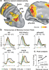
Profile of BOLD activity of intra- and extra-DMN regions during the memory task. a, Inflated representation of the left hemisphere showing regions of the lateral and medial parietal lobe centered on the peaks of positive memory search-related activity (process GLM). Regions (defined by black borders) are located either inside (intra-DMN: AG, PCC-PreCu) or outside (extra-DMN: aIPS, PoCS) the DMN (white borders) and are superimposed over the voxelwise map of positive search-related activity. A region of the medial temporal lobe with significant memory search-related activity (pParaHC) is also highlighted. b, Time courses of BOLD activity, aligned to the onset of the cue sentence (frame-by-frame, between-trial GLM), extracted from the five regions illustrated in a. Intra- and extra-DMN regions are located on the top and bottom rows, respectively. Time courses were grouped according to the retrieval time interval and refer to catch trials (gray), and early (1–4 s; green), middle (4–8 s; orange), and late (8–12 s; black) correct trials. The gray transparent vertical bar in each graph highlights the fifth and sixth frame (corresponding to ∼8–10 s after cue sentence onset) for comparison purpose. The horizontal line under the graphs illustrates the temporal duration of the corresponding retrieval intervals. c, The graph illustrates the relationship between peak latency of BOLD activity (frame of peak) and retrieval interval in extra-DMN regions (aIPS and PoCS; black line) compared with intra-DMN regions (AG and PCC-PreCu; gray line). Extra-DMN regions showed a greater increase in peak latency as subjects took more time to judge the correctness of cue sentences, indicating a more sustained involvement during retrieval.
Figure 4.
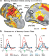
Sequence of BOLD responses from memory search to motor execution. a, Multiple-comparison corrected group _z_-map showing detection-related BOLD activations (red to yellow colors) with respect to the baseline in the memory search task. The detection parameter was obtained using all correct trials (process GLM) (see Materials and Methods). The DMN (white) and the parietal ROIs (black) are shown. An additional region [motor cortex (MC)] was created on the peak of detection-related response corresponding to the hand region of the motor cortex. b, Time courses of BOLD activity arranged so that different graphs represent the activity of different regions in each retrieval interval. The progressive involvement of different regions during the memory task is particularly evident in the late interval (right graph). Intra-DMN regions are presented in red (AG, PCC-PreCu), extra-DMN in light blue, pParaHC in gray, and motor cortex in black color. The green line and the letter “D” in each graph indicate the temporal interval in which detections occurred.
Figure 5.
Functional connectivity of intra- and extra-DMN parietal regions. a, Inflated representation of the left hemisphere showing the voxelwise rs-fcMRI statistical maps obtained in the same group of subjects using the left AG as seed. The group voxelwise _z_-map is shown for a one-sample, random-effects t test, corrected for multiple comparisons, of whether the Fisher _z_-transformed correlation values were different from zero. The black sphere represents the location of the 6 mm seed. b, c, Voxelwise rs-fcMRI statistical maps using PoCS and pParaHC gyrus as seeds, respectively. d, Cross-correlation matrix illustrating the correlation between pairs of regions among the sets of intra- and extra-DMN regions and the pParaHC region. The degree of correlation is color-coded so that green to blue colors represent increasing negative correlation and yellow to red colors represent increasing positive correlation.
Figure 6.
The relationship between the DMN and memory search-related activity. a, Whole-brain, multiple-comparison corrected group _z_-map showing search-related BOLD activations (red to yellow colors) and deactivations (light to dark blue colors) with respect to the baseline in the memory search task. The search parameter was obtained using Search-Corr-HC trials (process GLM) (see Materials and Methods). The borders of the DMN (in white) are superimposed over the activation map to reveal regions of overlap. b, Time courses of BOLD activity, aligned to the onset of the cue sentence (frame-by-frame, between-trial GLM), extracted from five regions showing significant positive and negative memory search-related responses that were located inside the DMN (defined by black borders on the flat map). Time courses were grouped according to the retrieval time interval and refer to catch trials (gray), and early (1–4 s; green), middle (4–8 s; orange), and late (8–12 s; black) correct trials. The horizontal line under the third graph illustrates the temporal duration of the corresponding retrieval intervals.
Figure 7.
BOLD-behavior relationship. BOLD percentage signal change for the search parameter (process GLM) corresponding to high (gray bar) and low (white bar) confidence correct trials in the memory task. These BOLD magnitudes were extracted from two posterior intra-DMN regions (AG, PCC; first and second graphs, respectively), two anterior intra-DMN regions (vmPFC, dmPFC; third and fourth graphs, respectively), and two parietal extra-DMN regions (aIPS, PoCS; fifth and sixth graphs, respectively). Error bars indicate SEM. The asterisks indicate the significance of the paired t test between high- and low-confidence correct search parameters.
Figure 8.
Functional organization of the left posterior parietal cortex. Inflated representation of the left hemisphere (lateral and medial views) showing parietal regions positively activated by memory (red color delimitated by dark red borders) and perceptual (green, delimitated by dark green borders) search as well as regions belonging to the DMN (blue, delimitated by dark blue borders), as defined by functional connectivity. Regions of overlap between memory search activity and the DMN are illustrated in purple color, whereas regions of overlap between memory and perceptual search are shown in yellow.
Similar articles
- Laterality effects in functional connectivity of the angular gyrus during rest and episodic retrieval.
Bellana B, Liu Z, Anderson JAE, Moscovitch M, Grady CL. Bellana B, et al. Neuropsychologia. 2016 Jan 8;80:24-34. doi: 10.1016/j.neuropsychologia.2015.11.004. Epub 2015 Nov 10. Neuropsychologia. 2016. PMID: 26559474 - Episodic Memory Retrieval Benefits from a Less Modular Brain Network Organization.
Westphal AJ, Wang S, Rissman J. Westphal AJ, et al. J Neurosci. 2017 Mar 29;37(13):3523-3531. doi: 10.1523/JNEUROSCI.2509-16.2017. Epub 2017 Feb 27. J Neurosci. 2017. PMID: 28242796 Free PMC article. - Functional heterogeneity in posterior parietal cortex across attention and episodic memory retrieval.
Hutchinson JB, Uncapher MR, Weiner KS, Bressler DW, Silver MA, Preston AR, Wagner AD. Hutchinson JB, et al. Cereb Cortex. 2014 Jan;24(1):49-66. doi: 10.1093/cercor/bhs278. Epub 2012 Sep 26. Cereb Cortex. 2014. PMID: 23019246 Free PMC article. - Top-down and bottom-up attention to memory: a hypothesis (AtoM) on the role of the posterior parietal cortex in memory retrieval.
Ciaramelli E, Grady CL, Moscovitch M. Ciaramelli E, et al. Neuropsychologia. 2008;46(7):1828-51. doi: 10.1016/j.neuropsychologia.2008.03.022. Epub 2008 Apr 8. Neuropsychologia. 2008. PMID: 18471837 Review. - Role of parietal regions in episodic memory retrieval: the dual attentional processes hypothesis.
Cabeza R. Cabeza R. Neuropsychologia. 2008;46(7):1813-27. doi: 10.1016/j.neuropsychologia.2008.03.019. Epub 2008 Apr 8. Neuropsychologia. 2008. PMID: 18439631 Free PMC article. Review.
Cited by
- From gut to brain: unveiling probiotic effects through a neuroimaging perspective-A systematic review of randomized controlled trials.
Crocetta A, Liloia D, Costa T, Duca S, Cauda F, Manuello J. Crocetta A, et al. Front Nutr. 2024 Sep 18;11:1446854. doi: 10.3389/fnut.2024.1446854. eCollection 2024. Front Nutr. 2024. PMID: 39360283 Free PMC article. - A Bayesian incorporated linear non-Gaussian acyclic model for multiple directed graph estimation to study brain emotion circuit development in adolescence.
Zhang A, Zhang G, Cai B, Wilson TW, Stephen JM, Calhoun VD, Wang YP. Zhang A, et al. Netw Neurosci. 2024 Oct 1;8(3):791-807. doi: 10.1162/netn_a_00384. eCollection 2024. Netw Neurosci. 2024. PMID: 39355441 Free PMC article. - Brain structural changes in diabetic retinopathy patients: a combined voxel-based morphometry and surface-based morphometry study.
Song Y, Xu T, Chen X, Wang N, Sun Z, Chen J, Xia J, Tian W. Song Y, et al. Brain Imaging Behav. 2024 Aug 22. doi: 10.1007/s11682-024-00905-7. Online ahead of print. Brain Imaging Behav. 2024. PMID: 39172355 - Neural signatures of default mode network subsystems in first-episode, drug-naive patients with major depressive disorder after 6-week thought induction psychotherapy treatment.
Lu F, Zhang J, Zhong Y, Hong L, Wang J, Du H, Fang J, Fan Y, Wang X, Yang Y, He Z, Jia C, Wang W, Lv X. Lu F, et al. Brain Commun. 2024 Aug 12;6(4):fcae263. doi: 10.1093/braincomms/fcae263. eCollection 2024. Brain Commun. 2024. PMID: 39171204 Free PMC article. - Cross-regional coordination of activity in the human brain during autobiographical self-referential processing.
Stieger JR, Pinheiro-Chagas P, Fang Y, Li J, Lusk Z, Perry CM, Girn M, Contreras D, Chen Q, Huguenard JR, Spreng RN, Edlow BL, Wagner AD, Buch V, Parvizi J. Stieger JR, et al. Proc Natl Acad Sci U S A. 2024 Aug 6;121(32):e2316021121. doi: 10.1073/pnas.2316021121. Epub 2024 Jul 30. Proc Natl Acad Sci U S A. 2024. PMID: 39078679
References
- Aguirre GK, Zarahn E, D'Esposito M. The inferential impact of global signal covariates in functional neuroimaging analyses. Neuroimage. 1998;8:302–306. - PubMed
- Amodio DM, Frith CD. Meeting of minds: the medial frontal cortex and social cognition. Nat Rev Neurosci. 2006;7:268–277. - PubMed
- Baddeley A. The episodic buffer: a new component of working memory? Trends Cogn Sci. 2000;4:417–423. - PubMed
Publication types
MeSH terms
Substances
LinkOut - more resources
Full Text Sources
Other Literature Sources


