Eosinophils sustain adipose alternatively activated macrophages associated with glucose homeostasis - PubMed (original) (raw)
Eosinophils sustain adipose alternatively activated macrophages associated with glucose homeostasis
Davina Wu et al. Science. 2011.
Abstract
Eosinophils are associated with helminth immunity and allergy, often in conjunction with alternatively activated macrophages (AAMs). Adipose tissue AAMs are necessary to maintain glucose homeostasis and are induced by the cytokine interleukin-4 (IL-4). Here, we show that eosinophils are the major IL-4-expressing cells in white adipose tissues of mice, and, in their absence, AAMs are greatly attenuated. Eosinophils migrate into adipose tissue by an integrin-dependent process and reconstitute AAMs through an IL-4- or IL-13-dependent process. Mice fed a high-fat diet develop increased body fat, impaired glucose tolerance, and insulin resistance in the absence of eosinophils, and helminth-induced adipose tissue eosinophilia enhances glucose tolerance. Our results suggest that eosinophils play an unexpected role in metabolic homeostasis through maintenance of adipose AAMs.
Conflict of interest statement
Competing interests statement. The authors declare that they have no competing
Figures
Fig. 1
IL-4-expressing cells in adipose tissue. (A) GFP-positive cells in perigonadal adipose from 4get mice on normal chow. Gating criteria delineated in Supplemental methods and fig. S1B. Data pooled from 3 independent experiments with 2 or more mice per group. (B) Hematoxylin and eosin stain of wildtype (WT) paraffin-embedded adipose (scale bar 20 μm), representative of 2 independent experiments. (C) Eosinophil numbers as ascertained by flow cytometry in perigonadal adipose from 8 wk-old male mice from ΔdblGATA, WT, and IL-5tg mice on normal chow diet. Data is representative of 3 or more independent experiments. (D) Representative immunofluorescent images of Siglec-F+ cells in perigonadal adipose from the strains indicated (scale bar 50 μm). Siglec-F, green; nuclei counterstain with DAPI, blue, representative of 2 experiments. (E–H) WT male C57BL/6 mice were fed high-fat diet for 10–14 wk and compared with normal chow WT C57BL/6 controls. Perigonadal adipose eosinophils were quantified by flow cytometry per g adipose tissue (E) or percent of total stromal vascular fraction (SVF) cells (F). Correlation is shown between mouse weight on high-fat chow and adipose eosinophil numbers (G, H), utilizing Pearson’s correlation coefficient. (E–H) Results are pooled data from two independent experiments with 20 total mice. *p<0.05, **p<0.01
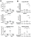
Fig. 2
Eosinophil migration to adipose tissue is integrin-mediated. (A) Eosinophils in the left lobe of the lung, spleen and perigonadal adipose tissue 3 and 7 days after adoptive transfer into eosinophil-deficient ΔdblGATA mice on normal chow. Data shown is pooled from two independent experiments. (B) ΔdblGATA mice received antibodies to α4 and αL integrins (100 μg each) or control isotypes (IgG2a and IgG2b) 2 hrs prior to adoptive transfer of eosinophils. Tissues were harvested 16 hrs later. Data shown is representative of two independent experiments. *p<0.05, **p<0.01 as determined using Student’s t-test. n.s. = not significant.
Fig. 3
Adipose macrophage alternative activation is impaired in the absence of IL-4/IL-13 or eosinophils. (A) Flow cytometric analysis of adipose from indicated mice on normal chow diet. Gates show YFP-positive cells as a percent of total CD11bhighF4/80high macrophages (A) and are quantitated (B) Results are pooled data from two or more independent experiments with 2–4 animals per experiment. *p<0.05, **p<0.01 as determined using ANOVA with Bonferonni’s post-test correction for multiple comparisons. (C) ΔdblGATA × YARG mice were sublethally irradiated and reconstituted with bone marrow cells from 4get × IL-5tg mice. After 4–6 weeks, perigonadal adipose tissues were analyzed for eosinophils (left gate; eosinophils were GFP-positive and side-scatterhigh, not shown) and macrophages (CD11bhigh F4/80high, right gate). Macrophages were then analyzed for YFP. Eosinophil-reconstituted mice (red); non-reconstituted mice (blue); WT control (non-reporter) mice (gray). (D) Statistical correlation (Spearman’s rank correlation) between the total numbers of eosinophils reconstituting perigonadal adipose tissues in eosinophil-deficient mice and the total numbers of AAM expressing the marker arginase-1 allele. Results are pooled data from 5 independent experiments with 2–4 animals per experiment. (E) Mice reconstituted with IL-5tg bone marrow lacking IL-4 and IL-13 (IL-5tg × 4/13 DKO) display significantly fewer total YARG+ AAM (E) or total YARG+AAM per 1,000 tissue eosinophils (IL-5tg 2.5 ± 1.1; IL-5tg × 4/13 DKO 2.4×10⁻⁴ ± 6.2×10⁻⁵). Results are pooled data from two or more independent experiments with 2–5 animals per experiment. *p<0.05, **p<0.01 as determined using Student’s t-test.
Fig. 4
Metabolic analysis of eosinophil-deficient and hypereosinophilic mice. (A) Perigonadal fat tissues (testis attached) from IL-5 transgenic (IL-5tg) and wildtype (WT) littermate controls. (B) Fasting male 8 wk-old WT or IL-5tg littermates maintained on normal chow (NC) diet were challenged with intraperitoneal glucose and blood was sampled for glucose at times indicated. Data compiled from two independent experiments with 6–7 mice in each group. (C,D) DEXA analysis of total, lean and fat tissue composition (C) or percentage adiposity (D) in ΔdblGATA and wildtype (WT) mice on normal chow (NC) or high-fat (HF) diet for 15 wk. Data compiled from two experiments with 5–8 mice in each group. (E) Intraperitoneal glucose tolerance test in male ΔdblGATA and WT mice on HF diet for 15 wk. Data compiled from 3 independent experiments with 5–8 mice in each group. (F) Fasting blood glucose in male WT and ΔdblGATA mice maintained on HF diet for 20–22 wk. Data compiled from 2 independent experiments with 5 mice in each group. (G, H) Insulin signaling, as measured by the ratio of serine phosphorylated AKT to total AKT in adipose, muscle and liver of mice aged 24 wk on HF diet (n = 4–6 mice per genotype, 4 representative mouse adipose samples shown). (I, J) Twelve-wk old wild-type C57BL/6 mice on HF diet for 6 wk were infected with N. brasiliensis (Nippo) or unchallenged (Control) and monitored for insulin tolerance (I) and glucose tolerance (J) at the indicated times. Insulin tolerance results are normalized to baseline fasting glucose, which was statistically different between cohorts (WT control 207 mg/dL ± 6; Nippo 179 mg/dL ± 7; p < 0.05). (K) Adipose tissue collected at days 40–45 post N. brasiliensis infection or from uninfected control mice and analyzed by flow cytometry for eosinophils per g adipose (K) or percent eosinophils (WT Control 2.9% ± 0.41; Nippo 10.1% ± 0.36, p<0.01). Data (I, J, K) are representative of two independent experiments with 20–30 total mice per cohort. *p<0.05, **p<0.01 as determined using Student’s t-test (B, E, F, H–K) or ANOVA with Bonferroni’s post-test correction for multiple comparisons (C–D); error bars = SEM; n.s. = not significant.
Comment in
- Immunology. Eosinophils forestall obesity.
Maizels RM, Allen JE. Maizels RM, et al. Science. 2011 Apr 8;332(6026):186-7. doi: 10.1126/science.1205313. Science. 2011. PMID: 21474746 No abstract available. - Granulocytes: a weighty role for eosinophils.
Minton K. Minton K. Nat Rev Immunol. 2011 May;11(5):299. doi: 10.1038/nri2976. Epub 2011 Apr 8. Nat Rev Immunol. 2011. PMID: 21494264 No abstract available.
Similar articles
- Innate lymphoid type 2 cells sustain visceral adipose tissue eosinophils and alternatively activated macrophages.
Molofsky AB, Nussbaum JC, Liang HE, Van Dyken SJ, Cheng LE, Mohapatra A, Chawla A, Locksley RM. Molofsky AB, et al. J Exp Med. 2013 Mar 11;210(3):535-49. doi: 10.1084/jem.20121964. Epub 2013 Feb 18. J Exp Med. 2013. PMID: 23420878 Free PMC article. - Macrophage-specific PPARgamma controls alternative activation and improves insulin resistance.
Odegaard JI, Ricardo-Gonzalez RR, Goforth MH, Morel CR, Subramanian V, Mukundan L, Red Eagle A, Vats D, Brombacher F, Ferrante AW, Chawla A. Odegaard JI, et al. Nature. 2007 Jun 28;447(7148):1116-20. doi: 10.1038/nature05894. Epub 2007 May 21. Nature. 2007. PMID: 17515919 Free PMC article. - Chronic helminth infection and helminth-derived egg antigens promote adipose tissue M2 macrophages and improve insulin sensitivity in obese mice.
Hussaarts L, García-Tardón N, van Beek L, Heemskerk MM, Haeberlein S, van der Zon GC, Ozir-Fazalalikhan A, Berbée JF, Willems van Dijk K, van Harmelen V, Yazdanbakhsh M, Guigas B. Hussaarts L, et al. FASEB J. 2015 Jul;29(7):3027-39. doi: 10.1096/fj.14-266239. Epub 2015 Apr 7. FASEB J. 2015. PMID: 25852044 - EoTHINophils: Eosinophils as key players in adipose tissue homeostasis.
Vohralik EJ, Psaila AM, Knights AJ, Quinlan KGR. Vohralik EJ, et al. Clin Exp Pharmacol Physiol. 2020 Aug;47(8):1495-1505. doi: 10.1111/1440-1681.13304. Epub 2020 Mar 24. Clin Exp Pharmacol Physiol. 2020. PMID: 32163614 Review. - Interleukin-4- and interleukin-13-mediated alternatively activated macrophages: roles in homeostasis and disease.
Van Dyken SJ, Locksley RM. Van Dyken SJ, et al. Annu Rev Immunol. 2013;31:317-43. doi: 10.1146/annurev-immunol-032712-095906. Epub 2013 Jan 3. Annu Rev Immunol. 2013. PMID: 23298208 Free PMC article. Review.
Cited by
- Isolation of adipose tissue immune cells.
Orr JS, Kennedy AJ, Hasty AH. Orr JS, et al. J Vis Exp. 2013 May 22;(75):e50707. doi: 10.3791/50707. J Vis Exp. 2013. PMID: 23728515 Free PMC article. - Invariant natural killer T cells in adipose tissue: novel regulators of immune-mediated metabolic disease.
Rakhshandehroo M, Kalkhoven E, Boes M. Rakhshandehroo M, et al. Cell Mol Life Sci. 2013 Dec;70(24):4711-27. doi: 10.1007/s00018-013-1414-1. Epub 2013 Jul 9. Cell Mol Life Sci. 2013. PMID: 23835837 Free PMC article. Review. - Eosinophil Deficiency Promotes Aberrant Repair and Adverse Remodeling Following Acute Myocardial Infarction.
Toor IS, Rückerl D, Mair I, Ainsworth R, Meloni M, Spiroski AM, Benezech C, Felton JM, Thomson A, Caporali A, Keeble T, Tang KH, Rossi AG, Newby DE, Allen JE, Gray GA. Toor IS, et al. JACC Basic Transl Sci. 2020 Jul 8;5(7):665-681. doi: 10.1016/j.jacbts.2020.05.005. eCollection 2020 Jul. JACC Basic Transl Sci. 2020. PMID: 32760855 Free PMC article. - Local proliferation initiates macrophage accumulation in adipose tissue during obesity.
Zheng C, Yang Q, Cao J, Xie N, Liu K, Shou P, Qian F, Wang Y, Shi Y. Zheng C, et al. Cell Death Dis. 2016 Mar 31;7(3):e2167. doi: 10.1038/cddis.2016.54. Cell Death Dis. 2016. PMID: 27031964 Free PMC article. - PVAT-conditioned media from Dahl S rats on high fat diet promotes inflammatory cytokine secretion by activated T cells prior to the development of hypertension.
Jin Y, Liu S, Guzmán KE, Kumar RK, Kaiser LM, Garver H, Bernard JJ, Bhattacharya S, Fink GD, Watts SW, Rockwell CE. Jin Y, et al. PLoS One. 2024 Oct 3;19(10):e0302503. doi: 10.1371/journal.pone.0302503. eCollection 2024. PLoS One. 2024. PMID: 39361560 Free PMC article.
References
- Hotamisligil GS. Inflammation and metabolic disorders. Nature. 2006;444:860–867. - PubMed
- Bouhlel MA, et al. PPARγ activation primes human monocytes into alternative M2 macrophages with anti-inflammatory properties. Cell Metab. 2007;6:137–143. - PubMed
- Jeninga EH, Gurnell M, Kalkhoven E. Functional implications of genetic variation in human PPARgamma. Trends Endocrinol Metab. 2009;20:380–387. - PubMed
Publication types
MeSH terms
Substances
Grants and funding
- R01 DK076760/DK/NIDDK NIH HHS/United States
- P30 DK063720/DK/NIDDK NIH HHS/United States
- AI026918/AI/NIAID NIH HHS/United States
- R01 DK081405/DK/NIDDK NIH HHS/United States
- DK063720/DK/NIDDK NIH HHS/United States
- 5F30DK083194-02/DK/NIDDK NIH HHS/United States
- F30 DK083194/DK/NIDDK NIH HHS/United States
- R37 AI026918-24/AI/NIAID NIH HHS/United States
- R37 AI026918/AI/NIAID NIH HHS/United States
- R01 AI030663/AI/NIAID NIH HHS/United States
- HHMI/Howard Hughes Medical Institute/United States
- R01 HL076746/HL/NHLBI NIH HHS/United States
- DP1 OD006415/OD/NIH HHS/United States
- F30 DK083194-03/DK/NIDDK NIH HHS/United States
- R01 AI026918/AI/NIAID NIH HHS/United States
LinkOut - more resources
Full Text Sources
Other Literature Sources
Medical
Molecular Biology Databases


