Pancreatic endocrine tumours: mutational and immunohistochemical survey of protein kinases reveals alterations in targetable kinases in cancer cell lines and rare primaries - PubMed (original) (raw)
Pancreatic endocrine tumours: mutational and immunohistochemical survey of protein kinases reveals alterations in targetable kinases in cancer cell lines and rare primaries
V Corbo et al. Ann Oncol. 2012 Jan.
Abstract
Background: Kinases represent potential therapeutic targets in pancreatic endocrine tumours (PETs).
Patients and methods: Thirty-five kinase genes were sequenced in 36 primary PETs and three PET cell lines: (i) 4 receptor tyrosine kinases (RTK), epithelial growth factor receptor (EGFR), human epidermal growth factor receptor 2 (HER2), tyrosine-protein kinase KIT (KIT), platelet-derived growth factor receptor alpha (PDGFRalpha); (ii) 6 belonging to the Akt/mTOR pathway; and (iii) 25 frequently mutated in cancers. The immunohistochemical expression of the four RTKs and the copy number of EGFR and HER2 were assessed in 140 PETs.
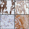
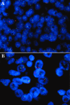
Results: Somatic mutations were found in KIT in one and ATM in two primary neoplasms. Among 140 PETs, EGFR was immunopositive in 18 (13%), HER2 in 3 (2%), KIT in 16 (11%), and PDGFRalpha in 135 (96%). HER2 amplification was found in 2/130 (1.5%) PETs. KIT membrane immunostaining was significantly associated with tumour aggressiveness and shorter patient survival. PET cell lines QGP1, CM and BON harboured mutations in FGFR3, FLT1/VEGFR1 and PIK3CA, respectively.
Conclusions: Only rare PET cases, harbouring either HER2 amplification or KIT mutation, might benefit from targeted drugs. KIT membrane expression deserves further attention as a prognostic marker. ATM mutation is involved in a proportion of PET. The finding of specific mutations in PET cell lines renders these models useful for preclinical studies involving pathway-specific therapies.
Figures
Figure 1.
Immunohistochemical staining for receptor tyrosine kinases in pancreatic endocrine tumours. Shown are positive staining for KIT (A), EGFR (B) and HER2 (C) in tumour cells; positive staining in tumour cells and in the stroma is shown for PDGFRalpha (D). Original magnification, x20.
Figure 2.
Fluorescent in situ hybridisation (FISH) analysis for EGFR and HER2 in pancreatic endocrine tumours. FISH analysis showing monosomy for EGFR (A), and gene amplification for HER2 (B). Original magnification, ×100. EGFR and HER2 signal red, centromeric probes signal green.
Figure 3.
Correlation between KIT membrane immunostaining and patients’ survival. Kaplan–Meier estimates of survival with regard to KIT membrane immunostaining (P < 0.001). Follow-up, months; KIT+, membrane-positive immunostaining; KIT−, membrane-negative immunostaining.
Similar articles
- Molecular markers for novel therapeutic strategies in pancreatic endocrine tumors.
Gilbert JA, Adhikari LJ, Lloyd RV, Halfdanarson TR, Muders MH, Ames MM. Gilbert JA, et al. Pancreas. 2013 Apr;42(3):411-21. doi: 10.1097/MPA.0b013e31826cb243. Pancreas. 2013. PMID: 23211371 Free PMC article. - Mutational profiling of kinases in human tumours of pancreatic origin identifies candidate cancer genes in ductal and ampulla of vater carcinomas.
Corbo V, Ritelli R, Barbi S, Funel N, Campani D, Bardelli A, Scarpa A. Corbo V, et al. PLoS One. 2010 Sep 8;5(9):e12653. doi: 10.1371/journal.pone.0012653. PLoS One. 2010. PMID: 20838624 Free PMC article. - Pancreatic tumours: molecular pathways implicated in ductal cancer are involved in ampullary but not in exocrine nonductal or endocrine tumorigenesis.
Moore PS, Orlandini S, Zamboni G, Capelli P, Rigaud G, Falconi M, Bassi C, Lemoine NR, Scarpa A. Moore PS, et al. Br J Cancer. 2001 Jan;84(2):253-62. doi: 10.1054/bjoc.2000.1567. Br J Cancer. 2001. PMID: 11161385 Free PMC article. - Molecular context of the EGFR mutations: evidence for the activation of mTOR/S6K signaling.
Conde E, Angulo B, Tang M, Morente M, Torres-Lanzas J, Lopez-Encuentra A, Lopez-Rios F, Sanchez-Cespedes M. Conde E, et al. Clin Cancer Res. 2006 Feb 1;12(3 Pt 1):710-7. doi: 10.1158/1078-0432.CCR-05-1362. Clin Cancer Res. 2006. PMID: 16467080 - Oncogene alterations in endometrial carcinosarcomas.
Biscuola M, Van de Vijver K, Castilla MÁ, Romero-Pérez L, López-García MÁ, Díaz-Martín J, Matias-Guiu X, Oliva E, Palacios Calvo J. Biscuola M, et al. Hum Pathol. 2013 May;44(5):852-9. doi: 10.1016/j.humpath.2012.07.027. Epub 2012 Nov 28. Hum Pathol. 2013. PMID: 23199529
Cited by
- Pancreatic neuroendocrine tumors G3 and pancreatic neuroendocrine carcinomas: Differences in basic biology and treatment.
Zhang MY, He D, Zhang S. Zhang MY, et al. World J Gastrointest Oncol. 2020 Jul 15;12(7):705-718. doi: 10.4251/wjgo.v12.i7.705. World J Gastrointest Oncol. 2020. PMID: 32864039 Free PMC article. Review. - Novel molecular targets for the treatment of gastroenteropancreatic endocrine tumors: answers and unsolved problems.
Capurso G, Fendrich V, Rinzivillo M, Panzuto F, Bartsch DK, Delle Fave G. Capurso G, et al. Int J Mol Sci. 2012 Dec 20;14(1):30-45. doi: 10.3390/ijms14010030. Int J Mol Sci. 2012. PMID: 23344019 Free PMC article. Review. - Reg3g Promotes Pancreatic Carcinogenesis in a Murine Model of Chronic Pancreatitis.
Yin G, Du J, Cao H, Liu X, Xu Q, Xiang M. Yin G, et al. Dig Dis Sci. 2015 Dec;60(12):3656-68. doi: 10.1007/s10620-015-3787-5. Epub 2015 Jul 17. Dig Dis Sci. 2015. PMID: 26182900 - Multigene mutational profiling of cholangiocarcinomas identifies actionable molecular subgroups.
Simbolo M, Fassan M, Ruzzenente A, Mafficini A, Wood LD, Corbo V, Melisi D, Malleo G, Vicentini C, Malpeli G, Antonello D, Sperandio N, Capelli P, Tomezzoli A, Iacono C, Lawlor RT, Bassi C, Hruban RH, Guglielmi A, Tortora G, de Braud F, Scarpa A. Simbolo M, et al. Oncotarget. 2014 May 15;5(9):2839-52. doi: 10.18632/oncotarget.1943. Oncotarget. 2014. PMID: 24867389 Free PMC article. - Prognostic and predictive role of the PI3K-AKT-mTOR pathway in neuroendocrine neoplasms.
Gajate P, Alonso-Gordoa T, Martínez-Sáez O, Molina-Cerrillo J, Grande E. Gajate P, et al. Clin Transl Oncol. 2018 May;20(5):561-569. doi: 10.1007/s12094-017-1758-3. Epub 2017 Nov 9. Clin Transl Oncol. 2018. PMID: 29124519 Review.
References
- Antonello D, Gobbo S, Corbo V, et al. Update on the molecular pathogenesis of pancreatic tumors other than common ductal adenocarcinoma. Pancreatology. 2009;9:25–33. - PubMed
- Capelli P, Martignoni G, Pedica F, et al. Endocrine neoplasms of the pancreas: pathologic and genetic features. Arch Pathol Lab Med. 2009;133:350–364. - PubMed
- Corbo V, Dalai I, Scardoni M, et al. MEN1 in pancreatic endocrine tumors: analysis of gene and protein status in 169 sporadic neoplasms reveals alterations in the vast majority of cases. Endocr Relat Cancer. 2010;17:771–783. - PubMed
- Kloppel G, Perren A, Heitz PU. The gastroenteropancreatic neuroendocrine cell system and its tumors: the WHO classification. Ann N Y Acad Sci. 2004;1014:13–27. - PubMed
- Bettini R, Mantovani W, Boninsegna L, et al. Primary tumour resection in metastatic nonfunctioning pancreatic endocrine carcinomas. Dig Liver Dis. 2009;41:49–55. - PubMed
Publication types
MeSH terms
Substances
LinkOut - more resources
Full Text Sources
Medical
Research Materials
Miscellaneous


