The requirement for enhanced CREB1 expression in consolidation of long-term synaptic facilitation and long-term excitability in sensory neurons of Aplysia - PubMed (original) (raw)
The requirement for enhanced CREB1 expression in consolidation of long-term synaptic facilitation and long-term excitability in sensory neurons of Aplysia
Rong-Yu Liu et al. J Neurosci. 2011.
Abstract
Accumulating evidence suggests that the transcriptional activator cAMP response element-binding protein 1 (CREB1) is important for serotonin (5-HT)-induced long-term facilitation (LTF) of the sensorimotor synapse in Aplysia. Moreover, creb1 is among the genes activated by CREB1, suggesting a role for this protein beyond the induction phase of LTF. The time course of the requirement for CREB1 synthesis in the consolidation of long-term facilitation was examined using RNA interference techniques in sensorimotor cocultures. Injection of CREB1 small-interfering RNA (siRNA) immediately or 10 h after 5-HT treatment blocked LTF when measured at 24 and 48 h after treatment. In contrast, CREB1 siRNA did not block LTF when injected 16 h after 5-HT treatment. These results demonstrate that creb1 expression must be sustained for a relatively long time to support the consolidation of LTF. In addition, LTF is also accompanied by a long-term increase in the excitability (LTE) of sensory neurons (SNs). Because LTE was observed in the isolated SN after 5-HT treatment, this long-term change was intrinsic to that element of the circuit. LTE was blocked when CREB1 siRNA was injected into isolated SNs immediately after 5-HT treatment. These data suggest that 5-HT-induced CREB1 synthesis is required for consolidation of both LTF and LTE.
Figures
Figure 1.
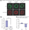
Serotonin-induced changes in tCREB1 and CRE-mediated gene expression. A, 5-HT treatment induced tCREB1 expression for at least 24 h in cultured SNs. A1, Representative confocal images of tCREB1 immunofluorescence in SNs at 2, 12, and 24 h after treatment with vehicle or 5-HT. Scale bar, 30 μm. A2, Summary data. CREB1 was elevated at 2 h, returned to basal level at 12 h, and increased again at 18 and 24 h after 5-HT treatment (*p < 0.05). B, CRE-mediated EGFP expression was enhanced 18–24 h after 5-HT treatment. B1, Representative images of sensory cells injected with CRE–EGFP reporter vector 18 h after 5-HT or vehicle treatment and fixed 6 h later. Texas Red–dextran was added to the injection buffer to confirm nuclear injection. Scale bar, 15 μm. B2, 5-HT-induced EGFP expression was calculated as the ratio of the 5-HT-treated to vehicle-treated samples for each experiment. EGFP expression was significantly increased in neurons after 5-HT treatment compared with vehicle-treated cells (*p < 0.05), demonstrating that CRE-dependent gene expression is still enhanced 18 h after 5-HT treatment.
Figure 2.
CREB1 siRNA injection blocked 5-HT-induced increase in tCREB1 expression without affecting basal levels. A1, Confocal images of neurons injected with CREB1 siRNA or control siRNA. siRNAs were injected immediately after vehicle or 5-HT treatment. Fluorescein–dextran was coinjected with siRNAs to monitor cytoplasmic injection (a, c, e). At 2 h after the end of treatment, cells were fixed and processed for immunostaining for CREB1 (b, d, f). Scale bar, 20 μm. A2, Data were expressed as the ratios of tCREB1 levels in 5-HT-treated cells to tCREB1 levels in vehicle-treated control siRNA cells. Summary data indicated that the 5-HT-induced increase in CREB1 can be blocked by CREB1 siRNA (*p < 0.05). B, CREB1 siRNA or control siRNA was injected into untreated SNs. Forty-eight or 72 h later, cells were processed for tCREB1 immunofluorescence. Summary data show that injection of CREB1 siRNA did not affect the basal level of tCREB1 48 h later but did significantly decrease the basal level 72 h later compared with the time-matched control siRNA-injected groups (*p < 0.05).
Figure 3.
CREB1 siRNA injection immediately after 5-HT treatment blocked LTF. A, Protocol for CREB1 or control siRNA injection and electrophysiological testing. siRNAs were injected into the cytosol of SNs immediately after the end of treatment with 5-HT or vehicle. B, Representative traces of EPSPs before (pre) and 24 and 48 h after treatment with 5-HT or vehicle (Veh). C, D, Summary data. Two-way ANOVA followed by post hoc tests indicated that injection of CREB siRNA blocked LTF measured at 24 (C) and 48 h (D) without significantly affecting basal synaptic transmission (*p < 0.05).
Figure 4.
CREB1 siRNA injection 10 h after 5-HT treatment blocked LTF. A, The 5-HT-induced increase in CREB1 protein levels at 24 h was blocked by CREB1 siRNA injected at 10 h after treatment (*p < 0.05). B1, Protocol for CREB1 siRNA or control siRNA injection and electrophysiological testing. siRNAs were injected into the cytosol of SNs 10 h after the end of treatment with 5-HT. B2, Representative traces of EPSPs before (pre) and 24 and 48 h after treatment with 5-HT or vehicle (Veh). C, D, Summary data. One-way ANOVA followed by post hoc tests indicated that injection of CREB1 siRNA at 10 h after treatment blocked LTF measured at 24 h (C) and 48 h (D) without significantly affecting basal transmission (*p < 0.05).
Figure 5.
Injection of CREB1 siRNA 16 h after 5-HT treatment did not block LTF. A, Protocol for CREB1 or control siRNA injection and electrophysiological testing. siRNAs were injected into SNs in coculture at 16–18 h after the end of treatment with 5-HT or vehicle. B, Representative traces of EPSPs recorded from cocultures before (pre) and 24 and 48 h after treatment with 5-HT or vehicle (Veh). C, D, Summary data. One-way ANOVA followed by post hoc tests indicated that injection of CREB1 siRNA did not block LTF measured at 24 h (C) and 48 h (D) (*p < 0.05; N.S., not significant).
Figure 6.
CREB1 siRNA blocked 5-HT-induced LTE in isolated SNs. A, Protocol for CREB1 or control siRNA injection and electrophysiological testing. siRNAs were injected into the cytosol of SNs immediately after the end of treatment with 5-HT or vehicle. B, Action potentials were recorded from cultured SNs before (pre) and 24 and 48 h after treatment with 5-HT or vehicle (Veh). C, Summary data (24 h after test). A significant increase in cell excitability was revealed in the 5-HT-treated, control siRNA-injected group. The 5-HT-induced changes in cell excitability were blocked by CREB1 siRNA injection (*p < 0.05). D, Summary data (48 h after test). The 5-HT-induced increase in cell excitability was no longer detected.
Similar articles
- cAMP response element-binding protein 1 feedback loop is necessary for consolidation of long-term synaptic facilitation in Aplysia.
Liu RY, Fioravante D, Shah S, Byrne JH. Liu RY, et al. J Neurosci. 2008 Feb 20;28(8):1970-6. doi: 10.1523/JNEUROSCI.3848-07.2008. J Neurosci. 2008. PMID: 18287513 Free PMC article. - Rescue of impaired long-term facilitation at sensorimotor synapses of Aplysia following siRNA knockdown of CREB1.
Zhou L, Zhang Y, Liu RY, Smolen P, Cleary LJ, Byrne JH. Zhou L, et al. J Neurosci. 2015 Jan 28;35(4):1617-26. doi: 10.1523/JNEUROSCI.3330-14.2015. J Neurosci. 2015. PMID: 25632137 Free PMC article. - cJun and CREB2 in the postsynaptic neuron contribute to persistent long-term facilitation at a behaviorally relevant synapse.
Hu JY, Levine A, Sung YJ, Schacher S. Hu JY, et al. J Neurosci. 2015 Jan 7;35(1):386-95. doi: 10.1523/JNEUROSCI.3284-14.2015. J Neurosci. 2015. PMID: 25568130 Free PMC article. - Postsynaptic regulation of the development and long-term plasticity of Aplysia sensorimotor synapses in cell culture.
Glanzman DL. Glanzman DL. J Neurobiol. 1994 Jun;25(6):666-93. doi: 10.1002/neu.480250608. J Neurobiol. 1994. PMID: 8071666 Review. - Transcriptional regulation of long-term memory in the marine snail Aplysia.
Lee YS, Bailey CH, Kandel ER, Kaang BK. Lee YS, et al. Mol Brain. 2008 Jun 17;1:3. doi: 10.1186/1756-6606-1-3. Mol Brain. 2008. PMID: 18803855 Free PMC article. Review.
Cited by
- Pattern and predictability in memory formation: from molecular mechanisms to clinical relevance.
Philips GT, Kopec AM, Carew TJ. Philips GT, et al. Neurobiol Learn Mem. 2013 Oct;105:117-24. doi: 10.1016/j.nlm.2013.05.003. Epub 2013 May 28. Neurobiol Learn Mem. 2013. PMID: 23727358 Free PMC article. Review. - Role of p90 ribosomal S6 kinase in long-term synaptic facilitation and enhanced neuronal excitability.
Liu RY, Zhang Y, Smolen P, Cleary LJ, Byrne JH. Liu RY, et al. Sci Rep. 2020 Jan 17;10(1):608. doi: 10.1038/s41598-020-57484-y. Sci Rep. 2020. PMID: 31953461 Free PMC article. - Molecular correlates of separate components of training that contribute to long-term memory formation after learning that food is inedible in Aplysia.
Briskin-Luchinsky V, Levy R, Halfon M, Susswein AJ. Briskin-Luchinsky V, et al. Learn Mem. 2018 Jan 16;25(2):90-99. doi: 10.1101/lm.046326.117. Print 2018 Feb. Learn Mem. 2018. PMID: 29339560 Free PMC article. - Persistent long-term facilitation at an identified synapse becomes labile with activation of short-term heterosynaptic plasticity.
Hu JY, Schacher S. Hu JY, et al. J Neurosci. 2014 Apr 2;34(14):4776-85. doi: 10.1523/JNEUROSCI.0098-14.2014. J Neurosci. 2014. PMID: 24695698 Free PMC article. - Doxorubicin attenuates serotonin-induced long-term synaptic facilitation by phosphorylation of p38 mitogen-activated protein kinase.
Liu RY, Zhang Y, Coughlin BL, Cleary LJ, Byrne JH. Liu RY, et al. J Neurosci. 2014 Oct 1;34(40):13289-300. doi: 10.1523/JNEUROSCI.0538-14.2014. J Neurosci. 2014. PMID: 25274809 Free PMC article.
References
- Alberini CM, Ghirardi M, Metz R, Kandel ER. C/EBP is an immediate-early gene required for the consolidation of long-term facilitation in Aplysia. Cell. 1994;76:1099–1114. - PubMed
- Artinian J, McGauran AM, De Jaeger X, Mouledous L, Frances B, Roullet P. Protein degradation, as with protein synthesis, is required during not only long-term spatial memory consolidation but also reconsolidation. Eur J Neurosci. 2008;27:3009–3019. - PubMed
Publication types
MeSH terms
Substances
LinkOut - more resources
Full Text Sources
Research Materials
Miscellaneous


