Anatomical properties of the arcuate fasciculus predict phonological and reading skills in children - PubMed (original) (raw)
Anatomical properties of the arcuate fasciculus predict phonological and reading skills in children
Jason D Yeatman et al. J Cogn Neurosci. 2011 Nov.
Abstract
For more than a century, neurologists have hypothesized that the arcuate fasciculus carries signals that are essential for language function; however, the relevance of the pathway for particular behaviors is highly controversial. The primary objective of this study was to use diffusion tensor imaging to examine the relationship between individual variation in the microstructural properties of arcuate fibers and behavioral measures of language and reading skills. A second objective was to use novel fiber-tracking methods to reassess estimates of arcuate lateralization. In a sample of 55 children, we found that measurements of diffusivity in the left arcuate correlate with phonological awareness skills and arcuate volume lateralization correlates with phonological memory and reading skills. Contrary to previous investigations that report the absence of the right arcuate in some subjects, we demonstrate that new techniques can identify the pathway in every individual. Our results provide empirical support for the role of the arcuate fasciculus in the development of reading skills.
Figures
Figure 1
Method of identifying the AF in a single subject. Whole-brain streamlines tracing technique (STT) tractography produced a large collection of estimated tracks (not shown). An ROI in the white matter was drawn comprising the voxels with an anterior–posterior (green) PDD that are located adjacent and lateral to the cortical spinal tract. The cortical spinal tract can be identified because its PDD is in the inferior–superior (blue) direction. (A) The PDD at each voxel in a typical axial slice (Z = 26). The white outline shows the region selected in this slice. The ROI is large so that all of the AF fibers will be included. (B) The complete ROI for this subject, which is selected from several adjacent planes. (C) Estimated fiber tracks passing through the ROI. We identify the AF fibers from the group by selecting the fibers that (1) project anterior to the central sulcus and (2) continue posterior and inferior into the temporal lobe (red lines). These waypoints were manually identified for each subject in an interactive fiber tract viewing and segmentation tool available for download at vistalab.stanford.edu/software. (D) The estimated left AF for this subject.
Figure 2
Cortical endpoints of the left AF in 55 children. Manually identified left AF fiber groups for three representative children are shown on the left. The frontal lobe endpoints are generally focused in the precentral gyrus and Brodmann’s area 44 of the inferior frontal gyrus, whereas the temporal lobe endpoints are spread over significantly more surface area. The heat map on the right shows the number of subjects with left AF endpoints at each region of the cortex. Endpoints for each subject were registered onto the cortical surface of an individual child.
Figure 3
Microstructural properties of the left AF correlate with phonological awareness standard scores. FA, RD, and AD are plotted against age-standardized measures of phonological awareness. Solid black circles show mean phonological awareness scores (±1 SE) at evenly spaced intervals of FA, RD, and AD. FA is significantly negatively correlated (r = −.33), RD is significantly positively correlated (r = .30), and AD is not correlated with phonological awareness.
Figure 4
FA varies along the trajectory of the AF because of crossing fibers and tract curvature. FA was calculated at 30 nodes along a core section of the AF (A). The 30 nodes for the subject that was most similar to the group mean are displayed as small, overlapping spheres and color-coded red to yellow based on the FA values in that region of the AF (scale bar at the right). Each subject showed a decrease in FA in the arcing region of the tract and an increase in FA in an anterior region of the tract medial to the central sulcus and before the location where fibers branch toward their cortical destinations. (B) Fibers were tracked from 3-mm spheres centered around the node of the highest FA and the node of the lowest FA (asterisks). The AF is shown in blue, non-AF fibers tracked from the high-FA node are shown in orange, and non-AF fibers tracked from the low-FA node are shown in green. The low-FA node contains a mix of AF fibers and fibers that turn laterally and continue to the lateral parietal lobe. The high-FA node contains AF fibers and coherently oriented fibers of the SLF that are destined for the inferior frontal cortex. (C) A coronal slice through the high-FA node shows that, in this region, some voxels along the border of the AF and the SLF are partial volumed between the two tracts; however, a majority (66%) of AF-containing voxels do not contain any SLF fibers.
Figure 5
Phonological awareness is correlated with AF microstructure in the core region of the AF after individual tracts are aligned. (A) The curved portion of the AF where crossing fibers cause decreased FA is not in register across subjects. (B) Aligning AF trajectories minimizes the confounds of crossing fiber tracts and allows for comparison of microstructural properties in anatomically equivalent regions of the tract across subjects. (C) In a 1-cm region of the tract anterior to the curved portion and posterior to the region where fibers branch toward cortex, FA is significantly negatively correlated with phonological awareness (r = −.36). (D) This correlation is driven by a positive correlation between RD and phonological awareness (r = .35).
Figure 6
Probabilistic tractography identifies the right AF in every subject. (A) The top scored 1000 fibers connecting an ROI in the posterior temporal lobe to an ROI in the inferior frontal lobe (top) are intersected with the waypoint ROIs typically used to define the arcuate (bottom) for a subject who had no identifiable right arcuate with deterministic tractography (STT). (B) A montage of right AF fiber groups tracked with a probabilistic algorithm for two subjects who appeared to be missing the right AF based on STT (top) and two subjects who had right AFs identified with STT (bottom). The AF fiber groups have the same shape and relative position for the two groups of subjects. (C) Comparison of FA along the right AF in two groups of subjects. One group of subjects (blue) had an identifiable AF using deterministic tractography. In the second group of subjects (red), deterministic methods could not identify the right arcuate (although probabilistic methods could). Tracts are aligned with the method shown in Figure 5, and FA is plotted at each node (±1 SE).
Figure 7
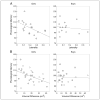
Scatter plots and regression lines show the relationship between laterality estimates (A) and age standardized phonological memory scores for women (r = −.55) and men (r = −.14). Women with less lateralized tracts had greater phonological memory scores. The absolute volume difference between left and right hemisphere tracts shows the same correlation with phonological memory (B). Unfilled shapes represent subjects without an identifiable right arcuate using deterministic tracking (−RAF), and filled shapes represent subjects with right and left arcuates (+RAF).
Similar articles
- Lateralization of the arcuate fasciculus from childhood to adulthood and its relation to cognitive abilities in children.
Lebel C, Beaulieu C. Lebel C, et al. Hum Brain Mapp. 2009 Nov;30(11):3563-73. doi: 10.1002/hbm.20779. Hum Brain Mapp. 2009. PMID: 19365801 Free PMC article. - White matter properties associated with pre-reading skills in 6-year-old children born preterm and at term.
Dodson CK, Travis KE, Borchers LR, Marchman VA, Ben-Shachar M, Feldman HM. Dodson CK, et al. Dev Med Child Neurol. 2018 Jul;60(7):695-702. doi: 10.1111/dmcn.13783. Epub 2018 May 3. Dev Med Child Neurol. 2018. PMID: 29722009 Free PMC article. - A tractography study in dyslexia: neuroanatomic correlates of orthographic, phonological and speech processing.
Vandermosten M, Boets B, Poelmans H, Sunaert S, Wouters J, Ghesquière P. Vandermosten M, et al. Brain. 2012 Mar;135(Pt 3):935-48. doi: 10.1093/brain/awr363. Epub 2012 Feb 10. Brain. 2012. PMID: 22327793 - Tracking the roots of reading ability: white matter volume and integrity correlate with phonological awareness in prereading and early-reading kindergarten children.
Saygin ZM, Norton ES, Osher DE, Beach SD, Cyr AB, Ozernov-Palchik O, Yendiki A, Fischl B, Gaab N, Gabrieli JD. Saygin ZM, et al. J Neurosci. 2013 Aug 14;33(33):13251-8. doi: 10.1523/JNEUROSCI.4383-12.2013. J Neurosci. 2013. PMID: 23946384 Free PMC article. - Controversy over the temporal cortical terminations of the left arcuate fasciculus: a reappraisal.
Giampiccolo D, Duffau H. Giampiccolo D, et al. Brain. 2022 May 24;145(4):1242-1256. doi: 10.1093/brain/awac057. Brain. 2022. PMID: 35142842 Review.
Cited by
- Reduced Volume of the Arcuate Fasciculus in Adults with High-Functioning Autism Spectrum Conditions.
Moseley RL, Correia MM, Baron-Cohen S, Shtyrov Y, Pulvermüller F, Mohr B. Moseley RL, et al. Front Hum Neurosci. 2016 May 12;10:214. doi: 10.3389/fnhum.2016.00214. eCollection 2016. Front Hum Neurosci. 2016. PMID: 27242478 Free PMC article. - Neonatal white matter tract microstructure and 2-year language outcomes after preterm birth.
Dubner SE, Rose J, Bruckert L, Feldman HM, Travis KE. Dubner SE, et al. Neuroimage Clin. 2020;28:102446. doi: 10.1016/j.nicl.2020.102446. Epub 2020 Sep 29. Neuroimage Clin. 2020. PMID: 33035964 Free PMC article. - Radiomic tractometry reveals tract-specific imaging biomarkers in white matter.
Neher P, Hirjak D, Maier-Hein K. Neher P, et al. Nat Commun. 2024 Jan 5;15(1):303. doi: 10.1038/s41467-023-44591-3. Nat Commun. 2024. PMID: 38182594 Free PMC article. - Association Between White Matter Microstructure and Verbal Fluency in Patients With Multiple Sclerosis.
Blecher T, Miron S, Schneider GG, Achiron A, Ben-Shachar M. Blecher T, et al. Front Psychol. 2019 Jul 18;10:1607. doi: 10.3389/fpsyg.2019.01607. eCollection 2019. Front Psychol. 2019. PMID: 31379663 Free PMC article. - Hidden word learning capacity through orthography in aphasia.
Tuomiranta LM, Càmara E, Froudist Walsh S, Ripollés P, Saunavaara JP, Parkkola R, Martin N, Rodríguez-Fornells A, Laine M. Tuomiranta LM, et al. Cortex. 2014 Jan;50:174-91. doi: 10.1016/j.cortex.2013.10.003. Epub 2013 Oct 24. Cortex. 2014. PMID: 24262200 Free PMC article.
References
- Akers D. CINCH: A cooperatively designed marking interface for 3D pathway selection. Paper presented at the User Interface Software and Technology (UIST); Montreux, Switzerland. 2006.
- Baldo JV, Klostermann EC, Dronkers NF. It’s either a cook or a baker: Patients with conduction aphasia get the gist but lose the trace. Brain and Language. 2008;105:134–140. - PubMed
- Basser PJ, Pajevic S, Pierpaoli C, Duda J, Aldroubi A. In vivo fiber tractography using DT-MRI data. Magnetic Resonance in Medicine. 2000;44:625–632. - PubMed
- Basser PJ, Pierpaoli C. Microstructural and physiological features of tissues elucidated by quantitative-diffusion-tensor MRI. Journal of Magnetic Resonance, Series B. 1996;111:209–219. - PubMed
Publication types
MeSH terms
LinkOut - more resources
Full Text Sources


