Identification and characterization of neuroblasts in the subventricular zone and rostral migratory stream of the adult human brain - PubMed (original) (raw)
. 2011 Nov;21(11):1534-50.
doi: 10.1038/cr.2011.83. Epub 2011 May 17.
Fang Liu, Ying-Ying Liu, Cai-Hong Zhao, Yan You, Lei Wang, Jingxiao Zhang, Bin Wei, Tong Ma, Qiangqiang Zhang, Yue Zhang, Rui Chen, Hongjun Song, Zhengang Yang
Affiliations
- PMID: 21577236
- PMCID: PMC3365638
- DOI: 10.1038/cr.2011.83
Identification and characterization of neuroblasts in the subventricular zone and rostral migratory stream of the adult human brain
Congmin Wang et al. Cell Res. 2011 Nov.
Abstract
It is of great interest to identify new neurons in the adult human brain, but the persistence of neurogenesis in the subventricular zone (SVZ) and the existence of the rostral migratory stream (RMS)-like pathway in the adult human forebrain remain highly controversial. In the present study, we have described the general configuration of the RMS in adult monkey, fetal human and adult human brains. We provide evidence that neuroblasts exist continuously in the anterior ventral SVZ and RMS of the adult human brain. The neuroblasts appear singly or in pairs without forming chains; they exhibit migratory morphologies and co-express the immature neuronal markers doublecortin, polysialylated neural cell adhesion molecule and βIII-tubulin. Few of these neuroblasts appear to be actively proliferating in the anterior ventral SVZ but none in the RMS, indicating that neuroblasts distributed along the RMS are most likely derived from the ventral SVZ. Interestingly, no neuroblasts are found in the adult human olfactory bulb. Taken together, our data suggest that the SVZ maintains the ability to produce neuroblasts in the adult human brain.
Figures
Figure 1
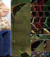
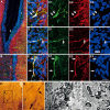
Neuroblasts in the SVZ and RMS of the adult rhesus monkey brain co-express Dcx and PSA-NCAM. (A) Photograph of the adult rhesus monkey brain (bottom view) showing the olfactory tract and OB. (B) The brain was cut coronally into 1.0–2.0 cm slabs. (C) DAPI staining of the coronal section shows the RMS. (D) The same section as C, double-immunostained for Dcx and PSA-NCAM. (E-I) Higher magnification of the boxed areas in D showing that the vast majority of Dcx+ cells express PSA-NCAM and vice versa. (J-L) Higher magnification of boxed areas in E, F and I. (M) Dcx/NeuN double immunostaining showing that Dcx+ cells migrate from the RMS to the anterior olfactory nucleus, which is located beneath (D). (N) Higher magnification of the boxed area in M. (O) Dcx/MCM2 double immunostaining in the coronal sections of the adult monkey SVZ. (P, Q) Higher magnification of boxed areas in O. (R-S') Higher magnification of boxed areas in P and Q. Note MCM2+/Dcx+ cells (arrows) and MCM2+ cells (arrowhead) in the SVZ. Scale bars represent 5 mm (A, B); 1 mm (C); 500 μm (D, M and O); 100 μm (in N applies to E-I, N, P and Q) and 20 μm (in J applies to J-L and R-S').
Figure 2
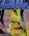
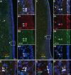
The RMS is prominent in the fetal human brain. (A1-A7) Representative coronal sections from the anterior SVZ to the anterior commissure (AC) stained for DAPI showing the RMS in the fetal human brain. Note that the intense DAPI staining, indicative of high cell density, distinguishes the SVZ and RMS from other regions of the forebrain. (B-D) Dcx/Tuj1 double-immunostained sections from the SVZ/RMS, olfactory tract and OB. (E-H) Higher magnification of the boxed areas in B-D. (I) Dcx/PSA-NCAM double immunostaining in the SVZ/RMS. (J) Higher magnification of the boxed area in I. Note that a lateral ventricular extension in B and I and chains of migrating neuroblasts (arrows in E) are seen in the RMS. Scale bars represent 1 mm (A7); 500 μm (in D applies to B-D and I); 100 μm (in J applies to E and J); 50 μm (in H applies to F-H).
Figure 3
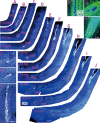
The RMS-like pathway exists in the adult human forebrain. (A-J) Serial coronal sections stained for DAPI showing the RMS-like pathway in the adult human brain. Note that the RMS starts to appear at the ventral floor of the anterior horn, which is about 6.0 mm ahead of the ventral extension of the lateral ventricle that contains small canals formed by ependymal cells. (F', G', I') Higher magnification of the RMS in F, G, I. Note that the RMS in F is very thin. (B', D') GFAP expression in the RMS. The regions of GFAP and DAPI staining in B' and D' correspond to the same location of the RMS in B and D. Note the glial tube formed by GFAP+ cells. (H') GFAP immunostaining in the ventral floor of the lateral ventricle. Scale bars represent 1 mm (in J applies to A-J); 200 μm (in I' applies to F', G' and I') and 100 μm (in H' applies to B', D' and H').
Figure 4
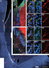
Dcx/GFAP double immunostaining of adult human brain sections indicating Dcx+ cells in the RMS. (A) One representative section stained for DAPI showing the ventral SVZ and RMS. The section broke at the ventral SVZ (dashed line) during processing. (B1, B2) The photomicrographs showing GFAP+ astrocytic cells surrounding individual Dcx+ cells in the RMS. (C1-E2) Higher magnification of Dcx+ cells in B1 and B2. Scale bars represent 1 mm (A); 100 μm (B1, B2) and 20 μm (in E2 applies to C1-E2).
Figure 5
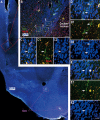
Neuroblasts in the adult human SVZ and RMS co-express Dcx and PSA-NCAM. (A) DAPI staining shows the anterior ventral SVZ and descending limb of the RMS. (B, C) Dcx+ cells express PSA-NCAM. Note that many PSA-NCAM+ cells in the SVZ, caudate nucleus and RMS do not express Dcx. (D-G) Higher magnification of boxed areas in B and C. Scale bars represent 1 mm (A); 100 μm (in C applies to B, C) and 20 μm (in G2 applies to D-G2).
Figure 6
Neuroblasts in the adult human SVZ and RMS co-express Dcx and Tuj1. (A, B) Dcx+/Tuj1+ cells in the SVZ and RMS of the adult human brain. (C-E) Higher magnification of boxed areas in A, B. Note that many Tuj1+ cells were found in the SVZ, caudate nucleus and RMS, but they do not express Dcx. Arrows indicate neuroblast cell bodies or nuclei. (F, G) Dcx+ cells are identified by light microscopy. (H, I) One Dcx+ cell and process are identified by electron microscopy (arrows). Scale bars represent 100 μm (A, B); 20 μm (in E3 applies to C-E3 and F); 10 μm (G) and 1 μm (H, I).
Figure 7
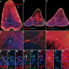
The ventral-lateral SVZ of adult human brains contains a few proliferative cells. (A, C) Representative sections double-immunostained for MCM2/Ki67. (B, D) Higher magnification of boxed areas in A and C showing MCM2+/Ki67+ cells (arrows). (E-H) The majority of MCM2+/Ki67+ cells appear in pairs. Scale bars represent 50 μm (A, C); 10 μm (in H applies to B-B3 and D-H).
Figure 8
There is a very small number of MCM2+/Dcx+ cells in the ventral-lateral SVZ of the adult human brain. (A-C) MCM2/Dcx double-labeling combined with 3-D reconstructions showing 2 Dcx+ cells in the ventral-lateral SVZ expressing MCM2 (arrows). (D1-F2) Confocal Z sectioning was performed at 0.6-μm intervals using PlanApo 60× oil-immersion (NA = 1.42) objectives. Photomicrographs depict three confocal images in the Z-dimension showing two MCM2+/Dcx+ cells (arrows) and one MCM2+ cell (arrowhead). (G-J) Dcx+ cells do not express MCM2 or Ki67 in the RMS (arrows). Scale bars represent 1 mm (A); 100 μm (in B applies to B and G) and 20 μm (in F2 applies to C-F2 and H-J).
Figure 9
Dcx+ cells in the adult human SVZ and RMS are immature neuroblasts. (A) One representative section stained for DAPI showing the ventral extension of the lateral ventricle and RMS. Note that many ependymal cells form small canals in this region. (B) The photomicrograph showing Dcx/NeuN double immunostaining in the boxed area in A. (C-E2) Higher magnification of boxed areas in B showing that Dcx+ cells do not express NeuN. Scale bars represent 1 mm (A), 100 μm (B) and 20 μm (in E2 applies to C-E2).
Figure 10
Dcx+ cells are extremely rare in the adult human olfactory tract. (A, B) Dcx/PSA-NCAM double immunostaining in coronal sections of the olfactory tract. (C) Dcx/GFAP double immunostaining in a coronal section of the olfactory tract. (D-F) Higher magnification of the boxed areas in A-C, respectively. (G-G2) Higher-magnification photomicrograph of 1 Dcx+/PSA-NCAM+ process (arrow). (H-H2) Higher-magnification photomicrograph of 1 Dcx+/PSA-NCAM+ cell (arrow). (I-I2) Higher-magnification photomicrograph of 1 Dcx+ process (arrow). Note that the Dcx+ process does not express GFAP. Scale bars represent 200 μm (in C applies to A-C); 50 μm (in F applies to D-F) and 10 μm (in I2 applies to G-I2).
Figure 11
Schematic depicting neuroblasts in the SVZ and RMS. While neuroblasts exist continuously in the adult human ventral SVZ and RMS, proliferating neuroblasts are only found in the ventral SVZ, indicating that the SVZ maintains the ability to produce neuroblasts in the adult human brain.
Comment in
- Migrating neuroblasts in the adult human brain: a stream reduced to a trickle.
van Strien ME, van den Berge SA, Hol EM. van Strien ME, et al. Cell Res. 2011 Nov;21(11):1523-5. doi: 10.1038/cr.2011.101. Epub 2011 Jun 21. Cell Res. 2011. PMID: 21691300 Free PMC article. No abstract available.
Similar articles
- Distribution of doublecortin expressing cells near the lateral ventricles in the adult mouse brain.
Yang HK, Sundholm-Peters NL, Goings GE, Walker AS, Hyland K, Szele FG. Yang HK, et al. J Neurosci Res. 2004 May 1;76(3):282-95. doi: 10.1002/jnr.20071. J Neurosci Res. 2004. PMID: 15079857 - Doublecortin expression in the adult rat telencephalon.
Nacher J, Crespo C, McEwen BS. Nacher J, et al. Eur J Neurosci. 2001 Aug;14(4):629-44. doi: 10.1046/j.0953-816x.2001.01683.x. Eur J Neurosci. 2001. PMID: 11556888 - Cytoarchitecture of the lateral ganglionic eminence and rostral extension of the lateral ventricle in the human fetal brain.
Guerrero-Cázares H, Gonzalez-Perez O, Soriano-Navarro M, Zamora-Berridi G, García-Verdugo JM, Quinoñes-Hinojosa A. Guerrero-Cázares H, et al. J Comp Neurol. 2011 Apr 15;519(6):1165-80. doi: 10.1002/cne.22566. J Comp Neurol. 2011. PMID: 21344407 Free PMC article. - Dynamic changes in the transcriptional profile of subventricular zone-derived postnatally born neuroblasts.
Khodosevich K, Alfonso J, Monyer H. Khodosevich K, et al. Mech Dev. 2013 Jun-Aug;130(6-8):424-32. doi: 10.1016/j.mod.2012.11.003. Epub 2012 Dec 5. Mech Dev. 2013. PMID: 23220001 Review. - Relationship between Blood Vessels and Migration of Neuroblasts in the Olfactory Neurogenic Region of the Rodent Brain.
Martončíková M, Alexovič Matiašová A, Ševc J, Račeková E. Martončíková M, et al. Int J Mol Sci. 2021 Oct 25;22(21):11506. doi: 10.3390/ijms222111506. Int J Mol Sci. 2021. PMID: 34768936 Free PMC article. Review.
Cited by
- Nose-to-brain delivery of stem cells in stroke: the role of extracellular vesicles.
Borlongan CV, Lee JY, D'Egidio F, de Kalbermatten M, Garitaonandia I, Guzman R. Borlongan CV, et al. Stem Cells Transl Med. 2024 Nov 12;13(11):1043-1052. doi: 10.1093/stcltm/szae072. Stem Cells Transl Med. 2024. PMID: 39401332 Free PMC article. Review. - Which neurodevelopmental processes continue in humans after birth?
Sorrells SF. Sorrells SF. Front Neurosci. 2024 Sep 6;18:1434508. doi: 10.3389/fnins.2024.1434508. eCollection 2024. Front Neurosci. 2024. PMID: 39308952 Free PMC article. - Adult Neurogenesis, Learning and Memory.
Šimončičová E, Henderson Pekarik K, Vecchiarelli HA, Lauro C, Maggi L, Tremblay MÈ. Šimončičová E, et al. Adv Neurobiol. 2024;37:221-242. doi: 10.1007/978-3-031-55529-9_13. Adv Neurobiol. 2024. PMID: 39207695 Review. - Ventricular-subventricular zone stem cell niche adaptations in a mouse model of post-infectious hydrocephalus.
Herman J, Rittenhouse N, Mandino F, Majid M, Wang Y, Mezger A, Kump A, Kadian S, Lake EMR, Verardi PH, Conover JC. Herman J, et al. Front Neurosci. 2024 Jul 31;18:1429829. doi: 10.3389/fnins.2024.1429829. eCollection 2024. Front Neurosci. 2024. PMID: 39145299 Free PMC article. - The Principle of Cortical Development and Evolution.
Yang Z. Yang Z. Neurosci Bull. 2024 Jul 18. doi: 10.1007/s12264-024-01259-2. Online ahead of print. Neurosci Bull. 2024. PMID: 39023844 Review.
References
- Ming GL, Song H. Adult neurogenesis in the mammalian central nervous system. Annu Rev Neurosci. 2005;28:223–250. - PubMed
- Suh H, Deng W, Gage FH. Signaling in adult neurogenesis. Annu Rev Cell Dev Biol. 2009;25:253–275. - PubMed
- Doetsch F, Caille I, Lim DA, Garcia-Verdugo JM, Alvarez-Buylla A. Subventricular zone astrocytes are neural stem cells in the adult mammalian brain. Cell. 1999;97:703–716. - PubMed
Publication types
MeSH terms
Substances
LinkOut - more resources
Full Text Sources
