Increased very low density lipoprotein (VLDL) secretion, hepatic steatosis, and insulin resistance - PubMed (original) (raw)
Review
Increased very low density lipoprotein (VLDL) secretion, hepatic steatosis, and insulin resistance
Sung Hee Choi et al. Trends Endocrinol Metab. 2011 Sep.
Abstract
Insulin resistance (IR) affects not only the regulation of carbohydrate metabolism but all aspects of lipid and lipoprotein metabolism. IR is associated with increased secretion of VLDL and increased plasma triglycerides, as well as with hepatic steatosis, despite the increased VLDL secretion. Here we link IR with increased VLDL secretion and hepatic steatosis at both the physiologic and molecular levels. Increased VLDL secretion, together with the downstream effects on high density lipoprotein (HDL) cholesterol and low density lipoprotein (LDL) size, is proatherogenic. Hepatic steatosis is a risk factor for steatohepatitis and cirrhosis. Understanding the complex inter-relationships between IR and these abnormalities of liver lipid homeostasis will provide insights relevant to new therapies for these increasing clinical problems.
Copyright © 2011 Elsevier Ltd. All rights reserved.
Figures
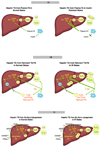
Fig 1
Hepatic TG derives from three sources. (a) The major source is from lipolysis of adipose tissue TG. FA is released, bound to albumin, and circulates to all organs, with the large majority taken up by the liver through several pathways. The major regulation of adipose tissue lipolysis is via insulin signaling (left). In IR, there is increased release of FA throughout the day, with increased delivery to the liver. Studies in individuals with hepatic steatosis indicate that about 80% of hepatic and VLDL TG come from plasma FA. Studies in mice suggest that plasma FA can stimulate apoB secretion, even when the delivery of FA is not great enough to stimulate TG secretion. The result of IR-mediated perturbations is secretion of more VLDL that may also carry more TG (right). (b) VLDL and chylomicrons (not shown) deliver TGFA to adipose tissue via the action of LpL. Approximately 80% of TG in these lipoproteins is removed during this process. The resulting remnant lipoproteins can deliver their remaining TGFA to the liver using several pathways, mainly the LDL receptor. The TGFA enters the lysosome and FA are released (left). In IR, LpL activity may be modestly reduced, leading to TGFA-enriched remnants that deliver more FA to the liver. The result of this IR-mediated perturbation is secretion of more VLDL that can also be larger. (right). (c) When the liver has more energy than it requires, glucose is converted to FA by DNL. Insulin plays the major regulatory role in this process via stimulation of SREBP-1c expression and maturation. Insulin may inhibit MTP expression, thus down-regulating VLDL assembly, as well as targeting apoB for degradation. ChREBP and PPARγ may also stimulate DNL (left). In IR, insulin levels are increased and, although the liver does not respond to insulin in terms of suppression of glucose production and release, DNL is increased. It is not clear if insulin’s negative actions on MTP and apoB are maintained in IR. ChREPB and PPARγ probably add to the stimulation of DNL. The result of increased DNL is the secretion of the same number of larger VLDL (right).
Fig 2
TG that is synthesized in the liver can be disposed of by three processes. FA can be released from TG and oxidized: FA are a major source of energy for the liver. FA can be released from TG and then re-esterified into TG and assembled, with apoB, into VLDL for secretion. Recent studies in mice suggest that hepatocytes can also secrete FA into the blood; there is no evidence for this in humans. Finally, TG can be stored in lipid droplets (left). In IR, increased TG storage in lipid droplets results from increased availability of FA from plasma, remnants, and DNL. There is no evidence that steatosis results from decreased FA oxidation. In fact, the modest evidence that exists in humans suggests that FA oxidation is increased in people with IR. Secretion of TGFA is also increased in IR (right).
Fig 3
ER stress and the associated UPR were first described as responses to inadequate energy in the cell leading to misfolding of proteins in the ER. Recent studies have demonstrated the ER stress can result from “over-nutrition” in the cell, particularly in terms of the delivery of FA. ER stress itself may stimulate DNL, leading to more FA in the hepatocytes. When ER stress is mild or moderate, the increased availability of TG and FA will stimulate both hepatic storage of TG and VLDL secretion (left). If ER stress is more severe, the UPR may actually inhibit VLDL secretion via either increasing co-translational degradation of apoB by the proteasome or stimulating post-ER degradation of apoB and VLDL, possibly via autophagy. In this scenario, which is often coincident with IR, VLDL secretion may be increased, but not enough to prevent significant steatosis, even though DNL may no longer be stimulated (right).
Similar articles
- Circulating very-low-density lipoprotein characteristics resulting from fatty liver in an insulin resistance rat model.
Zago V, Lucero D, Macri EV, Cacciagiú L, Gamba CA, Miksztowicz V, Berg G, Wikinski R, Friedman S, Schreier L. Zago V, et al. Ann Nutr Metab. 2010;56(3):198-206. doi: 10.1159/000276596. Epub 2010 Mar 4. Ann Nutr Metab. 2010. PMID: 20203480 - Methionine restriction prevents the progression of hepatic steatosis in leptin-deficient obese mice.
Malloy VL, Perrone CE, Mattocks DA, Ables GP, Caliendo NS, Orentreich DS, Orentreich N. Malloy VL, et al. Metabolism. 2013 Nov;62(11):1651-61. doi: 10.1016/j.metabol.2013.06.012. Epub 2013 Aug 5. Metabolism. 2013. PMID: 23928105 - GLP-1 Elicits an Intrinsic Gut-Liver Metabolic Signal to Ameliorate Diet-Induced VLDL Overproduction and Insulin Resistance.
Khound R, Taher J, Baker C, Adeli K, Su Q. Khound R, et al. Arterioscler Thromb Vasc Biol. 2017 Dec;37(12):2252-2259. doi: 10.1161/ATVBAHA.117.310251. Epub 2017 Oct 26. Arterioscler Thromb Vasc Biol. 2017. PMID: 29074588 - [Patomechanisms of hepatic steatosis].
Fülöp P, Paragh G. Fülöp P, et al. Orv Hetil. 2010 Feb 28;151(9):323-9. doi: 10.1556/OH.2010.28816. Orv Hetil. 2010. PMID: 20159747 Review. Hungarian. - VLDL Biogenesis and Secretion: It Takes a Village.
van Zwol W, van de Sluis B, Ginsberg HN, Kuivenhoven JA. van Zwol W, et al. Circ Res. 2024 Jan 19;134(2):226-244. doi: 10.1161/CIRCRESAHA.123.323284. Epub 2024 Jan 18. Circ Res. 2024. PMID: 38236950 Review.
Cited by
- ILDR2: an endoplasmic reticulum resident molecule mediating hepatic lipid homeostasis.
Watanabe K, Watson E, Cremona ML, Millings EJ, Lefkowitch JH, Fischer SG, LeDuc CA, Leibel RL. Watanabe K, et al. PLoS One. 2013 Jun 24;8(6):e67234. doi: 10.1371/journal.pone.0067234. Print 2013. PLoS One. 2013. PMID: 23826244 Free PMC article. - Silicon as a Functional Meat Ingredient Improves Jejunal and Hepatic Cholesterol Homeostasis in a Late-Stage Type 2 Diabetes Mellitus Rat Model.
Hernández-Martín M, Garcimartín A, Bocanegra A, Redondo-Castillejo R, Quevedo-Torremocha C, Macho-González A, García Fernández RA, Bastida S, Benedí J, Sánchez-Muniz FJ, López-Oliva ME. Hernández-Martín M, et al. Foods. 2024 Jun 7;13(12):1794. doi: 10.3390/foods13121794. Foods. 2024. PMID: 38928736 Free PMC article. - A nexus of lipid and _O-_Glcnac metabolism in physiology and disease.
Lockridge A, Hanover JA. Lockridge A, et al. Front Endocrinol (Lausanne). 2022 Aug 30;13:943576. doi: 10.3389/fendo.2022.943576. eCollection 2022. Front Endocrinol (Lausanne). 2022. PMID: 36111295 Free PMC article. Review. - Multi-organ Coordination of Lipoprotein Secretion by Hormones, Nutrients and Neural Networks.
Stahel P, Xiao C, Nahmias A, Tian L, Lewis GF. Stahel P, et al. Endocr Rev. 2021 Nov 16;42(6):815-838. doi: 10.1210/endrev/bnab008. Endocr Rev. 2021. PMID: 33743013 Free PMC article. Review.
References
- Reaven GM. Why Syndrome X? from Harold Himsworth to the insulin resistance syndrome. Cell Metab. 2005;1:9–14. - PubMed
- Haffner SM, Lehto S, Ronnemaa T, Pyorala K, Laakso M. Mortality from coronary heart disease in subjects with Type 2 diabetes and in nondiabetic subjects with and without prior myocardial infarction. N Engl J Med. 1998;339:229–234. - PubMed
- Chahil TJ, Ginsberg GN. Diabetic dyslipidemia. Endocrinol Metab Clin N Am. 2006;35:491–510. - PubMed
- Barter PJ, Brewer HB, Chapman MJ, Hennekens CH, Rader DJ, Tall AR. Cholesteryl ester transfer protein: a novel target for raising HDL and inhibiting atherosclerosis. Arterio. Thromb. & Vasc. Biol. 2003;23:160–167. - PubMed
Publication types
MeSH terms
Substances
Grants and funding
- R01 HL055638/HL/NHLBI NIH HHS/United States
- R01 HL055638-15/HL/NHLBI NIH HHS/United States
- R01 HL073030/HL/NHLBI NIH HHS/United States
- R01 HL073030-09/HL/NHLBI NIH HHS/United States
LinkOut - more resources
Full Text Sources
Other Literature Sources


