Enhancement of CA3 hippocampal network activity by activation of group II metabotropic glutamate receptors - PubMed (original) (raw)
Enhancement of CA3 hippocampal network activity by activation of group II metabotropic glutamate receptors
Jeanne Ster et al. Proc Natl Acad Sci U S A. 2011.
Abstract
Impaired function or expression of group II metabotropic glutamate receptors (mGluRIIs) is observed in brain disorders such as schizophrenia. This class of receptor is thought to modulate activity of neuronal circuits primarily by inhibiting neurotransmitter release. Here, we characterize a postsynaptic excitatory response mediated by somato-dendritic mGluRIIs in hippocampal CA3 pyramidal cells and in stratum oriens interneurons. The specific mGluRII agonists DCG-IV or LCCG-1 induced an inward current blocked by the mGluRII antagonist LY341495. Experiments with transgenic mice revealed a significant reduction of the inward current in mGluR3(-/-) but not in mGluR2(-/-) mice. The excitatory response was associated with periods of synchronized activity at theta frequency. Furthermore, cholinergically induced network oscillations exhibited decreased frequency when mGluRIIs were blocked. Thus, our data indicate that hippocampal responses are modulated not only by presynaptic mGluRIIs that reduce glutamate release but also by postsynaptic mGluRIIs that depolarize neurons and enhance CA3 network activity.
Conflict of interest statement
The authors declare no conflict of interest.
Figures
Fig. 1.
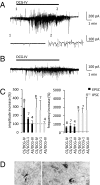
Stimulation of mGluRIIs increases synaptic activity recorded from CA3 pyramidal cells in acute as well as in cultured hippocampal slices. (A) DCG-IV (2 μM) increases amplitude and frequency of EPSCs and IPSCs in slice culture. (Lower) Expanded traces of spontaneous activity in the absence (1) and in the presence of DCG-IV (2). (B) Effects of DCG-IV in an acute slice. (C) Quantification of spontaneous activity (black: EPSCs; gray: IPSCs) induced by DCG-IV and LCCG-1 in cultured slices (CS) or acute slices (AS). (D) Subcellular localization of mGluR2/3 in the CA3 area of hippocampal slice cultures. mGluR2/3 is expressed in the preterminal region and along the length of axons (arrow). Asterisks indicate postsynaptic mGluR2/3 in dendrites in CA3 neurons. (Scale bars: 0.2 μm.)
Fig. 2.
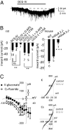
DCG-IV applied to CA3 pyramidal cells induces inward current associated with a decrease in potassium and an increase in cationic conductance. (A) Upper trace shows inward current induced by DCG-IV (2 μM, 10 min) in a CA3 pyramidal cell voltage clamped at −70 mV in the presence of TTX and picrotoxin. (B) Quantification of inward current induced by DCG-IV, by DCG-IV in the presence of the group II antagonist LY341495 or an NMDAR antagonist D-AP5, by LCCG-1, and by LCCG-1 in presence of D-AP5 or LY341495 (Left); quantification of inward current induced by DCG-IV in wild type (WT), mGluR2−/−, and mGluR3−/− mice (Right). (C) Subtracted I/V relationships (Left) in rat pyramidal cells obtained with voltage steps incrementing by 10-mV steps indicating (filled circles) a reduction in K conductance when measured with a K+-based intracellular medium and (open circles) an increase in a cationic conductance with a Cs+-based intracellular medium (n = 6). (Right) Current traces from a representative experiment obtained in the presence of DCG-IV (black line) or in the presence of DCG-IV and CPA (gray line) with a K+-based intracellular medium (Top) or with a Cs+-based intracellular medium (Bottom).
Fig. 3.
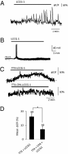
Calcium responses mediated by mGluRIIs. (A) A barrage of calcium transients evoked by LCCG-1 in a CA3 pyramidal cell. (B) In current clamp mode, LCCG-1 induces a depolarization resulting in action potential firing, accompanied by an increase in synaptic noise. (C) Addition of TTX blocks calcium spikes but not the maintained calcium rise induced by LCCG-1. Emptying calcium stores with CPA reduces the calcium rise. (D) Histogram represents the mean Δ_F_/F (%) to illustrate the effect of CPA.
Fig. 4.
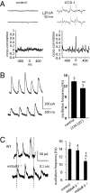
Activating mGluRIIs synchronizes network activity. (A) (Upper) LCCG1 (10 μM, 10 min) in rat slice cultures increases synaptic activity in two unconnected pyramidal cells leading to synchronized synaptic events. (Lower) Cross-correlograms of spontaneous synaptic activity in the two cells during control (Left) and in the presence of LCCG-1 (Right). (B) Methacholine (500 nM) induces oscillations that are significantly decreased by the group II mGluR antagonist LY341495 (n = 5). (C) LCCG1 (10 μM, 10 min) induces oscillations of the same frequency in wild type (WT) and in mGluR2−/−, but at lower frequency in mGluR3−/− mice.
Similar articles
- Reduction of excitatory postsynaptic responses by persistently active metabotropic glutamate receptors in the hippocampus.
Losonczy A, Somogyi P, Nusser Z. Losonczy A, et al. J Neurophysiol. 2003 Apr;89(4):1910-9. doi: 10.1152/jn.00842.2002. J Neurophysiol. 2003. PMID: 12686572 - Group II metabotropic glutamate receptors depress synaptic transmission onto subicular burst firing neurons.
Kintscher M, Breustedt J, Miceli S, Schmitz D, Wozny C. Kintscher M, et al. PLoS One. 2012;7(9):e45039. doi: 10.1371/journal.pone.0045039. Epub 2012 Sep 11. PLoS One. 2012. PMID: 22984605 Free PMC article. - Group II metabotropic glutamate receptors inhibit glutamate release at thalamocortical synapses in the developing somatosensory cortex.
Mateo Z, Porter JT. Mateo Z, et al. Neuroscience. 2007 May 25;146(3):1062-72. doi: 10.1016/j.neuroscience.2007.02.053. Epub 2007 Apr 6. Neuroscience. 2007. PMID: 17418955 Free PMC article. - Enhanced excitatory synaptic network activity following transient group I metabotropic glutamate activation.
Pan YZ, Rutecki PA. Pan YZ, et al. Neuroscience. 2014 Sep 5;275:22-32. doi: 10.1016/j.neuroscience.2014.05.062. Epub 2014 Jun 11. Neuroscience. 2014. PMID: 24928353 - mGluRs modulate strength and timing of excitatory transmission in hippocampal area CA3.
Cosgrove KE, Galván EJ, Barrionuevo G, Meriney SD. Cosgrove KE, et al. Mol Neurobiol. 2011 Aug;44(1):93-101. doi: 10.1007/s12035-011-8187-z. Epub 2011 May 11. Mol Neurobiol. 2011. PMID: 21559753 Review.
Cited by
- TrkB receptor interacts with mGlu2 receptor and mediates antipsychotic-like effects of mGlu2 receptor activation in the mouse.
Philibert CE, Disdier C, Lafon PA, Bouyssou A, Oosterlaken M, Galant S, Pizzoccaro A, Tuduri P, Ster J, Liu J, Kniazeff J, Pin JP, Rondard P, Marin P, Vandermoere F. Philibert CE, et al. Sci Adv. 2024 Jan 26;10(4):eadg1679. doi: 10.1126/sciadv.adg1679. Epub 2024 Jan 26. Sci Adv. 2024. PMID: 38277461 Free PMC article. - Effects of post-training administration of LY341495, as an mGluR2/3 antagonist on spatial memory deficit in rats fed with high-fat diet.
Moridi H, Sarihi A, Habibitabar E, Shateri H, Salehi I, Komaki A, Karimi J, Karimi SA. Moridi H, et al. IBRO Rep. 2020 Sep 25;9:241-246. doi: 10.1016/j.ibror.2020.09.001. eCollection 2020 Dec. IBRO Rep. 2020. PMID: 33024878 Free PMC article. - Metabotropic glutamate receptors-guardians and gatekeepers in neonatal hypoxic-ischemic brain injury.
Mielecki D, Bratek-Gerej E, Salińska E. Mielecki D, et al. Pharmacol Rep. 2024 Dec;76(6):1272-1285. doi: 10.1007/s43440-024-00653-x. Epub 2024 Sep 17. Pharmacol Rep. 2024. PMID: 39289333 Free PMC article. Review. - Modulation of Hippocampal Network Oscillation by PICK1-Dependent Cell Surface Expression of mGlu3 Receptors.
Tuduri P, Bouquier N, Girard B, Moutin E, Thouaye M, Perroy J, Bertaso F, Ster J. Tuduri P, et al. J Neurosci. 2022 Nov 23;42(47):8897-8911. doi: 10.1523/JNEUROSCI.0063-22.2022. Epub 2022 Oct 6. J Neurosci. 2022. PMID: 36202617 Free PMC article. - The G Protein-Coupled Glutamate Receptors as Novel Molecular Targets in Schizophrenia Treatment-A Narrative Review.
Kryszkowski W, Boczek T. Kryszkowski W, et al. J Clin Med. 2021 Apr 2;10(7):1475. doi: 10.3390/jcm10071475. J Clin Med. 2021. PMID: 33918323 Free PMC article. Review.
References
- Manzoni OJ, Castillo PE, Nicoll RA. Pharmacology of metabotropic glutamate receptors at the mossy fiber synapses of the guinea pig hippocampus. Neuropharmacology. 1995;34:965–971. - PubMed
- Scanziani M, Salin PA, Vogt KE, Malenka RC, Nicoll RA. Use-dependent increases in glutamate concentration activate presynaptic metabotropic glutamate receptors. Nature. 1997;385:630–634. - PubMed
- Capogna M. Distinct properties of presynaptic group II and III metabotropic glutamate receptor-mediated inhibition of perforant pathway-CA1 EPSCs. Eur J Neurosci. 2004;19:2847–2858. - PubMed
- Yokoi M, et al. Impairment of hippocampal mossy fiber LTD in mice lacking mGluR2. Science. 1996;273:645–647. - PubMed
Publication types
MeSH terms
Substances
LinkOut - more resources
Full Text Sources
Molecular Biology Databases
Miscellaneous


