The mTOR-regulated phosphoproteome reveals a mechanism of mTORC1-mediated inhibition of growth factor signaling - PubMed (original) (raw)
. 2011 Jun 10;332(6035):1317-22.
doi: 10.1126/science.1199498.
Seong A Kang, Jonathan Rameseder, Yi Zhang, Kathleen A Ottina, Daniel Lim, Timothy R Peterson, Yongmun Choi, Nathanael S Gray, Michael B Yaffe, Jarrod A Marto, David M Sabatini
Affiliations
- PMID: 21659604
- PMCID: PMC3177140
- DOI: 10.1126/science.1199498
The mTOR-regulated phosphoproteome reveals a mechanism of mTORC1-mediated inhibition of growth factor signaling
Peggy P Hsu et al. Science. 2011.
Abstract
The mammalian target of rapamycin (mTOR) protein kinase is a master growth promoter that nucleates two complexes, mTORC1 and mTORC2. Despite the diverse processes controlled by mTOR, few substrates are known. We defined the mTOR-regulated phosphoproteome by quantitative mass spectrometry and characterized the primary sequence motif specificity of mTOR using positional scanning peptide libraries. We found that the phosphorylation response to insulin is largely mTOR dependent and that mTOR exhibits a unique preference for proline, hydrophobic, and aromatic residues at the +1 position. The adaptor protein Grb10 was identified as an mTORC1 substrate that mediates the inhibition of phosphoinositide 3-kinase typical of cells lacking tuberous sclerosis complex 2 (TSC2), a tumor suppressor and negative regulator of mTORC1. Our work clarifies how mTORC1 inhibits growth factor signaling and opens new areas of investigation in mTOR biology.
Figures
Fig. 1. Identification of the mTOR-regulated phosphoproteome
(A) Phosphopeptide abundances were determined from two sets of samples: HEK-293E cells serum starved for 4 hrs, treated with 100 nM rapamycin, 250 nM Torin1, or vehicle control for 1 hr, and then stimulated with 150 nM insulin for 20 min and TSC2+/+ and TSC2−/− MEFs treated with 100 nM Torin1 or vehicle control for 1 hr. (B and C) Distributions of robust z-scores (median absolute deviations (MADs) away from the median (B) log2(Torin1/Insulin) for HEK-293Es or (C) log2(TSC2−/− Torin1/TSC2−/− vehicle) for MEFs). p-values associated with enrichment for known mTOR-modulated sites among the −2.5 MAD Torin1-sensitive phosphopeptides were determined by Fisher’s exact test. Phosphopeptides detected in both replicates had to meet the −2.5 MAD threshold both times to be considered mTOR-regulated. (D, E, and F) Correspondence between (D) Torin1 treatment and serum deprivation in HEK-293Es, (E) Torin1 and rapamycin treatment in HEK-293Es, and (F) Torin1 treatment and upregulation in TSC2−/− MEFs. The relevant robust z-scores for both replicates, phosphopeptides corresponding to known mTOR-modulated sites, Spearman’s rank correlation coefficient (ρ), and associated p-values are indicated. Outliers were excluded to aid in visualization.
Fig. 2. Characterization of a consensus mTOR phosphorylation motif
(A) The position-specific scoring matrix (PSSM) resulting from quantification of the in vitro phosphorylation of a position scoring peptide library (PSPL) by mTORC1. (B) The visualized mTOR consensus motif. Letter height is proportional to the PSSM score. Only those selected residues with scores greater than a standard deviation from the average PSSM score within a row are shown. (C and D) Classification of the mTOR-regulated phosphopeptides in (C) HEK-293E and (D) MEFs organized by rapamycin sensitivity (−2.5 MAD (log2 (Rapamycin/Insulin)) or TSC2 upregulation (+2.5 MAD log2(TSC2−/− vehicle/TSC2+/+ vehicle)), consistency with the mTOR motif (5th percentile by Scansite), or presence of an AGC motif ((R/K)X(R/K)XX(S*/T*)). The numbers represent the number of unique phosphopeptides or proteins. Refer to Figs. S5, S6 and Table S4 for more details.
Fig. 3. Grb10 as an mTORC1 substrate with rapamycin-sensitive and -insensitive sites
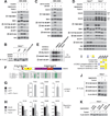
(A) HEK-293E cells were deprived of serum for 4 hrs, treated with 100 nM rapamycin or 250 nM Torin1 for 1 hr, and then stimulated with 150 nM insulin for 15 min. Cell lysates were analyzed by immunoblotting. (B) TSC2+/+ MEFs stably expressing FLAG-Grb10 were serum deprived for 4 hours, treated with 250 nM Torin1 for 1 hr, and then stimulated with 150 nM insulin for 15 min. All FLAG-tagged Grb10 constructs correspond to isoform c of human Grb10. FLAG-immunoprecipitates were incubated in buffer, CIP, or heat-inactivated CIP and analyzed by immunoblotting. (C) HEK-293E cells were deprived of amino acids or both amino acids and serum for 50 min, and then stimulated with either amino acids or serum for 10 min and analyzed by immunoblotting. (D) TSC2+/+ and TSC2−/− MEFs were treated and analyzed as in (A). (E) mTORC1 in vitro kinase assays with substrates in the presence of the indicated inhibitors and radiolabeled ATP were analyzed by autoradiography. (F) Schematic representation of Grb10 protein structure with the phosphorylation sites from vertebrate orthologs aligned below. Numbering is according to human isoform a. (G) The phosphorylation state of Grb10 from kinase assays performed similarly to (E) were analyzed by targeted mass spectrometry (MS) and phosphorylation ratios determined from chromatographic peak intensities. (H) FLAG-immunoprecipitates from HEK-293E cells stably expressing FLAG-Grb10 treated as in (A) were analyzed as in (G). Data are means ± s.e.m (n=2–6). *Mann-Whitney t-test p-values < 0.05 for differences between stimulated and treated conditions. (I) A summary of (F), (G), and (H) for each Grb10 phosphorylation site. (J) FLAG-immunoprecipitates from TSC2−/− MEFs stably expressing FLAG-Grb10 treated with 100 nM rapamycin or 250 nM Torin1 for 1 hr were analyzed by immunoblotting with Grb10 phospho-specific antibodies. (K) TSC2−/− MEFs stably expressing FLAG-Grb10, 5A (S150A T155A S158A S474A S476A), or 9A (5A + S104A S426A S428A S431A) mutants treated with 250 nM Torin1 for 1 hr were analyzed by immunoblotting.
Fig. 4. mTORC1 inhibits PI3K-Akt signaling by regulating Grb10 function and stability
(A) S6K1−/− S6K2−/− or control cells expressing short hairpin RNA (shRNA) constructs against GFP or raptor were treated with 250 nM Torin1 for 1 hr, and lysates were analyzed by immunoblotting. (B) TSC2−/− MEFs expressing shRNAs against GFP or Grb10 were deprived of serum for 4 hrs and then stimulated with 100 nM insulin for 15 min as indicated and analyzed by immunoblotting. (C) TSC2−/− MEFs expressing a control shRNA or shRNA against Grb10 were treated as in (B). IRS1 and IRS2 immunoprecipitates and cell lysates were analyzed by immunoblotting. (D) TSC2−/− MEFs coexpressing an shRNA against the mouse Grb10 3’UTR and an empty vector, FLAG-Grb10, or 5A cDNA expression construct were treated and analyzed as in (B). (E) TSC2−/− MEFs stably expressing FLAG-Grb10 were labeled for 2 hours with [35S]cysteine and methionine and then chased for the indicated times in the presence of vehicle control, 100 nM rapamycin, or 100 nM Torin1. FLAG-immunoprecipitates were analyzed by autoradiography. Data are means ± s.e.m (n=3). *Two-way ANOVA p-values < 0.05 for differences between vehicle and inhibitor treatment. (F) TSC2−/− MEFs stably expressing FLAG-Grb10 or 9A mutant were treated and analyzed as in (E) but without inhibitor treatment. (G) mTORC1 orchestrates feedback inhbition of PI3K-Akt signaling by activating and stabilizing Grb10 while inhibiting and destabilizing IRS proteins.
Comment in
- Cell signaling. New mTOR targets Grb attention.
Yea SS, Fruman DA. Yea SS, et al. Science. 2011 Jun 10;332(6035):1270-1. doi: 10.1126/science.1208071. Science. 2011. PMID: 21659593 No abstract available.
Similar articles
- Phosphoproteomic analysis identifies Grb10 as an mTORC1 substrate that negatively regulates insulin signaling.
Yu Y, Yoon SO, Poulogiannis G, Yang Q, Ma XM, Villén J, Kubica N, Hoffman GR, Cantley LC, Gygi SP, Blenis J. Yu Y, et al. Science. 2011 Jun 10;332(6035):1322-6. doi: 10.1126/science.1199484. Science. 2011. PMID: 21659605 Free PMC article. - Alkaline intracellular pH (pHi) increases PI3K activity to promote mTORC1 and mTORC2 signaling and function during growth factor limitation.
Kazyken D, Lentz SI, Wadley M, Fingar DC. Kazyken D, et al. J Biol Chem. 2023 Sep;299(9):105097. doi: 10.1016/j.jbc.2023.105097. Epub 2023 Jul 26. J Biol Chem. 2023. PMID: 37507012 Free PMC article. - RhoA modulates signaling through the mechanistic target of rapamycin complex 1 (mTORC1) in mammalian cells.
Gordon BS, Kazi AA, Coleman CS, Dennis MD, Chau V, Jefferson LS, Kimball SR. Gordon BS, et al. Cell Signal. 2014 Mar;26(3):461-7. doi: 10.1016/j.cellsig.2013.11.035. Epub 2013 Dec 3. Cell Signal. 2014. PMID: 24316235 Free PMC article. - The Role of Mammalian Target of Rapamycin (mTOR) in Insulin Signaling.
Yoon MS. Yoon MS. Nutrients. 2017 Oct 27;9(11):1176. doi: 10.3390/nu9111176. Nutrients. 2017. PMID: 29077002 Free PMC article. Review. - LKB1 and AMP-activated protein kinase control of mTOR signalling and growth.
Shaw RJ. Shaw RJ. Acta Physiol (Oxf). 2009 May;196(1):65-80. doi: 10.1111/j.1748-1716.2009.01972.x. Epub 2009 Feb 19. Acta Physiol (Oxf). 2009. PMID: 19245654 Free PMC article. Review.
Cited by
- The functions and regulation of the PTEN tumour suppressor.
Song MS, Salmena L, Pandolfi PP. Song MS, et al. Nat Rev Mol Cell Biol. 2012 Apr 4;13(5):283-96. doi: 10.1038/nrm3330. Nat Rev Mol Cell Biol. 2012. PMID: 22473468 Review. - Diabetic cardiomyopathy and metabolic remodeling of the heart.
Battiprolu PK, Lopez-Crisosto C, Wang ZV, Nemchenko A, Lavandero S, Hill JA. Battiprolu PK, et al. Life Sci. 2013 Mar 28;92(11):609-15. doi: 10.1016/j.lfs.2012.10.011. Epub 2012 Oct 30. Life Sci. 2013. PMID: 23123443 Free PMC article. Review. - Staying alive: metabolic adaptations to quiescence.
Valcourt JR, Lemons JM, Haley EM, Kojima M, Demuren OO, Coller HA. Valcourt JR, et al. Cell Cycle. 2012 May 1;11(9):1680-96. doi: 10.4161/cc.19879. Epub 2012 May 1. Cell Cycle. 2012. PMID: 22510571 Free PMC article. Review. - The butterfly effect in cancer: a single base mutation can remodel the cell.
Hart JR, Zhang Y, Liao L, Ueno L, Du L, Jonkers M, Yates JR 3rd, Vogt PK. Hart JR, et al. Proc Natl Acad Sci U S A. 2015 Jan 27;112(4):1131-6. doi: 10.1073/pnas.1424012112. Epub 2015 Jan 12. Proc Natl Acad Sci U S A. 2015. PMID: 25583473 Free PMC article. - Dynamic regulation of the translation initiation helicase complex by mitogenic signal transduction to eukaryotic translation initiation factor 4G.
Dobrikov MI, Dobrikova EY, Gromeier M. Dobrikov MI, et al. Mol Cell Biol. 2013 Mar;33(5):937-46. doi: 10.1128/MCB.01441-12. Epub 2012 Dec 21. Mol Cell Biol. 2013. PMID: 23263986 Free PMC article.
References
- Dowling RJ, Topisirovic I, Fonseca BD, Sonenberg N. Biochim Biophys Acta. 2010;1804:433. - PubMed
- Ross PL, et al. Mol Cell Proteomics. 2004;3:1154. - PubMed
Publication types
MeSH terms
Substances
Grants and funding
- ES015339/ES/NIEHS NIH HHS/United States
- R01 ES015339/ES/NIEHS NIH HHS/United States
- HHMI/Howard Hughes Medical Institute/United States
- T32 GM007753/GM/NIGMS NIH HHS/United States
- CA112967/CA/NCI NIH HHS/United States
- R37 AI047389/AI/NIAID NIH HHS/United States
- R01 CA129105/CA/NCI NIH HHS/United States
- P50 GM068762/GM/NIGMS NIH HHS/United States
- AI47389/AI/NIAID NIH HHS/United States
- R01 AI047389/AI/NIAID NIH HHS/United States
- R01 CA103866/CA/NCI NIH HHS/United States
- CA103866/CA/NCI NIH HHS/United States
- GM68762/GM/NIGMS NIH HHS/United States
- U54 CA112967/CA/NCI NIH HHS/United States
LinkOut - more resources
Full Text Sources
Other Literature Sources
Molecular Biology Databases
Research Materials
Miscellaneous


