Sequential assembly of flagellar radial spokes - PubMed (original) (raw)
Sequential assembly of flagellar radial spokes
Dennis R Diener et al. Cytoskeleton (Hoboken). 2011 Jul.
Abstract
The unicellular alga Chlamydomonas can assemble two 10 μm flagella in 1 h from proteins synthesized in the cell body. Targeting and transporting these proteins to the flagella are simplified by preassembly of macromolecular complexes in the cell body. Radial spokes are flagellar complexes that are partially assembled in the cell body before entering the flagella. On the axoneme, radial spokes are "T" shaped structures with a head of five proteins and a stalk of 18 proteins that sediment together at 20S. In the cell body, radial spokes are partially assembled; about half of the radial spoke proteins (RSPs) form a 12S complex. In mutants lacking a single RSP, smaller spoke subassemblies were identified. When extracts from two such mutants were mixed in vitro the 12S complex was assembled from several smaller complexes demonstrating that portions of the stepwise assembly of radial spoke assembly can be carried out in vitro to elucidate the order of spoke assembly in the cell body.
Copyright © 2011 Wiley-Liss, Inc.
Figures
Figure 1
RSP1-6 colelute from a molecular sieve column. Fractions of cell body extract eluted from a Sephadex S500 column were analyzed on immunoblots probed for RSPs as indicated on the right. The rightmost lane contains axonemes (Ax) as a standard. Column fractions are listed above the blots. Note the peak of RSP1-6 around fraction 39 (diffusion coefficient approximately 1.4 × 10⁻⁷ cm2/sec). RSP16 eluted separately, later than the other RSPs. RSP3 is known to have multiple phosphorylation forms [Piperno et al, 1981; Williams et al., 1989] and so appears as multiple bands in this and subsequent blots.
Figure 2
The 12S radial spoke complex is composed of at least 11 RSPs. Wild-type and pf14 cells were metabolically labeled with 35S during flagellar regeneration and the 12S radial spoke complex was isolated from cell body extracts. Autoradiography of 2-dimensional gels shows 11 RSPs were present in the complex. These RSPs are reduced in extracts from pf14 cells, which are almost devoid of the 12S complex. RSP3 labeling is low even in wild-type extracts, presumably because it contains no cysteine residues although it contains 11 methionine residues.
Figure 3
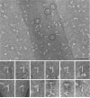
The 12S spoke complex is shaped like a “7”. The 12S fractions from a gradient of soluble flagellar proteins were pooled and the RSPs were further purified by ionic exchange chromatography. Negative stained preparations showed a stalk with a projection at one end resembling a “7” or “L”. The four encircled structures are shown at twice the magnification below along with 5 other examples of the 12S complex. For comparison the last three panels show images from the 20S spoke fraction after similar purification; two images are of the intact, “T”-shaped spoke and the final image shows a complex similar to that seen in the 12S fraction. The scale bar is 100 nm.
Figure 4
The 12S radial spoke complex does not assemble in the absence of RSP3 or 4. These immunoblots of gradients of cell body extracts from pf14 (reduced amount of a truncated RSP3) and pf1 (lacking RSP4) cells show the sedimentation of the RSPs from these mutant cells. The small amount of the 12S complex in pf14 (third panel on the left) contains truncated RSP3 (3m, arrows) in both phosphorylated (upper) and not phosphorylated (lower) states. RSP4 and 6 are designated in the fourth row. The asterisks mark a peak that contains spoke head proteins RSP4, 6, 9 and 10. Pairs of adjacent gradient fractions were pooled and loaded in each lane of these gels for a more compact presentation. The axonemal markers (Ax) are from wild-type flagella and can be used to differentiate the specific RSP bands from several specious bands present in the blots. In the RSP4/6 panels, for example, the upper band (without an arrow) is not an RSP.
Figure 5
The 12S complex can assemble in vitro in a mixture of extracts from pf24 and pf1. These immunoblots show cytoplasmic extracts analyzed as in Fig. 4. The extracts from the individual mutants do not contain the 12S complex, but after mixing all of the RSPs have a secondary peak at 12S (box) indicating that this complex was assembled in vitro.
Figure 6
Radial spoke heads can assemble onto radial spoke stalks in vitro. Cell body extracts from pf1 and pf14 cells were analyzed on sucrose gradients either separately or after mixing. Immunoblots were probed for RSP1 and 3. Note that the 16S (*) and 6.4S (**) peaks of RSP3 present in the pf1 extract disappear in the mixed extracts concomitant with the appearance of the 20S peak.
Figure 7
This diagram illustrates the various forms of radial spoke complexes from the cytoplasm of wild-type and three radial spoke mutants. The composition of the 6.4S complex in pf1 and pf24 is not known except that it contains RSP3. The 20S and 16S complexes were not always detectable in cell extracts of wild-type and pf1 cells. The flagellar forms of spoke complexes are shown on the right, bound to an outer doublet microtubule. The stalks in the flagella of pf24 have reduced amounts of RSP16 and 23 along with RSP2 and the head proteins [Yang et al., 2005].
Figure 8
This diagram illustrates the minimum assembly required to generate the 12S and 20S complexes in vitro from cytoplasmic extracts of radial spoke mutants as shown in Figs. 5 and 6.
Figure 9
The 12S radial spoke complex is assembled in the cell body and transported by IFT to the flagellar tip where it combines with the other RSPs to form the 20S mature spoke. Little is known about the assembly state of RSP8, 13-23 or when they attach to the 12S complex. The 20S complex is shown as a dimer of the 12S complex to illustrate how the asymmetric head of the 12S complex could give rise to the symmetric head of the mature spoke. The stalks of the two 12S complexes are shown intertwined to represent the helical quality sometimes seen in the stalk [Qin et al., 2004]. The 12S complexes may bind to a docking complex already present on the axonemal microtubules, possibly the CSC [Dymek and Smith, 2007].
Similar articles
- The Chlamydomonas mutant pf27 reveals novel features of ciliary radial spoke assembly.
Alford LM, Mattheyses AL, Hunter EL, Lin H, Dutcher SK, Sale WS. Alford LM, et al. Cytoskeleton (Hoboken). 2013 Dec;70(12):804-18. doi: 10.1002/cm.21144. Cytoskeleton (Hoboken). 2013. PMID: 24124175 Free PMC article. - Assembly of flagellar radial spoke proteins in Chlamydomonas: identification of the axoneme binding domain of radial spoke protein 3.
Diener DR, Ang LH, Rosenbaum JL. Diener DR, et al. J Cell Biol. 1993 Oct;123(1):183-90. doi: 10.1083/jcb.123.1.183. J Cell Biol. 1993. PMID: 8408197 Free PMC article. - Dimeric novel HSP40 is incorporated into the radial spoke complex during the assembly process in flagella.
Yang C, Compton MM, Yang P. Yang C, et al. Mol Biol Cell. 2005 Feb;16(2):637-48. doi: 10.1091/mbc.e04-09-0787. Epub 2004 Nov 24. Mol Biol Cell. 2005. PMID: 15563613 Free PMC article. - Functional diversity of axonemal dyneins as studied in Chlamydomonas mutants.
Kamiya R. Kamiya R. Int Rev Cytol. 2002;219:115-55. doi: 10.1016/s0074-7696(02)19012-7. Int Rev Cytol. 2002. PMID: 12211628 Review. - Flagellar radial spoke: a model molecular genetic system for studying organelle assembly.
Curry AM, Rosenbaum JL. Curry AM, et al. Cell Motil Cytoskeleton. 1993;24(4):224-32. doi: 10.1002/cm.970240403. Cell Motil Cytoskeleton. 1993. PMID: 8477455 Review. No abstract available.
Cited by
- FAP206 is a microtubule-docking adapter for ciliary radial spoke 2 and dynein c.
Vasudevan KK, Song K, Alford LM, Sale WS, Dymek EE, Smith EF, Hennessey T, Joachimiak E, Urbanska P, Wloga D, Dentler W, Nicastro D, Gaertig J. Vasudevan KK, et al. Mol Biol Cell. 2015 Feb 15;26(4):696-710. doi: 10.1091/mbc.E14-11-1506. Epub 2014 Dec 24. Mol Biol Cell. 2015. PMID: 25540426 Free PMC article. - Novel mutations in LRRC23 cause asthenozoospermia in a nonconsanguineous family.
Tang SX, Liu SY, Xiao H, Zhang X, Xiao Z, Zhou S, Ding YL, Yang P, Chen Q, Huang HL, Chen X, Lin X, Zhou HL, Liu MX. Tang SX, et al. Asian J Androl. 2024 Sep 1;26(5):484-489. doi: 10.4103/aja202435. Epub 2024 Jul 26. Asian J Androl. 2024. PMID: 39054792 Free PMC article. - Heme-binding protein CYB5D1 is a radial spoke component required for coordinated ciliary beating.
Zhao L, Xie H, Kang Y, Lin Y, Liu G, Sakato-Antoku M, Patel-King RS, Wang B, Wan C, King SM, Zhao C, Huang K. Zhao L, et al. Proc Natl Acad Sci U S A. 2021 Apr 27;118(17):e2015689118. doi: 10.1073/pnas.2015689118. Proc Natl Acad Sci U S A. 2021. PMID: 33875586 Free PMC article. - The mouse radial spoke protein 3 is a nucleocytoplasmic shuttling protein that promotes neurogenesis.
Yan R, Hu X, Zhang W, Song L, Wang J, Yin Y, Chen S, Zhao S. Yan R, et al. Histochem Cell Biol. 2015 Oct;144(4):309-19. doi: 10.1007/s00418-015-1338-y. Epub 2015 Jun 17. Histochem Cell Biol. 2015. PMID: 26082196 - The versatile molecular complex component LC8 promotes several distinct steps of flagellar assembly.
Gupta A, Diener DR, Sivadas P, Rosenbaum JL, Yang P. Gupta A, et al. J Cell Biol. 2012 Jul 9;198(1):115-26. doi: 10.1083/jcb.201111041. Epub 2012 Jul 2. J Cell Biol. 2012. PMID: 22753897 Free PMC article.
References
- Avidor-Reiss T, Maer AM, Koundakjian E, Polyanovsky A, Keil T, Subramaniam S, Zuker CS. Decoding cilia function: defining specialized genes required for compartmentalized cilia biogenesis. Cell. 2004;117(4):527–539. - PubMed
- Curry AM, Rosenbaum JL. Flagellar radial spoke: a model molecular genetic system for studying organelle assembly. Cell Motil Cytoskeleton. 1993;24:224–232. - PubMed
Publication types
MeSH terms
Substances
Grants and funding
- R01 GM051173/GM/NIGMS NIH HHS/United States
- R01 GM014642/GM/NIGMS NIH HHS/United States
- R15 GM090162/GM/NIGMS NIH HHS/United States
- R37 GM014642/GM/NIGMS NIH HHS/United States
- R37GM51173/GM/NIGMS NIH HHS/United States
- R37 GM051173/GM/NIGMS NIH HHS/United States
- GM90162/GM/NIGMS NIH HHS/United States
- GM14642/GM/NIGMS NIH HHS/United States
LinkOut - more resources
Full Text Sources
Other Literature Sources


