Genomic and protein structural maps of adaptive evolution of human influenza A virus to increased virulence in the mouse - PubMed (original) (raw)
doi: 10.1371/journal.pone.0021740. Epub 2011 Jun 30.
Liya Keleta, Nicole E Forbes, Samar Dankar, William Stecho, Shaun Tyler, Yan Zhou, Lorne Babiuk, Hana Weingartl, Rebecca A Halpin, Alex Boyne, Jayati Bera, Jessicah Hostetler, Nadia B Fedorova, Katie Proudfoot, Dan A Katzel, Tim B Stockwell, Elodie Ghedin, David J Spiro, Earl G Brown
Affiliations
- PMID: 21738783
- PMCID: PMC3128085
- DOI: 10.1371/journal.pone.0021740
Genomic and protein structural maps of adaptive evolution of human influenza A virus to increased virulence in the mouse
Jihui Ping et al. PLoS One. 2011.
Abstract
Adaptive evolution is characterized by positive and parallel, or repeated selection of mutations. Mouse adaptation of influenza A virus (IAV) produces virulent mutants that demonstrate positive and parallel evolution of mutations in the hemagglutinin (HA) receptor and non-structural protein 1 (NS1) interferon antagonist genes. We now present a genomic analysis of all 11 genes of 39 mouse adapted IAV variants from 10 replicate adaptation experiments. Mutations were mapped on the primary and structural maps of each protein and specific mutations were validated with respect to virulence, replication, and RNA polymerase activity. Mouse adapted (MA) variants obtained after 12 or 20-21 serial infections acquired on average 5.8 and 7.9 nonsynonymous mutations per genome of 11 genes, respectively. Among a total of 115 nonsynonymous mutations, 51 demonstrated properties of natural selection including 27 parallel mutations. The greatest degree of parallel evolution occurred in the HA receptor and ribonucleocapsid components, polymerase subunits (PB1, PB2, PA) and NP. Mutations occurred in host nuclear trafficking factor binding sites as well as sites of virus-virus protein subunit interaction for NP, NS1, HA and NA proteins. Adaptive regions included cap binding and endonuclease domains in the PB2 and PA polymerase subunits. Four mutations in NS1 resulted in loss of binding to the host cleavage and polyadenylation specificity factor (CPSF30) suggesting that a reduction in inhibition of host gene expression was being selected. The most prevalent mutations in PB2 and NP were shown to increase virulence but differed in their ability to enhance replication and demonstrated epistatic effects. Several positively selected RNA polymerase mutations demonstrated increased virulence associated with >300% enhanced polymerase activity. Adaptive mutations that control host range and virulence were identified by their repeated selection to comprise a defined model for studying IAV evolution to increased virulence in the mouse.
Conflict of interest statement
Competing Interests: The authors have declared that no competing interests exist.
Figures
Figure 1. Experimental design of parallel studies of mouse adaptation.
The parental strain of A/Hong Kong/1/68 (HK-wt) was clonally derived on MDCK cells and grown in chicken embryos before dilution in PBS to 1×10⁵ pfu/ 0.05 mL to initiate serial mouse passage; followed by 20 serial passages with 6 clones derived by plaque isolation from the passage 12 and 20 populations on MDCK cells. Replicate stocks of HK-wt (HK-#) were generated from individual infectious virions of HK-wt by plaque isolation. Each subclone was amplified in eggs and used without dilution to infect mice (≥1×10⁶ pfu/0.05 mL for each mouse) to initiate 9 parallel MA series as indicated in methods before isolating 3 clones from each of the passage 21 populations.
Figure 2. Mouse adaptive mutations on the primary structural maps of PB2, PB1, PA and NP proteins.
The amino acid location of mutations are numbered and indicated with arrowheads on the linear sequence, sites of positive selection are shown red and parallel mutations are additionally indicated with an asterisk and the number of populations in parenthesis. The locations of regions of interaction, or functions are indicated with rectangles and are labeled with respect to interacting viral proteins as indicated in Methods. Nuclear localization signals (NLS) are in black, and host protein sites are indicated for PB2, PA and NP; PB1 polymerase activity regions are in purple and PB2 cap binding regions are in orange. hCLE, the human transcription factor is positioned In PA according to The following mutations were mapped previously: PB2 D701N, PA Q556R, NP D34N, and NP D480N.
Figure 3. Mouse adaptive mutations on PB2 three dimensional maps.
Mutation sites are shown on ribbon structures of PB2 protein with space filling models of amino acids numbered in black for mutations found once, or red for positively selected mutations. (A) PB2 cap binding domain bound to m7GTP (in stick image); (B) PB2 C-terminal domain with the 627 site shown in stick image. (C) PB2 NLS with R753 (stick image) that forms a salt bridge with D701, NLS in red; (D) PB2 NLS in complex with human importin α5; NLS in red.
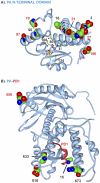
Figure 4. Mouse adaptive mutations on PA and PB1 three dimensional maps.
Images are shown as in Fig. 3. A) PA N-terminal domain with nuclease site active residues, H41, E80, D108, E119 and K134 shown in stick diagram; manganese ions are shown with purple spheres. (B) PA (blue) PB1 (red) complex; PA amino acids 1–14 are in direct contact with PB2.
Figure 5. Mouse adaptive mutations on NP protein trimer three dimensional maps.
A). The mutations are shown on the asymmetric NP trimer of subunits A (blue), B (grey) and C (gold). Mutations are shown in numbered space filling models on the ribbon backbone of subunit A with the exception to position 480 that is shown on both subunits A and B. Mutations are numbered and shown as described in Fig. 3. Mutation sites in contact with NP A and B subunits in the oligomer are: M426(B) to M448+E449(A); A428(B) to R261(A), V476(B) to D482+S483(A); and D480(A) to M481(B). B). Side view of trimmer showing the clustering of mutation on alternate faces that define adaptive domains.
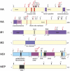
Figure 6. Mouse adaptive mutations on the primary structural maps of HA, NA, M1, M2, NS1 and NEP proteins.
Mutations are shown as indicated for Figure 3. The locations of protein binding and active sites are indicated; RNP ribonucleocapsid protein; NLS nuclear localization signal; NES nuclear export signal; dsRNA double-stranded RNA (aa 1–73); PABPI poly-A binding protein 1; PABPII poly-A binding protein II; RIG-I retinoic acid inducible gene I; E1B-AP5, E1B associated protein 5; CPSF cleavage and polyadenylation specificity factor; eIF4GI eukaryotic initiation factor 4GI; and PKR protein kinase R. The following mutations were mapped previously: HA1 G218W, HA2 T156N, NA P468H, M1 D232N, NS1 F103L + V23A, NEP K88R .
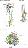
Figure 7. Mouse adaptive mutations in the crystal structure of the HA protein monomer, trimer and low-pH HA2 trimer.
Mutations are shown as described in Fig. 3. (A) the side view of the HA monomer composed of HA1 and HA2 (indicated with superscripts 1 and 2) with carbohydrate side chains shown (CHO) in stick diagram is included here for reference (a similar but unglycosylated map that was generated from an independent sequence analysis has been published [10]); (B) the low pH form of HA2; and (C) the HA trimer, top view with receptor sites indicated with arrows.
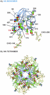
Figure 8. Mouse adaptive mutations on NA three dimensional maps.
Mutations are shown as described in Fig. 3. (A) Mutations are shown in the NA monomer with receptor site indicated with SA and carbohydrate (CHO) in stick diagram; (B) the tetrameric form of NA.
Figure 9. M2 tetramer and NEP three dimensional maps of mouse adaptive mutations.
Mutations are shown as described in Fig. 3. (A) on the M2 tetramer and (B) the NEP protein.
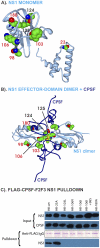
Figure 10. NS1 three dimensional maps of mouse adaptive mutations and effects on CPSF binding.
Mutations are shown as numbered space filling images as described in Fig. 3. (A) in the NS1 monomer; (B) the NS1 dimer effector domain (grey) bound to 2 CPSF F2F3 fragments (dark blue). Amino acid NS1 106 of each monomer is in direct contact in the dimer. CPSF30-F2F3 is in direct contact with NS1 amino acids 103, 106 and 180. (C) Coimmunoprecipitation of HK-wt and V23A, F103L, M106V, M106I, M106I+L98S, and V180A mutant NS1 proteins with FLAG-CPSF30-F2/F3. Recombinant NS1 proteins (2.0 µg) were mixed with equivalent amounts of FLAG-tagged CPSF30-F2/F3 before blotting in parallel using anti-NS1 or anti-FLAG monoclonal antibody respectively to demonstrate the input. Pull down samples were blotted in side-by-side comparisons for immunoglobulin (as a loading standard) and NS1 protein to demonstrate association of NS1 with CPSF30-F2/F3.
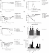
Figure 11. Assessment of the roles of PB2, PA and NP mutations in mouse models of virulence, replication and polymerase activity.
Body weight and survival were monitored for groups of mice infected with recombinant HK viruses that possessed mouse adaptive mutations. (A and B), r-HK-wt and PB2 mutants, (K482R, D701N, D740N, D701N+D740N) were used to infect groups of 4 mice with 5×10⁶ pfu of virus for body weight loss and with n = 7 for survival. (C and D) r-HK-wt and mutant viruses with NP mutations D34N, D290N, D209E, or PB2 D701N+NP D34N were used as indicated to infect groups of 4 mice with 5×10⁶ pfu of virus and with n = 7 for survival. (E and F) r-HK and r-HK + MA20-PA (Q556R) as well as r-MA20 and r-MA20 + HK-wt-PA were used to infect groups of 5 CD-1 mice with 1×10⁵ pfu of each with monitoring of weight loss and mortality. Weight loss was assessed relative to r-HK-wt infections using the single sample t-test at day 2 pi or using the paired t-test for 2–6 dpi; time to death was significant as indicated using student's t-test, P<0.05 indicated with *. (G) Replication in mouse lungs was monitored 1 dpi after infection of groups of 3 mice with 5×10³ pfu each. The values are means of infections yields ±SD for groups of 3 mice. (H) Polymerase activity was measured in mouse B82 cells for each of the indicated mutations relative to luciferase minigenome expression via a mouse POL1 polymerase by HK-wt plasmids expressing PB1, PB2, PA and NP and firefly luciferase driven by the NP promoter. Values are means ±SD for n = 3 samples. Statistical significance relative to HK-wt at the P≤0.05 and P≤0.01 are indicated with * and ** respectively.
Similar articles
- PB2 and hemagglutinin mutations are major determinants of host range and virulence in mouse-adapted influenza A virus.
Ping J, Dankar SK, Forbes NE, Keleta L, Zhou Y, Tyler S, Brown EG. Ping J, et al. J Virol. 2010 Oct;84(20):10606-18. doi: 10.1128/JVI.01187-10. Epub 2010 Aug 11. J Virol. 2010. PMID: 20702632 Free PMC article. - Multifunctional adaptive NS1 mutations are selected upon human influenza virus evolution in the mouse.
Forbes NE, Ping J, Dankar SK, Jia JJ, Selman M, Keleta L, Zhou Y, Brown EG. Forbes NE, et al. PLoS One. 2012;7(2):e31839. doi: 10.1371/journal.pone.0031839. Epub 2012 Feb 21. PLoS One. 2012. PMID: 22363747 Free PMC article. - Genetic analysis of mouse-adapted influenza A virus identifies roles for the NA, PB1, and PB2 genes in virulence.
Brown EG, Bailly JE. Brown EG, et al. Virus Res. 1999 May;61(1):63-76. doi: 10.1016/s0168-1702(99)00027-1. Virus Res. 1999. PMID: 10426210 - An overview of influenza A virus genes, protein functions, and replication cycle highlighting important updates.
Chauhan RP, Gordon ML. Chauhan RP, et al. Virus Genes. 2022 Aug;58(4):255-269. doi: 10.1007/s11262-022-01904-w. Epub 2022 Apr 26. Virus Genes. 2022. PMID: 35471490 Review. - Modulation of Innate Immune Responses by the Influenza A NS1 and PA-X Proteins.
Nogales A, Martinez-Sobrido L, Topham DJ, DeDiego ML. Nogales A, et al. Viruses. 2018 Dec 12;10(12):708. doi: 10.3390/v10120708. Viruses. 2018. PMID: 30545063 Free PMC article. Review.
Cited by
- Structural and functional characterization of K339T substitution identified in the PB2 subunit cap-binding pocket of influenza A virus.
Liu Y, Qin K, Meng G, Zhang J, Zhou J, Zhao G, Luo M, Zheng X. Liu Y, et al. J Biol Chem. 2013 Apr 19;288(16):11013-23. doi: 10.1074/jbc.M112.392878. Epub 2013 Feb 22. J Biol Chem. 2013. PMID: 23436652 Free PMC article. - Genomics of adaptation during experimental evolution of the opportunistic pathogen Pseudomonas aeruginosa.
Wong A, Rodrigue N, Kassen R. Wong A, et al. PLoS Genet. 2012 Sep;8(9):e1002928. doi: 10.1371/journal.pgen.1002928. Epub 2012 Sep 13. PLoS Genet. 2012. PMID: 23028345 Free PMC article. - Full-length genome sequences of the first H9N2 avian influenza viruses isolated in the Northeast of Algeria.
Barberis A, Boudaoud A, Gorrill A, Loupias J, Ghram A, Lachheb J, Alloui N, Ducatez MF. Barberis A, et al. Virol J. 2020 Jul 17;17(1):108. doi: 10.1186/s12985-020-01377-z. Virol J. 2020. PMID: 32680533 Free PMC article. - Establishment of a mouse- and egg-adapted strain for the evaluation of vaccine potency against H3N2 variant influenza virus in mice.
Bazarragchaa E, Hiono T, Isoda N, Hayashi H, Okamatsu M, Sakoda Y. Bazarragchaa E, et al. J Vet Med Sci. 2021 Oct 31;83(11):1694-1701. doi: 10.1292/jvms.21-0350. Epub 2021 Sep 14. J Vet Med Sci. 2021. PMID: 34526415 Free PMC article. - Integrated Analysis of microRNA-mRNA Expression in Mouse Lungs Infected With H7N9 Influenza Virus: A Direct Comparison of Host-Adapting PB2 Mutants.
Guo Y, Huang N, Tian M, Fan M, Liu Q, Liu Z, Sun T, Huang J, Xia H, Zhao Y, Ping J. Guo Y, et al. Front Microbiol. 2020 Jul 28;11:1762. doi: 10.3389/fmicb.2020.01762. eCollection 2020. Front Microbiol. 2020. PMID: 32849388 Free PMC article.
References
- Kuiken T, Holmes EC, McCauley J, Rimmelzwaan GF, Williams CS, et al. Host species barriers to influenza virus infections. Science. 2006;312:394–397. - PubMed
- Alizon S, Hurford A, Mideo N, Van BM. Virulence evolution and the trade-off hypothesis: history, current state of affairs and the future. J Evol Biol. 2009;22:245–259. JEB1658 [pii];10.1111/j.1420-9101.2008.01658.x [doi] - PubMed
Publication types
MeSH terms
Substances
LinkOut - more resources
Full Text Sources
Research Materials
Miscellaneous


