Quantitation of 7-ethylguanine in leukocyte DNA from smokers and nonsmokers by liquid chromatography-nanoelectrospray-high resolution tandem mass spectrometry - PubMed (original) (raw)
. 2011 Oct 17;24(10):1729-34.
doi: 10.1021/tx200262d. Epub 2011 Sep 14.
Affiliations
- PMID: 21859140
- PMCID: PMC3215090
- DOI: 10.1021/tx200262d
Quantitation of 7-ethylguanine in leukocyte DNA from smokers and nonsmokers by liquid chromatography-nanoelectrospray-high resolution tandem mass spectrometry
Silvia Balbo et al. Chem Res Toxicol. 2011.
Abstract
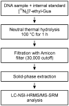
There is considerable evidence for the exposure of humans to an unknown ethylating agent, and some studies indicate that cigarette smoking may be one source of this exposure. Therefore, we have developed a liquid chromatography-nanoelectrospray-high resolution tandem mass spectrometry-selected reaction monitoring (LC-NSI-HRMS/MS-SRM) method for the analysis of 7-ethyl-Gua in human leukocyte DNA, a readily available source of DNA. [(15)N(5)]7-Ethyl-Gua was used as the internal standard. Leukocyte DNA was isolated and treated by thermal neutral hydrolysis. The hydrolysate was partially purified by solid-phase extraction. The fraction containing 7-ethyl-Gua was analyzed by LC-NSI-HRMS/MS-SRM using the transition m/z 180 [M + H](+)→ m/z 152.05669 [Gua + H](+) for 7-ethyl-Gua and m/z 185 → m/z 157.04187 for the internal standard. The detection limit was approximately 10 amol on column, while the limit of quantitation was about 8 fmol/μmol Gua starting with 180 μg DNA (corresponding to 36 μg DNA on-column). Leukocyte DNA samples from 30 smokers and 30 nonsmokers were analyzed. Clear peaks for 7-ethyl-Gua and the internal standard were observed in most of the samples. The mean (±SD) level of 7-ethyl-Gua measured in leukocyte DNA from smokers was 49.6 ± 43.3 (range 14.6-181) fmol/μmol Gua, while that from nonsmokers was 41.3 ± 34.9 (range 9.64-157) fmol/μmol Gua. Although a significant difference between smokers and nonsmokers was not observed, the method described here is unique in the use of high resolution mass spectrometry and establishes for the first time the presence of 7-ethyl-Gua in human leukocyte DNA.
Figures
Figure 1
Chromatograms obtained upon analysis of a standard mixture of 10 amol of 7-ethyl-Gua and 5 fmol [15N5]7-ethyl-Gua using LC-NSI-HRMS/MS-SRM. Panel (A) shows the results from the transition at m/z 180 [M + H]+ → m/z 152.05669 [Gua + H]+ for 7-ethyl-Gua. Panel (B) shows the corresponding transition m/z 185 [M + H]+ → m/z 157.04187 [Gua + H]+ for the internal standard. Results are shown with a 5 ppm mass tolerance.
Figure 2
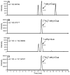
Chromatograms obtained upon LC-NSI-HRMS/MS-SRM analysis of human leukocyte DNA (129 μg, 12.9 μg on column) containing 59.4 fmol/μmol Gua. The relatively higher amount of analyte in this sample allowed confirmation of its identity by additional monitoring of the accurate mass of the molecular ion of 7-ethyl-Gua and the internal standard. Panel (A) shows the result from monitoring of the accurate mass of 7-ethyl-Gua (m/z 180.08799). Panel (B) shows the result from the monitoring of the accurate mass of [15N5]7-ethyl-Gua (m/z 185.07317). Panel (C) shows the results from the transition at m/z 180 [M + H]+ → m/z 152.05669 [Gua + H]+ for 7-ethyl-Gua and panel (D) shows the corresponding transition m/z 185 [M + H]+ → m/z 157.04187 [Gua + H]+ for the internal standard. Results are shown with a 5 ppm mass tolerance.
Figure 3
Relationship of added to detected 7-ethyl-Gua. Various amounts of the DNA adduct were added to calf thymus DNA (0.3 mg, 30 μg on column) and analyzed by the method described in the text; R2 = 0.99. 7-Ethyl-Gua present in the calf thymus DNA was subtracted from each value
Figure 4
Chromatograms obtained upon LC-NSI-HRMS/MS-SRM analysis of human leukocyte DNA (14 μg, 1,4 μg on column) for 7-ethyl-Gua by the method described in the text. This sample contained 70.2 fmol/μmol Gua of 7-ethyl-Gua corresponding to 130 amol injected on column (1 μl injected out of a 10 μl sample). Panel (A), analyte; panel (B), internal standard. Results are shown with a 5 ppm mass tolerance.
Scheme 1
Analytical scheme for determination of 7-ethyl-Gua in human leukocyte DNA
Similar articles
- Liquid chromatography-electrospray ionization tandem mass spectrometry analysis of 7-ethylguanine in human liver DNA.
Chen L, Wang M, Villalta PW, Hecht SS. Chen L, et al. Chem Res Toxicol. 2007 Oct;20(10):1498-502. doi: 10.1021/tx700147f. Epub 2007 Sep 22. Chem Res Toxicol. 2007. PMID: 17887725 - Quantitation by liquid chromatography-nanoelectrospray ionization-high resolution tandem mass spectrometry of DNA adducts derived from methyl glyoxal and carboxyethylating agents in leukocytes of smokers and non-smokers.
Cheng G, Reisinger SA, Shields PG, Hatsukami DK, Balbo S, Hecht SS. Cheng G, et al. Chem Biol Interact. 2020 Aug 25;327:109140. doi: 10.1016/j.cbi.2020.109140. Epub 2020 May 20. Chem Biol Interact. 2020. PMID: 32442416 Free PMC article. - Quantitation by Liquid Chromatography-Nanoelectrospray Ionization-High-Resolution Tandem Mass Spectrometry of Multiple DNA Adducts Related to Cigarette Smoking in Oral Cells in the Shanghai Cohort Study.
Cheng G, Guo J, Wang R, Yuan JM, Balbo S, Hecht SS. Cheng G, et al. Chem Res Toxicol. 2023 Feb 20;36(2):305-312. doi: 10.1021/acs.chemrestox.2c00393. Epub 2023 Jan 31. Chem Res Toxicol. 2023. PMID: 36719849 Free PMC article.
Cited by
- Metabolic Activation and DNA Interactions of Carcinogenic _N_-Nitrosamines to Which Humans Are Commonly Exposed.
Li Y, Hecht SS. Li Y, et al. Int J Mol Sci. 2022 Apr 20;23(9):4559. doi: 10.3390/ijms23094559. Int J Mol Sci. 2022. PMID: 35562949 Free PMC article. Review. - Detection of Benzo[a]pyrene Diol Epoxide Adducts to Histidine and Lysine in Serum Albumin In Vivo by High-Resolution-Tandem Mass Spectrometry.
Zurita J, Motwani HV, Ilag LL, Souliotis VL, Kyrtopoulos SA, Nilsson U, Törnqvist M. Zurita J, et al. Toxics. 2022 Jan 8;10(1):27. doi: 10.3390/toxics10010027. Toxics. 2022. PMID: 35051069 Free PMC article. - Mass Spectrometric Quantitation of Apurinic/Apyrimidinic Sites in Tissue DNA of Rats Exposed to Tobacco-Specific Nitrosamines and in Lung and Leukocyte DNA of Cigarette Smokers and Nonsmokers.
Guo J, Chen H, Upadhyaya P, Zhao Y, Turesky RJ, Hecht SS. Guo J, et al. Chem Res Toxicol. 2020 Sep 21;33(9):2475-2486. doi: 10.1021/acs.chemrestox.0c00265. Epub 2020 Sep 9. Chem Res Toxicol. 2020. PMID: 32833447 Free PMC article. - Ultrasensitive High-Resolution Mass Spectrometric Analysis of a DNA Adduct of the Carcinogen Benzo[a]pyrene in Human Lung.
Villalta PW, Hochalter JB, Hecht SS. Villalta PW, et al. Anal Chem. 2017 Dec 5;89(23):12735-12742. doi: 10.1021/acs.analchem.7b02856. Epub 2017 Nov 17. Anal Chem. 2017. PMID: 29111668 Free PMC article. - Occurrence, Biological Consequences, and Human Health Relevance of Oxidative Stress-Induced DNA Damage.
Yu Y, Cui Y, Niedernhofer LJ, Wang Y. Yu Y, et al. Chem Res Toxicol. 2016 Dec 19;29(12):2008-2039. doi: 10.1021/acs.chemrestox.6b00265. Epub 2016 Nov 7. Chem Res Toxicol. 2016. PMID: 27989142 Free PMC article. Review.
References
- U.S. Department of Health and Human Services. How Tobacco Smoke Causes Disease: The Biology and Behavioral Basis for Smoking-Attributable Disease: A Report of the Surgeon General. Atlanta, GA: U.S. Department of Health and Human Services, Centers for Disease Control and Prevention, National Center for Chronic Disease Prevention and Health Promotion, Office on Smoking and Health; 2010.
- Singh R, Kaur B, Farmer PB. Detection of DNA damage derived from a direct acting ethylating agent present in cigarette smoke by use of liquid chromatography-tandem mass spectrometry. Chem Res Toxicol. 2005;18:249–256. - PubMed
- Kopplin A, Eberle-Adamkiewicz G, Glusenkamp KH, Nehls P, Kirstein U. Urinary excretion of 3-methyladenine and 3-ethyladenine after controlled exposure to tobacco smoke. Carcinogenesis. 1995;16:2637–2641. - PubMed
- Chao MR, Wang CJ, Chang LW, Hu CW. Quantitative determination of urinary N7-ethylguanine in smokers and non-smokers using an isotope dilution liquid chromatography/tandem mass spectrometry with on-line analyte enrichment. Carcinogenesis. 2006;27:146–151. - PubMed
Publication types
MeSH terms
Substances
Grants and funding
- CA-77598/CA/NCI NIH HHS/United States
- P30 CA077598/CA/NCI NIH HHS/United States
- S10 RR024618/RR/NCRR NIH HHS/United States
- P30 CA077598-14/CA/NCI NIH HHS/United States
- N01PC64402/PC/NCI NIH HHS/United States
- N01 PC64402/PC/NCI NIH HHS/United States
- S10-RR-024618/RR/NCRR NIH HHS/United States
- N01-PC-64402/PC/NCI NIH HHS/United States
LinkOut - more resources
Full Text Sources


