Increased pathogenicity of a reassortant 2009 pandemic H1N1 influenza virus containing an H5N1 hemagglutinin - PubMed (original) (raw)
Increased pathogenicity of a reassortant 2009 pandemic H1N1 influenza virus containing an H5N1 hemagglutinin
Troy D Cline et al. J Virol. 2011 Dec.
Abstract
A novel H1N1 influenza virus emerged in 2009 (pH1N1) to become the first influenza pandemic of the 21st century. This virus is now cocirculating with highly pathogenic H5N1 avian influenza viruses in many parts of the world, raising concerns that a reassortment event may lead to highly pathogenic influenza strains with the capacity to infect humans more readily and cause severe disease. To investigate the virulence of pH1N1-H5N1 reassortant viruses, we created pH1N1 (A/California/04/2009) viruses expressing individual genes from an avian H5N1 influenza strain (A/Hong Kong/483/1997). Using several in vitro models of virus replication, we observed increased replication for a reassortant CA/09 virus expressing the hemagglutinin (HA) gene of HK/483 (CA/09-483HA) relative to that of either parental CA/09 virus or reassortant CA/09 expressing other HK/483 genes. This increased replication correlated with enhanced pathogenicity in infected mice similar to that of the parental HK/483 strain. The serial passage of the CA/09 parental virus and the CA/09-483HA virus through primary human lung epithelial cells resulted in increased pathogenicity, suggesting that these viruses easily adapt to humans and become more virulent. In contrast, serial passage attenuated the parental HK/483 virus in vitro and resulted in slightly reduced morbidity in vivo, suggesting that sustained replication in humans attenuates H5N1 avian influenza viruses. Taken together, these data suggest that reassortment between cocirculating human pH1N1 and avian H5N1 influenza strains will result in a virus with the potential for increased pathogenicity in mammals.
Figures
Fig. 1.
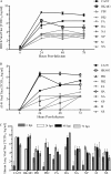
Replicative capacity of reassortant pH1N1-H5N1 viruses in vitro. MDCK (A) and A549 (B) cells were infected at an MOI of 0.01, cell culture supernatants were collected at 6, 24, 48, and 72 hpi, and viral titers were determined by TCID50 analysis. (C) NHBE cells were infected at an MOI of 0.03 for 2 h at 37°C. Following infection, the cells were maintained in culture at an air-liquid interface. At 6, 24, 48, and 72 hpi, medium was added to the apical surface for 30 min and collected, and viral titers were determined by TCID50 analysis in triplicate. Data are representative of at least two experiments. Error bars represent the standard errors of the means. Individual influenza virus genes indicate reassortant CA/09 virus expressing a single gene from HK/483 virus.
Fig. 2.
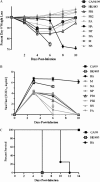
Pathogenicity of reassortant pH1N1-H5N1 viruses in vivo. Female 6- to 8-week-old BALB/c mice (n = 10) were intranasally infected with 103 TCID50 units of the indicated viruses, and weight loss (A) and survival (B) were monitored for 14 dpi. (C) At 3, 6, and 10 dpi, lungs were collected from three mice per group, and viral titers were determined in homogenates by TCID50 analysis. Data are representative of at least two experiments, and error bars represent the standard errors of the means. Individual influenza virus genes indicate reassortant CA/09 virus expressing a single gene from HK/483 virus.
Fig. 3.
Histological analysis of CA/09-483HA infection. Female 6- to 8-week-old BALB/c mice (n = 3) were inoculated with 103 TCID50 units of the indicated viruses. Lung tissue was collected at 3 or 6 dpi, fixed in 10% buffered formalin, processed, and embedded in paraffin. Four-micron-thick sections were stained with hematoxylin and eosin.
Fig. 4.
Replication of serially passaged viruses in vitro. (A) NHBE cells were inoculated with the indicated viruses at an MOI of 0.03 at 37°C for 2 h. At 24, 48, and 72 hpi medium was added to the apical surface for 30 min and collected, and viral titers were determined by TCID50 analysis in triplicate. This represents one passage (p1). Equal TCID50 doses of each virus from the sample of p1 at 72 h was used to infect another monolayer of NHBE cells at an MOI of 0.03. This procedure was repeated until p3 virus was collected. Error bars represent the standard errors of the means. (B) At 48 hpi, inserts from NHBE cells infected with the p2 viruses were fixed, stained for the viral NP (green) and nuclei by DAPI (blue), and visualized by confocal microscopy.
Fig. 5.
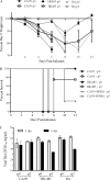
In vivo pathogenicity of serially passaged viruses. Female 6- to 8-week-old BALB/c mice (n = 10) were intranasally inoculated with 103 TCID50 units of the indicated viruses, and weight loss (A) and survival (B) were monitored for 12 dpi. (C) At 3 and 6 dpi, lungs were collected from three mice per group and viral titers were determined in homogenates by TCID50 analysis. Error bars represent the standard errors of the means.
Fig. 6.
Transmissibility of CA/09-483HA in ferrets. Male 9- to 15-week-old ferrets (n = 2) were intranasally inoculated with 104 TCID50 units of the indicated viruses. Twenty-four hpi, two naïve contact ferrets were introduced and housed together with the inoculated ferrets for the duration of the experiment. Nasal washes were collected, and viral titers were determined by TCID50 analysis at 1, 3, 5, and 7 dpi for the inoculated ferrets and at 3, 5, 7, and 10 days postexposure for the contact ferrets. Error bars represent standard errors of the means.
Similar articles
- Experimental adaptation of an influenza H5 HA confers respiratory droplet transmission to a reassortant H5 HA/H1N1 virus in ferrets.
Imai M, Watanabe T, Hatta M, Das SC, Ozawa M, Shinya K, Zhong G, Hanson A, Katsura H, Watanabe S, Li C, Kawakami E, Yamada S, Kiso M, Suzuki Y, Maher EA, Neumann G, Kawaoka Y. Imai M, et al. Nature. 2012 May 2;486(7403):420-8. doi: 10.1038/nature10831. Nature. 2012. PMID: 22722205 Free PMC article. - H5N1 hybrid viruses bearing 2009/H1N1 virus genes transmit in guinea pigs by respiratory droplet.
Zhang Y, Zhang Q, Kong H, Jiang Y, Gao Y, Deng G, Shi J, Tian G, Liu L, Liu J, Guan Y, Bu Z, Chen H. Zhang Y, et al. Science. 2013 Jun 21;340(6139):1459-63. doi: 10.1126/science.1229455. Epub 2013 May 2. Science. 2013. PMID: 23641061 - Reassortment between Avian H5N1 and human influenza viruses is mainly restricted to the matrix and neuraminidase gene segments.
Schrauwen EJ, Bestebroer TM, Rimmelzwaan GF, Osterhaus AD, Fouchier RA, Herfst S. Schrauwen EJ, et al. PLoS One. 2013;8(3):e59889. doi: 10.1371/journal.pone.0059889. Epub 2013 Mar 20. PLoS One. 2013. PMID: 23527283 Free PMC article. - Transmission of influenza A/H5N1 viruses in mammals.
Imai M, Herfst S, Sorrell EM, Schrauwen EJ, Linster M, De Graaf M, Fouchier RA, Kawaoka Y. Imai M, et al. Virus Res. 2013 Dec 5;178(1):15-20. doi: 10.1016/j.virusres.2013.07.017. Epub 2013 Aug 13. Virus Res. 2013. PMID: 23954580 Free PMC article. Review. - Progress in identifying virulence determinants of the 1918 H1N1 and the Southeast Asian H5N1 influenza A viruses.
Basler CF, Aguilar PV. Basler CF, et al. Antiviral Res. 2008 Sep;79(3):166-78. doi: 10.1016/j.antiviral.2008.04.006. Epub 2008 May 23. Antiviral Res. 2008. PMID: 18547656 Free PMC article. Review.
Cited by
- Polysaccharide Nanoparticles from Isatis indigotica Fort. Root Decoction: Diversity, Cytotoxicity, and Antiviral Activity.
Gao G, He C, Wang H, Guo J, Ke L, Zhou J, Chong PH, Rao P. Gao G, et al. Nanomaterials (Basel). 2021 Dec 23;12(1):30. doi: 10.3390/nano12010030. Nanomaterials (Basel). 2021. PMID: 35009980 Free PMC article. - Primary Swine Respiratory Epithelial Cell Lines for the Efficient Isolation and Propagation of Influenza A Viruses.
Meliopoulos V, Cherry S, Wohlgemuth N, Honce R, Barnard K, Gauger P, Davis T, Shult P, Parrish C, Schultz-Cherry S. Meliopoulos V, et al. J Virol. 2020 Nov 23;94(24):e01091-20. doi: 10.1128/JVI.01091-20. Print 2020 Nov 23. J Virol. 2020. PMID: 32967961 Free PMC article. - Respiratory Bacteria Stabilize and Promote Airborne Transmission of Influenza A Virus.
Rowe HM, Livingston B, Margolis E, Davis A, Meliopoulos VA, Echlin H, Schultz-Cherry S, Rosch JW. Rowe HM, et al. mSystems. 2020 Sep 1;5(5):e00762-20. doi: 10.1128/mSystems.00762-20. mSystems. 2020. PMID: 32873612 Free PMC article. - Direct interactions with influenza promote bacterial adherence during respiratory infections.
Rowe HM, Meliopoulos VA, Iverson A, Bomme P, Schultz-Cherry S, Rosch JW. Rowe HM, et al. Nat Microbiol. 2019 Aug;4(8):1328-1336. doi: 10.1038/s41564-019-0447-0. Epub 2019 May 20. Nat Microbiol. 2019. PMID: 31110359 Free PMC article. - Absence of β6 Integrin Reduces Influenza Disease Severity in Highly Susceptible Obese Mice.
Meliopoulos V, Livingston B, Van de Velde LA, Honce R, Schultz-Cherry S. Meliopoulos V, et al. J Virol. 2019 Jan 4;93(2):e01646-18. doi: 10.1128/JVI.01646-18. Print 2019 Jan 15. J Virol. 2019. PMID: 30381485 Free PMC article.
References
- Ashburn D. D., DeAntonio A., Reed M. J. 2010. Pulmonary system and obesity. Crit. Care Clin. 26:597–602 - PubMed
- Berhane Y., et al. 2010. Molecular characterization of pandemic H1N1 influenza viruses isolated from turkeys and pathogenicity of a human pH1N1 isolate in turkeys. Avian Dis. 54:1275–1285 - PubMed
Publication types
MeSH terms
Substances
LinkOut - more resources
Full Text Sources
Medical


