Initial evaluation of a direct detection device detector for single particle cryo-electron microscopy - PubMed (original) (raw)
Initial evaluation of a direct detection device detector for single particle cryo-electron microscopy
Anna-Clare Milazzo et al. J Struct Biol. 2011 Dec.
Abstract
We report on initial results of using a new direct detection device (DDD) for single particle reconstruction of vitreous ice embedded specimens. Images were acquired on a Tecnai F20 at 200keV and a nominal magnification of 29,000×. This camera has a significantly improved signal to noise ratio and modulation transfer function (MTF) at 200keV compared to a standard CCD camera installed on the same microscope. Control of the DDD has been integrated into Leginon, an automated data collection system. Using GroEL as a test specimen, we obtained images of ∼30K particles with the CCD and the DDD from the same specimen sample using essentially identical imaging conditions. Comparison of the maps reconstructed from the CCD images and the DDD images demonstrates the improved performance of the DDD. We also obtained a 3D reconstruction from ∼70K GroEL particles acquired using the DDD; the quality of the density map demonstrates the potential of this new recording device for cryoEM data acquisition.
Copyright © 2011 Elsevier Inc. All rights reserved.
Conflict of interest statement
Conflicts of Interest
Prior to the research for this publication, A.-C.M. received consulting fees from Direct Electron for unrelated professional services.
Figures
Figure 1
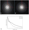
Comparison of detection limit between cameras installed on the same microscope using identical instrument conditions and almost matching pixel size. Fourier transforms and radially averaged power spectra of 3K × 3K cropped images from: (A) DDD (1.38Å/pixel) and (B) CCD (1.37Å/pixel). High tension 200 KeV, electron dose ~20e−/Å2, exposure time 400ms. The modulation transfer function is shown in (C) for the CCD and DDD cameras calculated using the edge method.
Figure 2
Representative cropped 2000 × 2000 pixel micrographs of GroEL. (A) DDD image (1.38Å/pixel), (B) CCD image (1.37Å/pixel). Defocus value is approximately 2μm for both images. Scale bar is 40nm.
Figure 3
GroEL reconstructions from ~30K particle data sets comparing DDD in (A) and the CCD camera in (B). The final volumes with no post processing are shown in (a) and (e). Maps shown in (b) and (f) were amplitude corrected with an X-ray scattering curve out to 8Å and low pass filtered to 7.5Å. Volumes shown in (c) and (g) were amplitude corrected with an X-ray scattering curve out to 5Å and low pass filtered to 6Å. The Fourier Shell Correlation plots for (a) and (e) are shown in (d) and (h).
Figure 4
A high resolution DDD reconstruction from a 72,316 particle dataset is shown in (A), low pass filtered to 4.5Å. In (B), the Fourier Shell Correlation, derived from reconstructed even-odd particle data sets, gives the FSC0.5 as 6.1Å. For comparison, we show an asymmetric subunit from the DDD map in (C) and previously published asymmetric subunits from a 4Å map acquired using film in (D) (Ludtke et al., 2008) and a 5.4Å map from a CCD camera in (E) (Stagg et al., 2008).
Similar articles
- Automated cryoEM data acquisition and analysis of 284742 particles of GroEL.
Stagg SM, Lander GC, Pulokas J, Fellmann D, Cheng A, Quispe JD, Mallick SP, Avila RM, Carragher B, Potter CS. Stagg SM, et al. J Struct Biol. 2006 Sep;155(3):470-81. doi: 10.1016/j.jsb.2006.04.005. Epub 2006 May 22. J Struct Biol. 2006. PMID: 16762565 - Assessing the capabilities of a 4kx4k CCD camera for electron cryo-microscopy at 300kV.
Booth CR, Jakana J, Chiu W. Booth CR, et al. J Struct Biol. 2006 Dec;156(3):556-63. doi: 10.1016/j.jsb.2006.08.019. Epub 2006 Sep 22. J Struct Biol. 2006. PMID: 17067819 - A test-bed for optimizing high-resolution single particle reconstructions.
Stagg SM, Lander GC, Quispe J, Voss NR, Cheng A, Bradlow H, Bradlow S, Carragher B, Potter CS. Stagg SM, et al. J Struct Biol. 2008 Jul;163(1):29-39. doi: 10.1016/j.jsb.2008.04.005. Epub 2008 May 6. J Struct Biol. 2008. PMID: 18534866 Free PMC article. - Cryo-Electron Tomography and Subtomogram Averaging.
Wan W, Briggs JA. Wan W, et al. Methods Enzymol. 2016;579:329-67. doi: 10.1016/bs.mie.2016.04.014. Epub 2016 Jun 22. Methods Enzymol. 2016. PMID: 27572733 Review. - Processing of Cryo-EM Movie Data.
Ripstein ZA, Rubinstein JL. Ripstein ZA, et al. Methods Enzymol. 2016;579:103-24. doi: 10.1016/bs.mie.2016.04.009. Epub 2016 Jun 1. Methods Enzymol. 2016. PMID: 27572725 Review.
Cited by
- Computational Methodologies for Real-Space Structural Refinement of Large Macromolecular Complexes.
Goh BC, Hadden JA, Bernardi RC, Singharoy A, McGreevy R, Rudack T, Cassidy CK, Schulten K. Goh BC, et al. Annu Rev Biophys. 2016 Jul 5;45:253-78. doi: 10.1146/annurev-biophys-062215-011113. Epub 2016 May 2. Annu Rev Biophys. 2016. PMID: 27145875 Free PMC article. Review. - Montage electron tomography of vitrified specimens.
Peck A, Carter SD, Mai H, Chen S, Burt A, Jensen GJ. Peck A, et al. J Struct Biol. 2022 Jun;214(2):107860. doi: 10.1016/j.jsb.2022.107860. Epub 2022 Apr 26. J Struct Biol. 2022. PMID: 35487464 Free PMC article. - Confessions of an icosahedral virus crystallographer.
Johnson JE. Johnson JE. Microscopy (Oxf). 2013 Feb;62(1):69-79. doi: 10.1093/jmicro/dfs097. Epub 2013 Jan 4. Microscopy (Oxf). 2013. PMID: 23291268 Free PMC article. Review. - Beam-induced motion of vitrified specimen on holey carbon film.
Brilot AF, Chen JZ, Cheng A, Pan J, Harrison SC, Potter CS, Carragher B, Henderson R, Grigorieff N. Brilot AF, et al. J Struct Biol. 2012 Mar;177(3):630-7. doi: 10.1016/j.jsb.2012.02.003. Epub 2012 Feb 16. J Struct Biol. 2012. PMID: 22366277 Free PMC article. - Molecular dynamics-based refinement and validation for sub-5 Å cryo-electron microscopy maps.
Singharoy A, Teo I, McGreevy R, Stone JE, Zhao J, Schulten K. Singharoy A, et al. Elife. 2016 Jul 7;5:e16105. doi: 10.7554/eLife.16105. Elife. 2016. PMID: 27383269 Free PMC article.
References
- Battaglia M, Contarato D, Denes P, Doering D, Giubilato P, et al. A rad-hard CMOS active pixel sensor for electron microscopy. Nucl Instrum Methods A. 2009;598:642–649.
- Cheng A, Fellmann D, Pulokas J, Potter CS, Carragher B. Does contamination buildup limit throughput for automated cryoEM? J Struct Biol. 2006;154:303–311. - PubMed
- Cheng Y, Walz T. The advent of near-atomic resolution in single-particle electron microscopy. Annu Rev Biochem. 2009;78:723–742. - PubMed
- Contarato Devis, Denes Peter, Doering Dionisio, Joseph John, Krieger Brad. Direct detection in Transmission Electron Microscopy with a 5 micron pitch CMOS pixel sensor. Nucl Instrum Methods Phys Res A. 2011;635:69–73.
Publication types
MeSH terms
Substances
Grants and funding
- P50GM073197/GM/NIGMS NIH HHS/United States
- RR004050/RR/NCRR NIH HHS/United States
- R01 RR018841/RR/NCRR NIH HHS/United States
- RR018841/RR/NCRR NIH HHS/United States
- P41 RR004050/RR/NCRR NIH HHS/United States
- P41 RR004050-23/RR/NCRR NIH HHS/United States
- P50 GM073197-08/GM/NIGMS NIH HHS/United States
- P41 RR017573-10/RR/NCRR NIH HHS/United States
- P41 RR017573/RR/NCRR NIH HHS/United States
- RR17573/RR/NCRR NIH HHS/United States
- P50 GM073197/GM/NIGMS NIH HHS/United States
- P41 RR004050-24/RR/NCRR NIH HHS/United States
- R01 RR018841-06/RR/NCRR NIH HHS/United States
LinkOut - more resources
Full Text Sources
Other Literature Sources
Research Materials
Miscellaneous


