Imaging of the bursae - PubMed (original) (raw)
Imaging of the bursae
Zameer Hirji et al. J Clin Imaging Sci. 2011.
Abstract
When assessing joints with various imaging modalities, it is important to focus on the extraarticular soft tissues that may clinically mimic joint pathology. One such extraarticular structure is the bursa. Bursitis can clinically be misdiagnosed as joint-, tendon- or muscle-related pain. Pathological processes are often a result of inflammation that is secondary to excessive local friction, infection, arthritides or direct trauma. It is therefore important to understand the anatomy and pathology of the common bursae in the appendicular skeleton. The purpose of this pictorial essay is to characterize the clinically relevant bursae in the appendicular skeleton using diagrams and corresponding multimodality images, focusing on normal anatomy and common pathological processes that affect them. The aim is to familiarize radiologists with the radiological features of bursitis.
Keywords: Bursae; computed tomography; imaging; interventions; magnetic resonance; ultrasound.
Conflict of interest statement
Conflict of Interest: None declared.
Figures
Figure 1
Diagram of normal bursae surrounding the shoulder joint: (1) subacromial-subdeltoid bursa, (2) subscapular recess, (3) subcoracoid bursa, (4) coracoclavicular bursa, (5) supra-acromial bursa and (6) medial extension of subacromial-subdeltoid bursa.
Figure 2
Coronal magnetic resonance arthrogram depicting gadolinium in the SASD bursa from a full-thickness rotator cuff tear.
Figure 3
Transverse ultrasound image of the rotator cuff depicting acute subacromial-subdeltoid bursitis.
Figure 4
(a) Line diagram and (b) corresponding magnetic resonance arthrogram of the subcoracoid bursa.
Figure 5
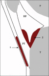
Line diagram showing the superficial and deep infrapatellar bursae. (1) Superficial infrapatellar bursa, (2) deep infrapatellar, femur (F), Hoffa's fat pad (HF), patella (P), patellar tendon (PT) and tibia (T).
Figure 6
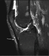
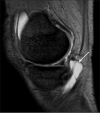
Sagittal magnetic resonance T2 fat sat image depicting the superficial infrapatellar bursa.
Figure 7
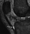
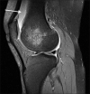
Sagittal magnetic resonance T2 gradient showing deep infrapatellar bursal fluid.
Figure 8
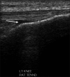
Ultrasound in the longitudinal plane showing deep infrapatellar bursal fluid.
Figure 9
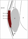
Line diagram showing compartmentalization of the prepatellar bursa. SCCT - subcutaneous cellular tissues; QT - quadriceps tendon; PT - patellar tendon; F - femur; P, patella; (1) superficial compartment; (2) intermediate compartment; (3) deep compartment.
Figure 10
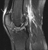
Sagittal magnetic resonance T2 fat sat image showing high-signal fluid intensity within the prepatellar bursitis.
Figure 11
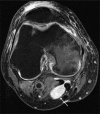
Axial magnetic resonance T2 fat sat image depicting Baker's cyst with its neck between the semimembranosus and the medial gastrocnemius tendons. Image also depicts prepatellar bursitis.
Figure 12
Longitudinal ultrasound image demonstrating fluid within the suprapatellar bursa.
Figure 13
Sagittal magnetic resonance T2 gradient image demonstrating fluid within the suprapatellar bursa.
Figure 14
Axial magnetic resonance T2 fat sat image showing semimembranosus bursa.
Figure 15
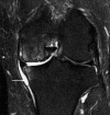
Coronal magnetic resonance T2 Short TI Inversion Recovery (STIR) image showing semimembranosus bursa.
Figure 16
Sagittal magnetic resonance gradient T2 image showing semimembranosus bursa.
Figure 17
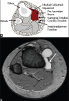
(a) Axial line diagram and (b) axial magnetic resonance image showing pes anserine bursitis
Figure 18
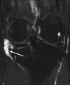
Coronal magnetic resonance T2 fat sat image showing fluid within the medial collateral ligament bursa.
Figure 19
Axial magnetic resonance T2 fat sat image showing fluid within the medial collateral ligament bursa.
Figure 20
Sagittal magnetic resonance T2 fat sat image showing retrocalcaneal bursitis with a thick synovial wall.
Figure 21
Longitudinal ultrasound image showing superficial retrocalcaneal bursa.
Figure 22
(a) Axial magnetic resonance T2 and (b) coronal magnetic resonance T2 STIR images of the left hip demonstrate a fluid-filled structure deep to the iliopsoas muscle in the expected location of the iliopsoas bursa. The iliopsoas tendon can be seen medial to the bursa and the femoral vessels can be seen further medially
Figure 23
Coronal line diagram of the trochanteric bursa overlying the posterior facet of the greater trochanter deep to the gluteus medius tendon.
Figure 24
Coronal magnetic resonance T2 STIR image of the right trochanteric bursitis.
Figure 25
Coronal magnetic resonance T2 STIR image of the bilateral subgluteus medius bursitis, larger on the right side.
Figure 26
Coronal magnetic resonance STIR image of the right subgluteus minimus bursitis.
Figure 27
Line diagram depicting location of the olecranon bursa.
Figure 28
Magnetic resonance T2 fat sat image with fluid in the olecranon bursa.
Figure 29
Longitudinal ultrasound image demonstrating fluid and debris in the olecranon bursa.
Figure 30
(a) Enhanced axial computed tomography of the right iliopsoas bursitis and (b) ultrasound-guided drainage.
Figure 31
(a) Ultrasound- and (b) fluoroscopy-guided trochanteric bursal injection.
Similar articles
- Beyond the greater trochanter: a pictorial review of the pelvic bursae.
Friedman MV, Stensby JD, Long JR, Currie SA, Hillen TJ. Friedman MV, et al. Clin Imaging. 2017 Jan-Feb;41:37-41. doi: 10.1016/j.clinimag.2016.09.010. Epub 2016 Sep 28. Clin Imaging. 2017. PMID: 27764718 Review. - Ultrasound evaluation of bursae: anatomy and pathological appearances.
Ruangchaijatuporn T, Gaetke-Udager K, Jacobson JA, Yablon CM, Morag Y. Ruangchaijatuporn T, et al. Skeletal Radiol. 2017 Apr;46(4):445-462. doi: 10.1007/s00256-017-2577-x. Epub 2017 Feb 11. Skeletal Radiol. 2017. PMID: 28190095 Review. - Sonographic assessment of the anatomy and common pathologies of clinically important bursae.
Ivanoski S, Nikodinovska VV. Ivanoski S, et al. J Ultrason. 2019 Nov;19(78):212-221. doi: 10.15557/JoU.2019.0032. Epub 2019 Sep 30. J Ultrason. 2019. PMID: 31807327 Free PMC article. Review. - Imaging of bursae around the shoulder joint.
Bureau NJ, Dussault RG, Keats TE. Bureau NJ, et al. Skeletal Radiol. 1996 Aug;25(6):513-7. doi: 10.1007/s002560050127. Skeletal Radiol. 1996. PMID: 8865483 Review. - Bicipitoradial bursitis: MR imaging findings in eight patients and anatomic data from contrast material opacification of bursae followed by routine radiography and MR imaging in cadavers.
Skaf AY, Boutin RD, Dantas RW, Hooper AW, Muhle C, Chou DS, Lektrakul N, Trudell DJ, Haghighi P, Resnick DL. Skaf AY, et al. Radiology. 1999 Jul;212(1):111-6. doi: 10.1148/radiology.212.1.r99jl49111. Radiology. 1999. PMID: 10405729
Cited by
- Ultrasound Imaging in Diagnosis and Management of Lower Limb Injuries: A Comprehensive Review.
Maruszczak K, Kochman M, Madej T, Gawda P. Maruszczak K, et al. Med Sci Monit. 2024 Sep 3;30:e945413. doi: 10.12659/MSM.945413. Med Sci Monit. 2024. PMID: 39223775 Free PMC article. Review. - Imaging of common hip pathologies in runners.
Friedman JM, Diaz LE, Roemer FW, Guermazi A. Friedman JM, et al. Jpn J Radiol. 2023 May;41(5):488-499. doi: 10.1007/s11604-022-01381-z. Epub 2023 Jan 6. Jpn J Radiol. 2023. PMID: 36607548 Review. - [Popliteal artery aneurysm: surgical and endovascular therapy].
Ghotbi R, Deilmann K. Ghotbi R, et al. Chirurg. 2013 Mar;84(3):243-54. doi: 10.1007/s00104-013-2501-4. Chirurg. 2013. PMID: 23494062 Review. German. - Understanding Scapulohumeral Periarthritis: A Comprehensive Systematic Review.
Iordan DA, Leonard S, Matei DV, Sardaru DP, Onu I, Onu A. Iordan DA, et al. Life (Basel). 2025 Jan 26;15(2):186. doi: 10.3390/life15020186. Life (Basel). 2025. PMID: 40003595 Free PMC article. Review. - Clinical utility of ozone therapy for musculoskeletal disorders.
Seyam O, Smith NL, Reid I, Gandhi J, Jiang W, Khan SA. Seyam O, et al. Med Gas Res. 2018 Sep 25;8(3):103-110. doi: 10.4103/2045-9912.241075. eCollection 2018 Jul-Sep. Med Gas Res. 2018. PMID: 30319765 Free PMC article. Review.
References
- Resnick D. 3rd ed. Philadelphia (PA): W.B. Saunders; 1995. Diagnosis of Bone and Joint Disorders; pp. 667–8.
- Farooki S, Ashman C, Lee J, Guttikonda S, Yu J. Common bursae around the body: A review of normal anatomy and magnetic resonance imaging findings. Radiologist. 2002;9:209–11.
- Bureau N, Dussault R, Keats T. Imaging of bursae around the shoulder joint. Skeletal Radiol. 1996;25:513–7. - PubMed
- Horwitz TM, Tocantins LM. An anatomical study of the role of long thoracic nerve and the related scapular bursae in the pathogenesis of local paralysis of the serratus anterior muscle. Anat Rec. 1938;71:375–86.
- Gray H. 35th ed. Philadelphia (PA): Saunders; 1973. Gray's Anatomy.
LinkOut - more resources
Full Text Sources


