Development and evolution of the muscles of the pelvic fin - PubMed (original) (raw)
Development and evolution of the muscles of the pelvic fin
Nicholas J Cole et al. PLoS Biol. 2011 Oct.
Abstract
Locomotor strategies in terrestrial tetrapods have evolved from the utilisation of sinusoidal contractions of axial musculature, evident in ancestral fish species, to the reliance on powerful and complex limb muscles to provide propulsive force. Within tetrapods, a hindlimb-dominant locomotor strategy predominates, and its evolution is considered critical for the evident success of the tetrapod transition onto land. Here, we determine the developmental mechanisms of pelvic fin muscle formation in living fish species at critical points within the vertebrate phylogeny and reveal a stepwise modification from a primitive to a more derived mode of pelvic fin muscle formation. A distinct process generates pelvic fin muscle in bony fishes that incorporates both primitive and derived characteristics of vertebrate appendicular muscle formation. We propose that the adoption of the fully derived mode of hindlimb muscle formation from this bimodal character state is an evolutionary innovation that was critical to the success of the tetrapod transition.
Conflict of interest statement
The authors have declared that no competing interests exist.
Figures
Figure 1. Evolution of the tetrapod pelvis and the known mechanisms of vertebrate appendicular muscle formation.
(A) In sarcopterygian fish such as the extinct Eusthenopteron, the pelvic girdle is only supported by the hypaxial musculature and consists of a pubis (pb) with a caudally oriented acetabulum (ac) (articulation to the fin) (redrawn from [46]). (B) By contrast in early tetrapods such as Acanthostega, the pelvic girdle consists of a pubis, an ischium (ish), and an ilium (il), which connects to the vertebral column through the sacral rib (sr). The acetabulum is placed laterally. (redrawn from [47]) (C) Chondrichthyans utilise the primitive mechanism of direct epithelial extension to generate the muscle of the pectoral fin. (D) Zebrafish utilise the long range migration of individual mesenchymal migratory myoblasts to make the muscle of the pectoral fin. (E) Amniote limb muscle formation also occurs by the long range migration of individual mesenchymal migratory myoblasts in both the fore and hind limbs. nt, neural tube; nc, notochord; mmp, migrating muscle precursors; L, limb; lm, limb muscle; my, myotome; F, fin; ME, myotomal extension; EB, epithelial bud.
Figure 2. Pectoral fin muscle formation in paddlefish (Polyodon spathula) and lungfish (Neoceratodus forsteri) utilises the fully derived mode of appendicular muscle formation and is not associated with an epithelial extension.
(A–E) Pectoral fin muscle formation in paddlefish (Polyodon spathula). (A) At 2 d post-hatching (dph) the pectoral fin is present as a bud (arrows). Scale bar 1 mm. (B) A 2 dph embryo transverse section stained for MHC (brown). The pectoral fin is initially present as a fin bud (arrowhead). (C) Magnification of the region boxed in (B) (pfb, pelvic fin bud). (D) Immunohistochemmistry of a section, serial to that in (C), with an anti-Lbx antibody reveals Lbx-positive fin myoblasts migrating as small groups of mesenchymal cells towards the pectoral fin. (E) At 4 dph the larvae possess differentiated muscle evident within the pectoral fins, and a gap (arrows) with no differentiated muscle between the fin and the myotome as the muscle precursors have migrated into the fin bud prior to differentiation (pf, pectoral fin). (F–L) Pectoral fin muscle formation in lungfish (Neoceratodus forsteri). (F) At stage 46 the pectoral fin is present as a bud (arrows). Pelvic fin buds are not yet present at this stage. Scale bar 1 mm. (G) At stage 47 pectoral fin muscle differentiation, stained for MHC (brown), is discrete with the fin with no evidence of a myotomal extension. (H) High magnification view of the region boxed in (G). Counterstain (H&E). (I) Stage 48 lungfish stained for MHC alone reveals the formation of the dorsal and ventral muscle masses of the pectoral fin, without an association of a myotmal extension. (J) Migratory _lbx1_-positive cells (purple, indicated with arrows) are present both between the developing pectoral fin and myotome and within the fin at stage 46. No myotomal extension is evident. (K) _lbx1_-positive cells (purple) are present within fin at stage 47. No myotomal extension is evident. Section level is at the anterior base of the pectoral fin. Boxed region magnified in (L) (_lbx1_-positive cells within the pectoral fin muscle masses are purple, indicated with arrows).
Figure 3. Pelvic fin muscle formation in the chondricthyans.
Pelvic fin muscle formation in Chiloscyllium punctatum (bamboo shark) (A–F) and Callorhinchus milii (Chimera) (G–L). (my, myotomes; nt, neural tube; nc, notochord; l, limb; mmp, migrating muscle pioneers; f, fin; ep, epithelial bud; me, myotome extension; pf, pelvic fin). Arrowheads in (D) and arrows in (M) denote differentiating muscle fibres detected with an antibody to Myosin Heavy Chain (Brown). Total length of the specimens is noted in mm.
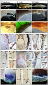
Figure 4. Pelvic fin muscle formation in bony fish.
Pelvic fin muscle formation in P. spathula (A–J), D. rerio (K–T), and N. forsteri (U–DD). Larvae at stages when pelvic fin muscles form (A, K, and U). Developing pelvic fin bud (B, L, and V). Muscle fibres in developing pelvic fin are separate and distinct from the muscle of the somite (C, M, and W). Immediately before pelvic fin formation epithelial buds (mb) head the myotomal extension (me) (D, E, F, N, O, X, Y). The pelvic fin muscles (pfm) have formed within the pelvic fin and are separate from the myotomal extension (me) (G, H, I, P, Q, Z, AA, BB). myoD is restricted to individual, post-migratory, differentiating pelvic fin muscles (R). lbx1 positive cells (purple) at the position of the forming pelvic fin muscles (pfm) in lungfish (DD). Lbx1 expression in 14 dph pelvic fin of P. spathula, inset is a cross-section of the pelvic fin at the same stage, revealing lbx expression in the muscle masses (J). Lbx1 positive (blue) precursors in the tip of the extension position of the future pelvic fin muscles in D. rerio at 8 mm TL (S) and 9 mm TL (T). _Lbx1_-positive precursors in stage 50 pelvic fin bud of N. forsteri (DD). (ep, epithelial bud; me, myotome extension; pf, pelvic fin; pfm, pelvic fin muscle).
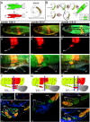
Figure 5. Transgenic somite transplantation in D. rerio reveals pelvic fin muscles derive from myotomal extension.
(A) Donor embryos are generated by crossing Tg(acta1∶mCherry)pc4 with Tg(acta1∶GFP)zf13. (B) Donor somites are surgically removed and transplanted into the host. (C–I) Myotomal extensions derived from somites 10 and 11 generate the pelvic fin muscles (Section through s10 (blue line in F) of host (5 wk post-operation) (G, H, and I)). (Comprehensive methods are provided in supplementary data, however surgery involving transplantation of two consecutive somites lead to a greater probability of transplanting the entire somite, including the ventral aspect required for pelvic fin muscle formation). (J–L) Contribution to the pelvic fin muscle requires the most ventral tip of the somite to be transplanted. Full transplantation of somite 9 contributes to ventral muscle anterior to pelvic fin. but a partial transplant of somite 10 does not result in contribution to the pelvic fin muscle. (M, N) Section through s10 (blue line in M) of the transplanted host (5 wk post-operation). (N) If the donor somite is not included in the most ventral tip of the extension, contribution to the fin does not occur . (O–T) The most ventral group of pelvic fin muscles are derived from somite 11 (s11+s12) section through s10 (S) (blue line in R) and s11 (T) (blue line in R) of host (5 wk post-operation). Pf, pelvic fin; rfp, red fluorescent protein; pfm, pelvic fin muscle; me, myotomal extension; s9,s10,s11,s12, somite numbered from anterior to posterior.
Figure 6. A phylogenetic framework for pelvic fin muscle evolution.
Evolution of pelvic fin and hindlimb developmental mechanisms. Pelvic fin and hindlimb developmental mechanisms mapped onto the vertebrate phylogeny.
Comment in
- Australian fish stretching their legs.
Vance E. Vance E. PLoS Biol. 2011 Oct;9(10):e1001167. doi: 10.1371/journal.pbio.1001167. Epub 2011 Oct 4. PLoS Biol. 2011. PMID: 21990961 Free PMC article. No abstract available.
Similar articles
- Shaping muscle bioarchitecture for the fin to limb transition.
Cole NJ, Currie P. Cole NJ, et al. Bioarchitecture. 2012 May 1;2(3):98-103. doi: 10.4161/bioa.20969. Bioarchitecture. 2012. PMID: 22880150 Free PMC article. - Evolution of Hindlimb Muscle Anatomy Across the Tetrapod Water-to-Land Transition, Including Comparisons With Forelimb Anatomy.
Molnar JL, Diogo R, Hutchinson JR, Pierce SE. Molnar JL, et al. Anat Rec (Hoboken). 2020 Feb;303(2):218-234. doi: 10.1002/ar.23997. Epub 2018 Nov 25. Anat Rec (Hoboken). 2020. PMID: 30365249 - Musculoskeletal anatomy of the pelvic fin of Polypterus: implications for phylogenetic distribution and homology of pre- and postaxial pelvic appendicular muscles.
Molnar JL, Johnston PS, Esteve-Altava B, Diogo R. Molnar JL, et al. J Anat. 2017 Apr;230(4):532-541. doi: 10.1111/joa.12573. Epub 2016 Dec 15. J Anat. 2017. PMID: 27976380 Free PMC article. - Reconstructing pectoral appendicular muscle anatomy in fossil fish and tetrapods over the fins-to-limbs transition.
Molnar JL, Diogo R, Hutchinson JR, Pierce SE. Molnar JL, et al. Biol Rev Camb Philos Soc. 2018 May;93(2):1077-1107. doi: 10.1111/brv.12386. Epub 2017 Nov 10. Biol Rev Camb Philos Soc. 2018. PMID: 29125205 Review. - The evolutionary history of the development of the pelvic fin/hindlimb.
Don EK, Currie PD, Cole NJ. Don EK, et al. J Anat. 2013 Jan;222(1):114-33. doi: 10.1111/j.1469-7580.2012.01557.x. Epub 2012 Aug 23. J Anat. 2013. PMID: 22913749 Free PMC article. Review.
Cited by
- Muscle development in the shark Scyliorhinus canicula: implications for the evolution of the gnathostome head and paired appendage musculature.
Ziermann JM, Freitas R, Diogo R. Ziermann JM, et al. Front Zool. 2017 Jun 21;14:31. doi: 10.1186/s12983-017-0216-y. eCollection 2017. Front Zool. 2017. PMID: 28649268 Free PMC article. - Zebrafish: A Model for the Study of Toxicants Affecting Muscle Development and Function.
Dubińska-Magiera M, Daczewska M, Lewicka A, Migocka-Patrzałek M, Niedbalska-Tarnowska J, Jagla K. Dubińska-Magiera M, et al. Int J Mol Sci. 2016 Nov 19;17(11):1941. doi: 10.3390/ijms17111941. Int J Mol Sci. 2016. PMID: 27869769 Free PMC article. Review. - The zebrafish HGF receptor met controls migration of myogenic progenitor cells in appendicular development.
Nord H, Dennhag N, Tydinger H, von Hofsten J. Nord H, et al. PLoS One. 2019 Jul 9;14(7):e0219259. doi: 10.1371/journal.pone.0219259. eCollection 2019. PLoS One. 2019. PMID: 31287821 Free PMC article. - On the peculiar morphology and development of the hypoglossal, glossopharyngeal and vagus nerves and hypobranchial muscles in the hagfish.
Oisi Y, Fujimoto S, Ota KG, Kuratani S. Oisi Y, et al. Zoological Lett. 2015 Jan 29;1:6. doi: 10.1186/s40851-014-0005-9. eCollection 2015. Zoological Lett. 2015. PMID: 26605051 Free PMC article.
References
- Ahlberg P. E, Clack J. A, Blom H. The axial skeleton of the Devonian tetrapod Ichthyostega. Nature. 2005;437:137–140. - PubMed
- Ahlberg P. E, Clack J. A, Luksevics E, Blom H, Zupins I. Ventastega curonica and the origin of tetrapod morphology. Nature. 2008;453:1199–1204. - PubMed
- Andrews S. M, Westoll T. S. The postcranial skeleton of Eusthenopteron foordi Whiteves. Transactions of the Royal Society of Edinburgh. 1970;68:207–329.
- Boisvert C. A. The pelvic fin and girdle of Panderichthys and the origin of tetrapod locomotion. Nature. 2005;438:1145–1147. - PubMed
- Callier V, Clack J. A, Ahlberg P. E. Contrasting developmental trajectories in the earliest known tetrapod forelimbs. Science. 2009;324:364–367. - PubMed
Publication types
MeSH terms
LinkOut - more resources
Full Text Sources
Molecular Biology Databases
Miscellaneous


