Soluble Aβ seeds are potent inducers of cerebral β-amyloid deposition - PubMed (original) (raw)
Soluble Aβ seeds are potent inducers of cerebral β-amyloid deposition
Franziska Langer et al. J Neurosci. 2011.
Abstract
Cerebral β-amyloidosis and associated pathologies can be exogenously induced by the intracerebral injection of small amounts of pathogenic Aβ-containing brain extract into young β-amyloid precursor protein (APP) transgenic mice. The probable β-amyloid-inducing factor in the brain extract has been identified as a species of aggregated Aβ that is generated in its most effective conformation or composition in vivo. Here we report that Aβ in the brain extract is more proteinase K (PK) resistant than is synthetic fibrillar Aβ, and that this PK-resistant fraction of the brain extract retains the capacity to induce β-amyloid deposition upon intracerebral injection in young, pre-depositing APP23 transgenic mice. After ultracentrifugation of the brain extract, <0.05% of the Aβ remained in the supernatant fraction, and these soluble Aβ species were largely PK sensitive. However, upon intracerebral injection, this soluble fraction accounted for up to 30% of the β-amyloid induction observed with the unfractionated extract. Fragmentation of the Aβ seeds by extended sonication increased the seeding capacity of the brain extract. In summary, these results suggest that multiple Aβ assemblies, with various PK sensitivities, are capable of inducing β-amyloid aggregation in vivo. The finding that small and soluble Aβ seeds are potent inducers of cerebral β-amyloidosis raises the possibility that such seeds may mediate the spread of β-amyloidosis in the brain. If they can be identified in vivo, soluble Aβ seeds in bodily fluids also could serve as early biomarkers for cerebral β-amyloidogenesis and eventually Alzheimer's disease.
Figures
Figure 1.
The β-amyloid-inducing activity is partly proteinase K-resistant. Brain extracts from an aged APP23 tg mouse (Tg extract), an aged non-tg mouse (Wt extract) spiked with synthetic Aβ fibrils (syn. Aβ fibrils in Wt extract; 10 μ
m
Aβ), and synthetic Aβ fibrils in PBS (syn. Aβ fibrils in PBS; 10 μ
m
) were treated with 50 μg/ml PK for 0 min, 30 min, 1 h, or 2 h at 37°C. A, Silver staining of a 12% Bis-Tris NuPage gel for total protein reveals the digestion of the majority of proteins in the brain extracts and of synthetic Aβ fibrils already after 30 min PK treatment. B, Immunoblot analysis with an antibody specific to human Aβ (6E10) shows that Aβ in the Tg extract is largely PK resistant for up to 2 h. Aβ in the spiked Wt extract revealed some, but significantly less PK resistance, whereas synthetic Aβ fibrils in PBS were almost completely digested by the PK treatment. C, Densitometric quantification of Aβ-immunoblots after PK treatment of five Tg extracts (each from a different animal) and three independent synthetic Aβ fibril preparations are shown. Each preparation was tested at least three times and the mean was taken. Aβ concentration at time point 0 was designated as 100%. Indicated is the mean ± SEM. Repeated-measures two-way ANOVA revealed a significant difference between the groups (F(2,34) = 112.2). Multiple Bonferroni post hoc tests showed significances after PK digestion for all group comparisons, p < 0.001. D–F, In vivo seeding activity of brain extracts with or without PK treatment (50 μg/ml PK for 30 min) was tested by intrahippocampal injections into young, pre-depositing 4-month-old APP23 mice. Brains were immunohistochemically analyzed for Aβ deposition 5 months later. Shown is the dentate gyrus of the hippocampus, which revealed robust β-amyloid induction by the Tg extract (D) and the PK-treated Tg extract (E), but not with Wt extract (F). Note that extracts were boiled before injection to deactivate residual PK activity. The small insert shows double staining for Aβ immunoreactivity and Congo red binding of the induced β-amyloid in an adjacent section. Scale bars: 200 μm and 20 μm. G, Stereological quantification of Aβ load in the hippocampus. Although PK treatment reduced the β-amyloid induction, the PK-resistant Tg material still showed potent seeding activity compared to the Wt extract (n = 5–6 mice per group; mean ± SEM: ANOVA followed by Bonferroni post hoc tests revealed a significant difference between the extracts (F(2,14) = 18.95, ***p < 0.001; *p < 0.05). Note that neither PK-treated nor untreated Wt extract induced any amyloid, and therefore these groups were combined.
Figure 2.
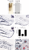
The β-amyloid-inducing activity is partly soluble. A, Brain extracts (10%, w/v) from aged APP23 mice (Tg extract) were subjected to 100,000 × g ultracentrifugation (1 h, 4°C). Left panel, Silver staining of a 12% Bis-Tris gel shows somewhat less protein in the pellet fraction (P) compared to the supernatant (SN) fraction (this was confirmed by total protein measurements revealing ∼60% of the protein in the supernatant and 40% in the pellet, see results). Right panel, Immunoblotting for human Aβ (antibody 6E10) using 12% Bis-Tris gels reveals that most Aβ is retained in the pellet fraction. Aβ in the supernatant was below the level of detection by this immunoblot, but was estimated to be <0.05% of total Aβ concentration in the original Tg extract using ELISA (see Table 1). Loading control: synthetic Aβ, 15 ng/lane. B–D, In vivo seeding activity of the supernatant and pellet fractions was tested by intrahippocampal injections into young, pre-depositing 4-month-old APP23 mice. Brains were immunohistochemically analyzed for Aβ deposition 4 months after injection. Shown is the dentate gyrus of the hippocampus. While the Tg extract (B) induced robust Aβ deposition, the SN fraction (C) induced largely diffuse Congo red-negative Aβ deposits. Aβ deposition induced by the pellet fraction (D) is similar to that induced by the Tg extract. The small insert shows double staining of Aβ and Congo red in seeded β-amyloid in an adjacent section. Note that the β-amyloid-induction by the SN was largely Congo red negative after this 4 month incubation period, but was partly Congo red positive after a 6 month incubation (see G below). E, Stereological quantification of Aβ load in the hippocampus 4 months after injection (n = 5 mice per group; mean ± SEM; ANOVA followed by Bonferroni post hoc test (F(2,13) = 11.93; **p < 0.01; n.s., not significant). Note that an additive effect of the β-amyloid induction by the SN and P fractions cannot be assumed (Meyer-Luehmann et al., 2006). F, β-Amyloid induction by the SN fraction 6 months after injection. G, The majority of the SN fraction-induced β-amyloid-deposits at 6 months after injection were still Congo red negative; however, a subset of the induced deposits were congophilic (arrow) and surrounded by activated, darkly stained Iba-1-positive microglia, hypertrophic GFAP-positive astrocytes, and APP-positive dystrophic boutons and neurites (arrowheads). Shown are sections adjacent to F. Scale bars: 200 μm, 20 μm (B–D, F), and 50 μm (G).
Figure 3.
The soluble β-amyloid-inducing activity is mostly proteinase K sensitive. A, Brain extracts (10%, w/v) from aged APP23 mice (Tg extract) and the 100,000 × g supernatant (SN) of the Tg extract (ultracentrifugation, 1 h, 4°C) were treated with 50 μg PK/ml for 30 min at 37°C. Immunoblotting for human Aβ (antibody 6E10) using 12% Bis-Tris gels reveals the expected PK resistance of the Tg extract. In contrast, only a minor fraction of the SN was not cleaved by PK (see long exposure). Note that the Tg extract was diluted 10-fold, and that 10-fold more of the SN fraction was loaded compared to that in Figures 1_B_ and 2_A_, respectively. Also note that there is an empty lane between the two Tg extract lanes and the two lanes with the SN. B, Densitometric analysis of Aβ immunoblots after PK treatment [4 Tg extracts (each from a different animal) and the corresponding 100,000 × g supernatant were used; each preparation was tested at least 2 times and the mean was taken]. Aβ concentration at time point 0 was defined as 100%. Indicated is the mean ± SEM. C, D, In vivo seeding activity of brain extracts with or without PK treatment was tested by intrahippocampal injections into young, pre-depositing 3 month-old APP23 mice. Brains were immunohistochemically analyzed for Aβ-deposition 6 months later. Shown is the dentate gyrus of the hippocampus. SN of Tg brain extract was boiled for 5 min at 95°C (C). The small inset shows double labeling of Aβ immunoreactivity and Congo red binding of the induced β-amyloid in an adjacent section. D, PK-treated (30 min) and boiled SN of Tg brain extract. Scale bars: 200 μm and 20 μm. E, Stereological quantification of Aβ load in the hippocampus. PK treatment of the 100,000 × g supernatant of Tg brain extract diminished the seeding activity. t test revealed a significant difference between the two groups (n = 4–5 mice per group, mean ± SEM, t(7) = 4.713; **p < 0.01).
Figure 4.
Extended sonication increases β-amyloid-inducing activity. Brain extracts [10% (w/v)] from aged APP23 mice (Tg extract) were subjected to additional sonication (3 × 20 s). A, Immunoblot analysis with an antibody specific to human Aβ reveals no change in total Aβ between the original Tg extract (−) and the extract with extended sonication (+). However, more Aβ was found in the supernatant (SN) of the extra-sonicated extract after ultracentrifugation (100,000 × g; 1 h) (long exposure). B, C, In vivo seeding activity of the original and extra-sonicated extracts was tested by intrahippocampal injections into young, pre-depositing APP23 mice. Brains were immunohistochemically analyzed for Aβ deposition 4 months after injection. Shown is the dentate gyrus of the hippocampus. Injection of original Tg extract (B) induced robust congophilic Aβ deposition with a filamentous and dense pattern, while Aβ induction with the extra-sonicated Tg extract generated more punctate and small deposits (C). The inset shows double labeling of Aβ immunoreactivity and Congo red binding of the induced β-amyloid. Scale bars: 200 μm and 20 μm. D, Stereological quantification of Aβ load in the hippocampus revealed a significant increase in β-amyloid induction by extended sonication (n = 5–6 mice per group; mean ± SEM, t(9) = 3.188; *p < 0.05).
Similar articles
- Multiple factors contribute to the peripheral induction of cerebral β-amyloidosis.
Eisele YS, Fritschi SK, Hamaguchi T, Obermüller U, Füger P, Skodras A, Schäfer C, Odenthal J, Heikenwalder M, Staufenbiel M, Jucker M. Eisele YS, et al. J Neurosci. 2014 Jul 30;34(31):10264-73. doi: 10.1523/JNEUROSCI.1608-14.2014. J Neurosci. 2014. PMID: 25080588 Free PMC article. - Aβ seeds resist inactivation by formaldehyde.
Fritschi SK, Cintron A, Ye L, Mahler J, Bühler A, Baumann F, Neumann M, Nilsson KP, Hammarström P, Walker LC, Jucker M. Fritschi SK, et al. Acta Neuropathol. 2014 Oct;128(4):477-84. doi: 10.1007/s00401-014-1339-2. Epub 2014 Sep 6. Acta Neuropathol. 2014. PMID: 25193240 Free PMC article. - Conversion of Synthetic Aβ to In Vivo Active Seeds and Amyloid Plaque Formation in a Hippocampal Slice Culture Model.
Novotny R, Langer F, Mahler J, Skodras A, Vlachos A, Wegenast-Braun BM, Kaeser SA, Neher JJ, Eisele YS, Pietrowski MJ, Nilsson KP, Deller T, Staufenbiel M, Heimrich B, Jucker M. Novotny R, et al. J Neurosci. 2016 May 4;36(18):5084-93. doi: 10.1523/JNEUROSCI.0258-16.2016. J Neurosci. 2016. PMID: 27147660 Free PMC article. - Alzheimer's disease.
De-Paula VJ, Radanovic M, Diniz BS, Forlenza OV. De-Paula VJ, et al. Subcell Biochem. 2012;65:329-52. doi: 10.1007/978-94-007-5416-4_14. Subcell Biochem. 2012. PMID: 23225010 Review. - Pathogenic protein seeding in Alzheimer disease and other neurodegenerative disorders.
Jucker M, Walker LC. Jucker M, et al. Ann Neurol. 2011 Oct;70(4):532-40. doi: 10.1002/ana.22615. Ann Neurol. 2011. PMID: 22028219 Free PMC article. Review.
Cited by
- Potential mechanisms and implications for the formation of tau oligomeric strains.
Gerson JE, Mudher A, Kayed R. Gerson JE, et al. Crit Rev Biochem Mol Biol. 2016 Nov/Dec;51(6):482-496. doi: 10.1080/10409238.2016.1226251. Epub 2016 Sep 21. Crit Rev Biochem Mol Biol. 2016. PMID: 27650389 Free PMC article. Review. - Biophysical insights into how surfaces, including lipid membranes, modulate protein aggregation related to neurodegeneration.
Burke KA, Yates EA, Legleiter J. Burke KA, et al. Front Neurol. 2013 Mar 1;4:17. doi: 10.3389/fneur.2013.00017. eCollection 2013. Front Neurol. 2013. PMID: 23459674 Free PMC article. - The role of prion strain diversity in the development of successful therapeutic treatments.
Holec SAM, Block AJ, Bartz JC. Holec SAM, et al. Prog Mol Biol Transl Sci. 2020;175:77-119. doi: 10.1016/bs.pmbts.2020.07.001. Epub 2020 Aug 28. Prog Mol Biol Transl Sci. 2020. PMID: 32958242 Free PMC article. Review. - A native interactor scaffolds and stabilizes toxic ATAXIN-1 oligomers in SCA1.
Lasagna-Reeves CA, Rousseaux MW, Guerrero-Muñoz MJ, Park J, Jafar-Nejad P, Richman R, Lu N, Sengupta U, Litvinchuk A, Orr HT, Kayed R, Zoghbi HY. Lasagna-Reeves CA, et al. Elife. 2015 May 19;4:e07558. doi: 10.7554/eLife.07558. Elife. 2015. PMID: 25988806 Free PMC article. - Aβ Assemblies Promote Amyloidogenic Processing of APP and Intracellular Accumulation of Aβ42 Through Go/Gβγ Signaling.
Antonino M, Marmo P, Freites CL, Quassollo GE, Sánchez MF, Lorenzo A, Bignante EA. Antonino M, et al. Front Cell Dev Biol. 2022 Apr 4;10:852738. doi: 10.3389/fcell.2022.852738. eCollection 2022. Front Cell Dev Biol. 2022. PMID: 35445022 Free PMC article.
References
- Aguzzi A, Rajendran L. The transcellular spread of cytosolic amyloids, prions, and prionoids. Neuron. 2009;64:783–790. - PubMed
- Aguzzi A, Heikenwalder M, Polymenidou M. Insights into prion strains and neurotoxicity. Nat Rev Mol Cell Biol. 2007;8:552–561. - PubMed
- Calhoun ME, Burgermeister P, Phinney AL, Stalder M, Tolnay M, Wiederhold KH, Abramowski D, Sturchler-Pierrat C, Sommer B, Staufenbiel M, Jucker M. Neuronal overexpression of mutant amyloid precursor protein results in prominent deposition of cerebrovascular amyloid. Proc Natl Acad Sci U S A. 1999;96:14088–14093. - PMC - PubMed
Publication types
MeSH terms
Substances
LinkOut - more resources
Full Text Sources
Other Literature Sources


