Predicting MCI outcome with clinically available MRI and CSF biomarkers - PubMed (original) (raw)
Predicting MCI outcome with clinically available MRI and CSF biomarkers
D Heister et al. Neurology. 2011.
Abstract
Objective: To determine the ability of clinically available volumetric MRI (vMRI) and CSF biomarkers, alone or in combination with a quantitative learning measure, to predict conversion to Alzheimer disease (AD) in patients with mild cognitive impairment (MCI).
Methods: We stratified 192 MCI participants into positive and negative risk groups on the basis of 1) degree of learning impairment on the Rey Auditory Verbal Learning Test; 2) medial temporal atrophy, quantified from Food and Drug Administration-approved software for automated vMRI analysis; and 3) CSF biomarker levels(.) We also stratified participants based on combinations of risk factors. We computed Cox proportional hazards models, controlling for age, to assess 3-year risk of converting to AD as a function of risk group and used Kaplan-Meier analyses to determine median survival times.
Results: When risk factors were examined separately, individuals testing positive showed significantly higher risk of converting to AD than individuals testing negative (hazard ratios [HR] 1.8-4.1). The joint presence of any 2 risk factors substantially increased risk, with the combination of greater learning impairment and increased atrophy associated with highest risk (HR 29.0): 85% of patients with both risk factors converted to AD within 3 years, vs 5% of those with neither. The presence of medial temporal atrophy was associated with shortest median dementia-free survival (15 months).
Conclusions: Incorporating quantitative assessment of learning ability along with vMRI or CSF biomarkers in the clinical workup of MCI can provide critical information on risk of imminent conversion to AD.
Figures
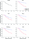
Figure 1. Survival curves according to risk category
Survival curves for the full mild cognitive impairment (MCI) cohort, and for negative and positive risk groups defined according to learning performance (Auditory Rey Verbal Learning Test [AVLT]), CSF T-tau, Aβ1–42, and the tau/Aβ1–42 ratio, as well as for medial temporal atrophy determined from the hippocampal occupancy score (HOC). Cox proportional hazard models controlled for age. The x-axis shows months to conversion to AD; the y-axis shows proportion of subjects who have not converted to Alzheimer disease. High risk is shown in red, low risk in blue.
Figure 2. Survival curves as a function of risk factor combinations
Survival curves are shown for patients with mild cognitive impairment stratified according to the combination of learning (Auditory Rey Verbal Learning Test [AVLT]) and atrophy (hippocampal occupancy score [HOC]) risk, learning and CSF risk, atrophy and CSF risk, and for individuals concordant on risk for all 3 measures. Cox proportional hazard model controlled for age. Green lines show those testing negative on all measures in the analysis, red lines show those testing positive on all measures. Blue and purple lines show survival for those with discordant risk factors.
Figure 3. Median survival times for those testing positive on each risk factor or combination of risk factors
Median survival time (in months) reflects the last time at which 50% of the subjects in the group retained the MCI diagnosis. AVLT = Auditory Rey Verbal Learning Test.
Similar articles
- Combined Biomarker Prognosis of Mild Cognitive Impairment: An 11-Year Follow-Up Study in the Alzheimer's Disease Neuroimaging Initiative.
Spencer BE, Jennings RG, Brewer JB; Alzheimer’s Disease Neuroimaging Initiative. Spencer BE, et al. J Alzheimers Dis. 2019;68(4):1549-1559. doi: 10.3233/JAD-181243. J Alzheimers Dis. 2019. PMID: 30958366 Free PMC article. - Progression to dementia in memory clinic patients with mild cognitive impairment and normal β-amyloid.
Rosenberg A, Solomon A, Jelic V, Hagman G, Bogdanovic N, Kivipelto M. Rosenberg A, et al. Alzheimers Res Ther. 2019 Dec 5;11(1):99. doi: 10.1186/s13195-019-0557-1. Alzheimers Res Ther. 2019. PMID: 31805990 Free PMC article. - Multimodal prediction of dementia with up to 10 years follow up: the Gothenburg MCI study.
Eckerström C, Olsson E, Klasson N, Berge J, Nordlund A, Bjerke M, Wallin A. Eckerström C, et al. J Alzheimers Dis. 2015;44(1):205-14. doi: 10.3233/JAD-141053. J Alzheimers Dis. 2015. PMID: 25201779 - Posterior atrophy predicts time to dementia in patients with amyloid-positive mild cognitive impairment.
Pyun JM, Park YH, Kim HR, Suh J, Kang MJ, Kim BJ, Youn YC, Jang JW, Kim S; Alzheimer’s Disease Neuroimaging Initiative. Pyun JM, et al. Alzheimers Res Ther. 2017 Dec 16;9(1):99. doi: 10.1186/s13195-017-0326-y. Alzheimers Res Ther. 2017. PMID: 29246250 Free PMC article. - Diffusion tensor imaging surpasses cerebrospinal fluid as predictor of cognitive decline and medial temporal lobe atrophy in subjective cognitive impairment and mild cognitive impairment.
Selnes P, Aarsland D, Bjørnerud A, Gjerstad L, Wallin A, Hessen E, Reinvang I, Grambaite R, Auning E, Kjærvik VK, Due-Tønnessen P, Stenset V, Fladby T. Selnes P, et al. J Alzheimers Dis. 2013;33(3):723-36. doi: 10.3233/JAD-2012-121603. J Alzheimers Dis. 2013. PMID: 23186987
Cited by
- MRI and cerebrospinal fluid biomarkers for predicting progression to Alzheimer's disease in patients with mild cognitive impairment: a diagnostic accuracy study.
Richard E, Schmand BA, Eikelenboom P, Van Gool WA; Alzheimer's Disease Neuroimaging Initiative. Richard E, et al. BMJ Open. 2013 Jun 20;3(6):e002541. doi: 10.1136/bmjopen-2012-002541. BMJ Open. 2013. PMID: 23794572 Free PMC article. - Health risk prediction models incorporating personality data: Motivation, challenges, and illustration.
Chapman BP, Lin F, Roy S, Benedict RHB, Lyness JM. Chapman BP, et al. Personal Disord. 2019 Jan;10(1):46-58. doi: 10.1037/per0000300. Personal Disord. 2019. PMID: 30604983 Free PMC article. - Decreased Vascular Pulsatility in Alzheimer's Disease Dementia Measured by Transcranial Color-Coded Duplex Sonography.
Ortner M, Hauser C, Schmaderer C, Muggenthaler C, Hapfelmeier A, Sorg C, Diehl-Schmid J, Kurz A, Förstl H, Ikenberg B, Kotliar K, Poppert H, Grimmer T. Ortner M, et al. Neuropsychiatr Dis Treat. 2019 Dec 20;15:3487-3499. doi: 10.2147/NDT.S225754. eCollection 2019. Neuropsychiatr Dis Treat. 2019. PMID: 31908463 Free PMC article. - Combined Biomarker Prognosis of Mild Cognitive Impairment: An 11-Year Follow-Up Study in the Alzheimer's Disease Neuroimaging Initiative.
Spencer BE, Jennings RG, Brewer JB; Alzheimer’s Disease Neuroimaging Initiative. Spencer BE, et al. J Alzheimers Dis. 2019;68(4):1549-1559. doi: 10.3233/JAD-181243. J Alzheimers Dis. 2019. PMID: 30958366 Free PMC article. - Using Optical Coherence Tomography to Screen for Cognitive Impairment and Dementia.
Galvin JE, Kleiman MJ, Walker M. Galvin JE, et al. J Alzheimers Dis. 2021;84(2):723-736. doi: 10.3233/JAD-210328. J Alzheimers Dis. 2021. PMID: 34569948 Free PMC article.
References
- Devanand DP, Pradhaban G, Liu X, et al. Hippocampal and entorhinal atrophy in mild cognitive impairment: prediction of Alzheimer disease. Neurology 2007;68:828–836 - PubMed
- Mattsson N, Zetterberg H, Hansson O, et al. CSF biomarkers and incipient Alzheimer disease in patients with mild cognitive impairment. JAMA 2009;302:385–393 - PubMed
Publication types
MeSH terms
Substances
Grants and funding
- UL1 TR000117/TR/NCATS NIH HHS/United States
- R01 AG022374/AG/NIA NIH HHS/United States
- P30 AG010129/AG/NIA NIH HHS/United States
- K01AG029218/AG/NIA NIH HHS/United States
- U19 AG010483/AG/NIA NIH HHS/United States
- UL1 RR033173/RR/NCRR NIH HHS/United States
- U01 AG024904/AG/NIA NIH HHS/United States
- R01 AG012101/AG/NIA NIH HHS/United States
- K02NS067427/NS/NINDS NIH HHS/United States
- K01 AG030514/AG/NIA NIH HHS/United States
- P30 AG019610/AG/NIA NIH HHS/United States
- UL1 TR001998/TR/NCATS NIH HHS/United States
LinkOut - more resources
Full Text Sources
Medical


