Domain topology of nucleoporin Nup98 within the nuclear pore complex - PubMed (original) (raw)
Domain topology of nucleoporin Nup98 within the nuclear pore complex
Guillaume Chatel et al. J Struct Biol. 2012 Jan.
Abstract
Nuclear pore complexes (NPCs) facilitate selective transport of macromolecules across the nuclear envelope in interphase eukaryotic cells. NPCs are composed of roughly 30 different proteins (nucleoporins) of which about one third are characterized by the presence of phenylalanine-glycine (FG) repeat domains that allow the association of soluble nuclear transport receptors with the NPC. Two types of FG (FG/FxFG and FG/GLFG) domains are found in nucleoporins and Nup98 is the sole vertebrate nucleoporin harboring the GLFG-type repeats. By immuno-electron microscopy using isolated nuclei from Xenopus oocytes we show here the localization of distinct domains of Nup98. We examined the localization of the C- and N-terminal domain of Nup98 by immunogold-labeling using domain-specific antibodies against Nup98 and by expressing epitope tagged versions of Nup98. Our studies revealed that anchorage of Nup98 to NPCs through its C-terminal autoproteolytic domain occurs in the center of the NPC, whereas its N-terminal GLFG domain is more flexible and is detected at multiple locations within the NPC. Additionally, we have confirmed the central localization of Nup98 within the NPC using super resolution structured illumination fluorescence microscopy (SIM) to position Nup98 domains relative to markers of cytoplasmic filaments and the nuclear basket. Our data support the notion that Nup98 is a major determinant of the permeability barrier of NPCs.
Copyright © 2011 Elsevier Inc. All rights reserved.
Figures
Figure 1
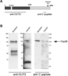
Domain-specific antibodies against Nup98. (A) A schematic presentation of human Nup98 is illustrated. A commercially available rat monoclonal antibody, which was raised against the GLFG domain (residues 1-466) of human Nup98 was used and a rabbit polyclonal antibody raised against a peptide within the C-terminal domain of Xenopus Nup98. Dark grey, FG/GLFG domain; light grey, C-terminal auteoproteolytic domain. GLEBS, residues 181-224. Cleavage site, between residues 863 and 864. (B) The anti-GLFG and the anti-C peptide antibodies detect a protein of about 110 kDa in both Xenopus and HeLa extracts. The positions of the markers in kilodalton are indicated.
Figure 2
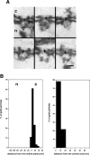
Localization of the C-terminal domain of Nup98 in isolated Xenopus nuclei. (A) Intact isolated nuclei were pre-immuno-labeled with the anti-C peptide antibody conjugated directly to 8-nm colloidal gold and prepared for EM by Epon embedding and thin-sectioning. A gallery of selected examples of gold-labeled NPCs in cross sections is shown. c, cytoplasm; n, nucleus. Scale bar, 100 nm. (B) Quantitative analysis of the gold particles associated with the NPCs after labeling with the anti-C antibody. Sixty-three gold particles were scored.
Figure 3
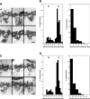
Immuno-localization of the GLFG domain of Nup98. (A) A gallery of selected examples of NPCs in cross sections labeled with the anti-GLFG antibody directly conjugated to 8-nm colloidal gold in isolated, intact Xenopus nuclei is shown. c, cytoplasm; n, nucleus. Scale bar, 100 nm. (B) Quantitative analysis of the gold particle distribution in NPCs labeled with the anti-GLFG antibody conjugated to 8-nm colloidal gold. One hundred twenty five gold particles were scored. (C)Immuno-localization of N-terminally myc-tagged Nup98 expressed in Xenopus oocytes with a monoclonal anti-myc antibody directly conjugated to 8-nm gold. The antibody recognized epitopes on both faces of the NPC. Selected examples of labeled NPCs in cross sections are shown. c, cytoplasm; n, nucleus. Scale bar, 100 nm. (D) Quantitation of the gold particle distribution associated with the NPC in Xenopus nuclei that have incorporated myc-Nup98 after labeling with an anti-myc antibody. One hundred one gold particles were scored.
Figure 4
Relative localization of Nup98 within the NPC by SIM. (A) Cartoon depicting domain organization of the nucleoporins used, along with position of epitopes for each antibody. Nup98: blue box within the Nup98 FG/GLFG domain is the binding site for Rae1/Gle2. Red line denotes autoproteolytic site. Nup358: RBD represents Ran binding domains.(B) Comparison of widefield, deconvolved widefield, and SIM images. Nup98 anti-GLFG is shown in red, Nup153 anti-ZnF in green. Scale bar, 5μm. Inset scale bar, 1 μm. (C) Regions of stained nuclear envelopes with dotted line indicating the position of the linescan, displayed to the right of each image. Each panel depicts one pair of fluorescent labels as indicated in the corresponding color on the image. The linescans show the normalized intensity of each channel as a function of distance along the line. The direction of all linescans is cytoplasm to nucleoplasm. In panels a though d, the position of the Nup98 peak was used to align graphs. In panels e-g, graphs were aligned to the position of the 414 peak. Line Scale bar, 1 μm.
Figure 5
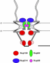
Schematic representation of the anchoring sites of FG nucleoporins within the 3-D architecture by elliptic location clouds. Nup153 is anchored near the nuclear ring moiety by its N-terminal domain and near the distal ring of the nuclear basket by its zinc-finger domain (Fahrenkrog et al., 2002), while Nup214 is anchored near the cytoplasmic ring moiety by its N-terminal and central domain (Paulillo et al., 2005). Nup62 (Schwarz-Herion et al., 2007) and Nup98 are anchored in the center of the NPC with Nup98 being slightly more central than Nup62. The positions of their respective FG domains are indicated by clouds in the according lighter clours. c, cytoplasm; n, nucleus. Scale bar: 50 nm.
Similar articles
- Nucleoporin domain topology is linked to the transport status of the nuclear pore complex.
Paulillo SM, Phillips EM, Köser J, Sauder U, Ullman KS, Powers MA, Fahrenkrog B. Paulillo SM, et al. J Mol Biol. 2005 Aug 26;351(4):784-98. doi: 10.1016/j.jmb.2005.06.034. J Mol Biol. 2005. PMID: 16045929 - Entry into the nuclear pore complex is controlled by a cytoplasmic exclusion zone containing dynamic GLFG-repeat nucleoporin domains.
Fiserova J, Spink M, Richards SA, Saunter C, Goldberg MW. Fiserova J, et al. J Cell Sci. 2014 Jan 1;127(Pt 1):124-36. doi: 10.1242/jcs.133272. Epub 2013 Oct 21. J Cell Sci. 2014. PMID: 24144701 - Impact of distinct FG nucleoporin repeats on Nup98 self-association.
Ibáñez de Opakua A, Pantoja CF, Cima-Omori MS, Dienemann C, Zweckstetter M. Ibáñez de Opakua A, et al. Nat Commun. 2024 May 7;15(1):3797. doi: 10.1038/s41467-024-48194-4. Nat Commun. 2024. PMID: 38714656 Free PMC article. - Functional architecture of the nuclear pore complex.
Grossman E, Medalia O, Zwerger M. Grossman E, et al. Annu Rev Biophys. 2012;41:557-84. doi: 10.1146/annurev-biophys-050511-102328. Annu Rev Biophys. 2012. PMID: 22577827 Review. - Structural dynamics of the nuclear pore complex.
Sakiyama Y, Panatala R, Lim RYH. Sakiyama Y, et al. Semin Cell Dev Biol. 2017 Aug;68:27-33. doi: 10.1016/j.semcdb.2017.05.021. Epub 2017 Jun 1. Semin Cell Dev Biol. 2017. PMID: 28579449 Review.
Cited by
- Physics of the Nuclear Pore Complex: Theory, Modeling and Experiment.
Hoogenboom BW, Hough LE, Lemke EA, Lim RYH, Onck PR, Zilman A. Hoogenboom BW, et al. Phys Rep. 2021 Jul 25;921:1-53. doi: 10.1016/j.physrep.2021.03.003. Epub 2021 Mar 24. Phys Rep. 2021. PMID: 35892075 Free PMC article. - Disordered proteinaceous machines.
Fuxreiter M, Tóth-Petróczy Á, Kraut DA, Matouschek A, Lim RY, Xue B, Kurgan L, Uversky VN. Fuxreiter M, et al. Chem Rev. 2014 Jul 9;114(13):6806-43. doi: 10.1021/cr4007329. Epub 2014 Apr 4. Chem Rev. 2014. PMID: 24702702 Free PMC article. Review. No abstract available. - Targeted nanodiamonds for identification of subcellular protein assemblies in mammalian cells.
Lake MP, Bouchard LS. Lake MP, et al. PLoS One. 2017 Jun 21;12(6):e0179295. doi: 10.1371/journal.pone.0179295. eCollection 2017. PLoS One. 2017. PMID: 28636640 Free PMC article. - Routing of Biomolecules and Transgenes' Vectors in Nuclei of Oocytes.
Malecki M, Malecki B. Malecki M, et al. J Fertili In Vitro. 2012 Apr 30;2012(2):108-118. doi: 10.4172/2165-74. J Fertili In Vitro. 2012. PMID: 22896814 Free PMC article. - Advances in the understanding of nuclear pore complexes in human diseases.
Li Y, Zhu J, Zhai F, Kong L, Li H, Jin X. Li Y, et al. J Cancer Res Clin Oncol. 2024 Jul 30;150(7):374. doi: 10.1007/s00432-024-05881-5. J Cancer Res Clin Oncol. 2024. PMID: 39080077 Free PMC article. Review.
References
- Arnaoutov A, Azuma Y, Ribbeck K, Joseph J, Boyarchuk Y, Karpova T, McNally J, Dasso M. Crm1 is a mitotic effector of Ran-GTP in somatic cells. Nat Cell Biol. 2005;7:626–632. - PubMed
- Bayliss R, Littlewood T, Stewart M. Structural basis for the interaction between FxFG nucleoporin repeats and importin-beta in nuclear trafficking. Cell. 2000;102:99–108. - PubMed
- Beck M, Lucic V, Forster F, Baumeister W, Medalia O. Snapshots of nuclear pore complexes in action captured by cryo-electron tomography. Nature. 2007;449:611–615. - PubMed
- Blevins MB, Smith AM, Phillips EM, Powers MA. Complex formation among the RNA export proteins Nup98, Rae1/Gle2, and TAP. J Biol Chem. 2003;278:20979–20988. - PubMed
Publication types
MeSH terms
Substances
Grants and funding
- P30 NS055077/NS/NINDS NIH HHS/United States
- R01 GM059975/GM/NIGMS NIH HHS/United States
- P30NS055077/NS/NINDS NIH HHS/United States
- R01 GM-059975/GM/NIGMS NIH HHS/United States
LinkOut - more resources
Full Text Sources


