Hypoplastic left heart syndrome: current considerations and expectations - PubMed (original) (raw)
Review
. 2012 Jan 3;59(1 Suppl):S1-42.
doi: 10.1016/j.jacc.2011.09.022.
D Woodrow Benson, Anne M Dubin, Meryl S Cohen, Dawn M Maxey, William T Mahle, Elfriede Pahl, Juan Villafañe, Ami B Bhatt, Lynn F Peng, Beth Ann Johnson, Alison L Marsden, Curt J Daniels, Nancy A Rudd, Christopher A Caldarone, Kathleen A Mussatto, David L Morales, D Dunbar Ivy, J William Gaynor, James S Tweddell, Barbara J Deal, Anke K Furck, Geoffrey L Rosenthal, Richard G Ohye, Nancy S Ghanayem, John P Cheatham, Wayne Tworetzky, Gerard R Martin
Affiliations
- PMID: 22192720
- PMCID: PMC6110391
- DOI: 10.1016/j.jacc.2011.09.022
Review
Hypoplastic left heart syndrome: current considerations and expectations
Jeffrey A Feinstein et al. J Am Coll Cardiol. 2012.
Erratum in
- J Am Coll Cardiol. 2012 Jan 31;59(5):544
Abstract
In the recent era, no congenital heart defect has undergone a more dramatic change in diagnostic approach, management, and outcomes than hypoplastic left heart syndrome (HLHS). During this time, survival to the age of 5 years (including Fontan) has ranged from 50% to 69%, but current expectations are that 70% of newborns born today with HLHS may reach adulthood. Although the 3-stage treatment approach to HLHS is now well founded, there is significant variation among centers. In this white paper, we present the current state of the art in our understanding and treatment of HLHS during the stages of care: 1) pre-Stage I: fetal and neonatal assessment and management; 2) Stage I: perioperative care, interstage monitoring, and management strategies; 3) Stage II: surgeries; 4) Stage III: Fontan surgery; and 5) long-term follow-up. Issues surrounding the genetics of HLHS, developmental outcomes, and quality of life are addressed in addition to the many other considerations for caring for this group of complex patients.
Copyright © 2012 American College of Cardiology Foundation. Published by Elsevier Inc. All rights reserved.
Figures
Figure 1. Fetal Echocardiograms
(A) Four-chamber view of a fetal echocardiogram at 20 weeks’ gestation demonstrates a dilated left ventricle with echo bright endocardium suggestive of endocardial fibroelastosis. The position of the atrial septum suggests abnormal left atrial to right atrial shunting in utero. (B) Four-chamber view of a fetal echocardiogram in the same fetus imaged at 33 weeks’ gestation demonstrates that the left ventricle has become hypoplastic. The echo bright endocardium is even more evident. LA = left atrium; LV = left ventricle; RA = right atrium; RV = right ventricle; Sp = spine.
Figure 2. Complication Risk Associated With Superior Venous Oximetry
Risk of complication according to post-operative superior venous oximetry saturation (SvO2) assessed hourly during first 48 h. *Significant difference from risk at lower SvO2 in time-series regression. CPR = cardiopulmonary resuscitation; ECMO = extracorporeal membrane oxygenator. Reprinted with permission from Tweddell et al. (80).
Figure 3. Hemodynamic Monitoring in the Immediate Post-Operative Period
Multichannel recording of the arterial saturation (SaO2), mean arterial blood pressure (MAP) and superior vena cava saturation (SvO2) during the first 4 h after the Norwood procedure. Two episodes of decreased SvO2 were identified. Fall in SvO2 was mirrored by changes in MAP. Fall in SvO2 was initially mirrored by changes in SaO2, but with a marked decline in SvO2, the SaO2 decreased as well. These changes indicate that acute changes in SvO2 can occur and are not reliably identified by changes in SaO2 or MAP. Reprinted with permission from Tweddell et al. (73).
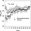
Figure 4. Superior Venous Saturation During the First 48 h After Norwood Procedure
The SvO2 was significantly higher during hours 1 to 10 in infants treated with phenoxybenzamine (0.25 mg · kg at commencement of cardiopulmonary bypass + selective use of continuous infusion 0.25 · mg · kg · day) than in those treated with milrinone (load 50 _μ_g · kg · min prior to separation from bypass + continuous infusion 0.5 _μ_g · kg · min after surgery). Reprinted with permission from Tweddell et al. (73).
Figure 5. Oxygen Saturation by Age at Surgery for Stage II Palliation
Patients undergoing early (<4 months) Stage II palliation initially had lower arterial saturation although they were not different than older patients at the time of hospital discharge. Reprinted with permission from Jaquiss et al. (193).
Figure 6. Fontan Surgical Techniques
The original atriopulmonary Fontan (A) has been replaced with the lateral tunnel (B) and extracardiac conduit (C) Fontan. Reprinted with permission from de Leval (488).
Figure 7. Computational Simulation as an Emerging Tool
Representative Fontan model showing angiography and computational simulation-derived measures of wall shear stress. The red areas indicate higher levels of shear (when compared with blue or green).
Similar articles
- Cardiac surgery 2002: staged repair of hypoplastic left heart syndrome.
Wright C. Wright C. Crit Care Nurs Q. 2002 Nov;25(3):72-8. doi: 10.1097/00002727-200211000-00009. Crit Care Nurs Q. 2002. PMID: 12450161 - Outcomes in Hypoplastic Left Heart Syndrome.
Metcalf MK, Rychik J. Metcalf MK, et al. Pediatr Clin North Am. 2020 Oct;67(5):945-962. doi: 10.1016/j.pcl.2020.06.008. Pediatr Clin North Am. 2020. PMID: 32888691 Review. - Hypoplastic Left Heart Syndrome Is Not a Predictor of Worse Intermediate Mortality Post Fontan.
Martin BJ, Mah K, Eckersley L, Harder J, Pockett C, Schantz D, Dyck J, Al Aklabi M, Rebeyka IM, Ross DB. Martin BJ, et al. Ann Thorac Surg. 2017 Dec;104(6):2037-2044. doi: 10.1016/j.athoracsur.2017.08.032. Epub 2017 Oct 31. Ann Thorac Surg. 2017. PMID: 29096870 - Magnetic resonance imaging catheter stress haemodynamics post-Fontan in hypoplastic left heart syndrome.
Pushparajah K, Wong JK, Bellsham-Revell HR, Hussain T, Valverde I, Bell A, Tzifa A, Greil G, Simpson JM, Kutty S, Razavi R. Pushparajah K, et al. Eur Heart J Cardiovasc Imaging. 2016 Jun;17(6):644-51. doi: 10.1093/ehjci/jev178. Epub 2015 Jul 18. Eur Heart J Cardiovasc Imaging. 2016. PMID: 26188193 Free PMC article. - Individualized approach in the management of patients with hypoplastic left heart syndrome (HLHS).
Bacha EA. Bacha EA. Semin Thorac Cardiovasc Surg Pediatr Card Surg Annu. 2013;16(1):3-6. doi: 10.1053/j.pcsu.2013.01.001. Semin Thorac Cardiovasc Surg Pediatr Card Surg Annu. 2013. PMID: 23561811 Review.
Cited by
- Retrospective Cohort Study of Additional Procedures and Transplant-Free Survival for Patients With Functionally Single Ventricle Disease Undergoing Staged Palliation in England and Wales.
Huang Q, Ridout D, Tsang V, Drury NE, Jones TJ, Bellsham-Revell H, Hadjicosta E, Seale AN, Mehta C, Pagel C, Crowe S, Espuny-Pujol F, Franklin RCG, Brown KL. Huang Q, et al. J Am Heart Assoc. 2024 Jul 16;13(14):e033068. doi: 10.1161/JAHA.123.033068. Epub 2024 Jul 3. J Am Heart Assoc. 2024. PMID: 38958142 Free PMC article. - Prenatal Genetic Diagnosis in Three Fetuses With Left Heart Hypoplasia (LHH) From Three Unrelated Families.
Luo S, Chen L, Wei W, Tan L, Zhang M, Duan Z, Cao J, Zhou Y, Zhou A, He X. Luo S, et al. Front Cardiovasc Med. 2021 Apr 9;8:631374. doi: 10.3389/fcvm.2021.631374. eCollection 2021. Front Cardiovasc Med. 2021. PMID: 33898534 Free PMC article. - In-Silico and In-Vitro Analysis of the Novel Hybrid Comprehensive Stage II Operation for Single Ventricle Circulation.
Das A, Hameed M, Prather R, Farias M, Divo E, Kassab A, Nykanen D, DeCampli W. Das A, et al. Bioengineering (Basel). 2023 Jan 19;10(2):135. doi: 10.3390/bioengineering10020135. Bioengineering (Basel). 2023. PMID: 36829630 Free PMC article. - Alterations in cerebral ventricle size in children with congenital heart disease.
Ackerman LL, Kralik SF, Daniels Z, Farrell A, Schamberger MS, Mastropietro CW. Ackerman LL, et al. Childs Nerv Syst. 2018 Nov;34(11):2233-2240. doi: 10.1007/s00381-018-3973-9. Epub 2018 Sep 12. Childs Nerv Syst. 2018. PMID: 30209597
References
- Schidlow DN, Anderson JB, Klitzner TS, et al. Variation in interstage outpatient care after the Norwood procedure: a report from the Joint Council on Congenital Heart Disease National Quality Improvement Collaborative. Congenit Heart Dis 2011;6:98–107. - PubMed
- Galindo A, Nieto O, Villagra´ S, Grañeras A, Herraiz I, Mendoza A. Hypoplastic left heart syndrome diagnosed in fetal life: associated findings, pregnancy outcome and results of palliative surgery. Ultrasound Obstet Gynecol 2009;33:560–6. - PubMed
- Feit LR, Copel JA, Kleinman CS. Foramen ovale size in the normal and abnormal human fetal heart: an indicator of transatrial flow physiology. Ultrasound Obstet Gynecol 1991;1:313–9. - PubMed
- Chin AJ, Weinberg PM, Barber G. Subcostal two-dimensional echocardiographic identification of anomalous attachment of septum primum in patients with left atrioventricular valve underdevelopment. J Am Coll Cardiol 1990;15:678–81. - PubMed
Publication types
MeSH terms
LinkOut - more resources
Full Text Sources
Medical


