Emergence of patterned activity in the developing zebrafish spinal cord - PubMed (original) (raw)
Emergence of patterned activity in the developing zebrafish spinal cord
Erica Warp et al. Curr Biol. 2012.
Abstract
Background: Developing neural networks display spontaneous and correlated rhythmic bursts of action potentials that are essential for circuit refinement. In the spinal cord, it is poorly understood how correlated activity is acquired and how its emergence relates to the formation of the spinal central pattern generator (CPG), the circuit that mediates rhythmic behaviors like walking and swimming. It is also unknown whether early, uncorrelated activity is necessary for the formation of the coordinated CPG.
Results: Time-lapse imaging in the intact zebrafish embryo with the genetically encoded calcium indicator GCaMP3 revealed a rapid transition from slow, sporadic activity to fast, ipsilaterally correlated, and contralaterally anticorrelated activity, characteristic of the spinal CPG. Ipsilateral correlations were acquired through the coalescence of local microcircuits. Brief optical manipulation of activity with the light-driven pump halorhodopsin revealed that the transition to correlated activity was associated with a strengthening of ipsilateral connections, likely mediated by gap junctions. Contralateral antagonism increased in strength at the same time. The transition to coordinated activity was disrupted by long-term optical inhibition of sporadic activity in motoneurons and ventral longitudinal descending interneurons and resulted in more neurons exhibiting uncoordinated activity patterns at later time points.
Conclusions: These findings show that the CPG in the zebrafish spinal cord emerges directly from a sporadically active network as functional connectivity strengthens between local and then more distal neurons. These results also reveal that early, sporadic activity in a subset of ventral spinal neurons is required for the integration of maturing neurons into the coordinated CPG network.
Copyright © 2012 Elsevier Ltd. All rights reserved.
Figures
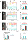
Figure 1. Spontaneous calcium activity in spinal neurons progresses from sporadic to locomotor-like during embryonic development
GCaMP3 activity in single neurons in one example embryo at 18 hpf (left) and 20 hpf (right). (A,D) Dorsal views of GCaMP3 baseline fluorescence with active regions circled (rostral left; imaged area somites 4–8). (B,E) Normalized intensity traces for active regions (identified on y-axis) for the left and right sides of the cord, with amplitude corresponding to standard deviations (s.d.) of fluorescence away from baseline. At 18 hpf (B) ipsilateral neurons have little correlated firing, though some synchronization is observed (e.g. cells 8 & 9). At 20 hpf (E) ipsilateral neurons are tightly synchronized, with few exceptions (e.g. cell 4; note elongated shape extending to the midline). (C,F) Raster plots of detected events for subsection of data in (B) and (E). At 18 hpf (C), population activity is uncoordinated. By 20 hpf (F), ipsilateral cells are synchronized, contralateral cells alternate, and a higher order left/right bursting organization is observed. See also Supplemental Fig. 1.
Figure 2. Pair-wise cell relationships progress from independent to ipsi-correlated/contra-anti-correlated during a short period of development
(A) Correlation matrices of single cell traces through the development of an example embryo. Each pixel represents a pair-wise comparison between 2 cells, with high correlation values in red and perfect auto-correlation along the diagonal. Cells are sorted left to right and top to bottom as shown for 17.5 hpf. Ticks mark border between left and right cord and bound a high degree of ipsilateral correlation observed at later time points. (B) Cross-correlation shows a strengthening of ipsilateral coupling between 18 and 20 hpf and acquisition of oscillatory rhythm by 20 hpf. Cross-correlation was calculated by averaging time-shifted correlation data for all ipsilateral and contralateral cell pairs in individual movies, pooled across 9 different fish. (C) Average pair-wise correlations for synchronous events comparing ipsilateral and contralateral cell pairs from individual time lapse movies acquired from fish ages 17.5 to 21 hpf and pooled across fish. We observed a significant difference between ipsilateral correlations in younger versus older embryos (18 hpf, r = 0.272 ± 0.082; 20 hpf, r = 0.761 ± 0.031; P < 10−3, paired Student’s t test; n = 9 fish), and a significant increase in the anti-correlation of contralateral cells (18 hpf, r = −0.207 ± 0.067; 20 hpf, r = −0.710 ± 0.028; P = < 10−3, paired Student’s t test; n = 9 fish). n=9 fish for 18–21 hpf; n=4 fish at 17.5 hpf. Error bars=s.e.m. See also Supplemental Fig. 2.
Figure 3. Ipsilateral correlation is acquired through the progressive synchronization of local subgroups of cells
Spatial maps of correlated groups in an example fish from 18 to 21 hpf show small local circuits containing a few cells at 18 and 18.5 hpf that expand into full correlation of each side at later stages. Correlations between all cell pairs were calculated and lines were drawn between cell pairs with correlations greater than 0.2, with thicker lines representing stronger correlation. Line color represents the log of the standard deviation of the lags between event start times of cell pairs and shows an overall increase in temporal precision between ipsilateral pairs as development progresses. See also Supplemental Fig. 3.
Figure 4. Optical manipulation of targeted network components with NpHR reveals changes in functional connectivity between ipsilateral neurons during development
(A,B) Single cell optical manipulation of spontaneous activity with NpHR at 18 hpf (A) and 20 hpf (B). 593nm light at 19mW/mm2 is targeted successively to two regions outlined in yellow (A1, left), while calcium population activity is simultaneously recorded (A1, right) in the illuminated cells (red) and in the other ipsilateral cells (teal), and here displayed as normalized traces (standard deviation, s.d.) with regions indicated on y-axis. (A1) At 18 hpf, application of yellow light to a single cell (during yellow highlight bar) inhibits only the illuminated cell, while other cells remain active. (A2) Pooled results (n=6 embryos) show inhibition during light-ON and activation at light-OFF to be limited to illuminated cells (red bars). (B) At 20 hpf, single cell illumination has no effect on activity of either the illuminated or non-illuminated cells (n=7 embryos). (C) At 20 hpf, illumination of one side of spinal cord in region spanning two somites (yellow outline in image, left) inhibits and rebound excites both the illuminated cells and other ipsilateral cells (n=7 embryos). (D) Reduction of activity and rebound due to NpHR activation at 20 hpf are observed in cells that are both rostral and caudal to the region illuminated. (E) Control application of light aimed to the side of the cord but within the embryo (C1) does not perturb activity (n=7 embryos), indicating that effect on un-illuminated ipsilateral cells is not due to light scattering though we acknowledge light scattering may have different properties in this region. Rostral, up in fluorescence images; * P<0.05; ** P<0.01, paired Student’s t test. See also Supplemental Figs. 4 and 5.
Figure 5. Bilateral activation with NpHR rebound reveals acquisition of contralateral antagonism during development
Raster plots of spontaneous events of left and right cells in a single embryo at 18 hpf (A) and 20 hpf (B) during and following bilateral NpHR inhibition with 593nm light (yellow bars) covering approximately four somites (C). (A) Bilateral activation following NpHR inhibition at 18 hpf results in near simultaneous activation of left (LT) and right (RT) cells following light offset. Arrows indicate the time when two or more cells participate in an event following light offset for one side of the cord (left side, blue; right side, red). (B) At 20 hpf, activation at light offset of bilateral illumination results in a burst of activity in which one side fires first, followed, after a delay, by firing on the other side and continuing in alternation of firing from side to side. In this example, the right side is active first in trial #1, but the left side is active first in the trial #2. (D) The delay following offset of bilateral illumination between synchronous events on the left and right sides of the cord (two or more cells participating) increases during development, suggesting an increase in left/right antagonism. n = 5 fish (4 trials per fish per condition); * P<0.05; ** P<0.01, paired Student’s t test. See also Supplemental Fig. 5.
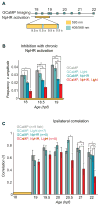
Figure 6. Inhibition of spontaneous events with NpHR from 18 to 19 hpf yields a subsequent decrease in ipsilateral correlation
(A) Experimental protocol for chronic inhibition experiments. GCaMP movies were acquired during the light manipulation (at 18, 18.5 and 19 hpf) with 488 nm light to determine the effectiveness of the light protocol, and at half hour/hour intervals thereafter (until 22 hpf) to asses subsequent changes in network dynamics. Stimulation of NpHR was performed from 18 to 19 hpf with continuous 593 nm light at 19nW/mm2 interspersed every 10 seconds with 500 msec long pulses of light simultaneously at two wavelengths: 405 nm to reduce desensitization of the NpHR and 568 nm to activate it. (B) The frequency of calcium events from 18 to 19 hpf was quantified for experimental fish expressing NpHR and receiving the yellow light protocol (GCaMP, NpHR, Light) as well as for three kinds of control fish: i) NpHR-negative fish without yellow/blue light (GCaMP), ii) NpHR-negative fish with yellow/blue light (GCaMP, Light), and iii) NpHR-positive fish without yellow/blue light (GCaMP, NpHR). Means were calculated per cell, n = 13–385 cells per group. There was a significant effect of group at 18, 18.5 and 19 hpf (one-way ANOVA at each time point, P<0.05), with greatest decreases in the experimental group (red bars; GCaMP, NpHR, Light). The reduction in activity in embryos that expressed NpHR but did not receive the light protocol can be attributed to the activation of NpHR by the 488 nm imaging light. (C) Average ipsilateral pair-wise correlations measured for experimental fish (n = 8) and the three control groups (n = 7 to 9) in movies acquired after the termination of the yellow/blue light protocol reveal a decrease in correlated activity in the experimental fish (GCaMP, NpHR, Light) at later time points compared to all of the controls. There was no difference between groups at 19, 19.5 and 20 hpf (one-way ANOVA at each time point, P>0.05), with significant differences at 20.5, 21 and 22 hpf (one-way ANOVA at each time point, P<0.05). Note that to avoid activation of NpHR in controls without yellow/blue light protocol, GCaMP imaging in this group was only done at 22 hpf. Bars=s.e.m. Asterisks in (B) and (C) mark pair-wise significance from post-hoc comparison with Bonferroni correction (* P<0.05; ** P<0.01)
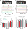
Figure 7. Light-inhibition decreases the number of cells joining the correlated network
(A,B) Baseline GCaMP fluorescence images with active regions circled (top, rostral left) and associated normalized intensity traces (bottom; amplitude plots standard deviation, s.d.) for example (A) control fish (without NpHR but illuminated with yellow/blue light protocol from 18–19 hpf) and (B) experimental fish (with NpHR and illuminated with yellow/blue light protocol from 18–19 hpf) at 22 hpf. Asterisks mark cells with long-duration, uncorrelated events, which increase in number in the experimental fish (B, bottom), and can be seen to reside in the medial spinal cord (b, top). (C) Average event duration through development was quantified using width at half maximum for experimental fish expressing NpHR and receiving the yellow light protocol (GCaMP, NpHR, Light) and for the three sets of control fish: i) lacking light and NpHR expression (GCaMP), ii) lacking NpHR expression (GCaMP, Light) or lacking light (GCaMP, NpHR). There was no difference between groups at 19, 19.5 20 and 21 hpf (one-way ANOVA at each time point, P>0.05), with significant differences at 20.5 and 22 hpf (one-way ANOVA at each time point, P<0.05), when experimental fish showed increases in event duration. (D) The distance from the cell center to the midline of the cord for active cells is reduced significantly in the experimental (GCaMP, NpHR, Light) fish compared to the three controls at all ages tested except for 20 hpf (one-way ANOVA at each time point, P<0.05). (C) and (D), means were calculated per cell (72–137 cells per group). Bars=s.e.m. Asterisks in (C) and (D) mark pair-wise significance from post-hoc comparison with Bonferroni correction (* P<0.05; ** P<0.01). See also Supplemental Fig. 6.
Comment in
- Motor development: activity matters after all.
Wenner P. Wenner P. Curr Biol. 2012 Jan 24;22(2):R47-8. doi: 10.1016/j.cub.2011.12.008. Curr Biol. 2012. PMID: 22280904
Similar articles
- Modeling spinal locomotor circuits for movements in developing zebrafish.
Roussel Y, Gaudreau SF, Kacer ER, Sengupta M, Bui TV. Roussel Y, et al. Elife. 2021 Sep 2;10:e67453. doi: 10.7554/eLife.67453. Elife. 2021. PMID: 34473059 Free PMC article. - A hybrid electrical/chemical circuit in the spinal cord generates a transient embryonic motor behavior.
Knogler LD, Ryan J, Saint-Amant L, Drapeau P. Knogler LD, et al. J Neurosci. 2014 Jul 16;34(29):9644-55. doi: 10.1523/JNEUROSCI.1225-14.2014. J Neurosci. 2014. PMID: 25031404 Free PMC article. - Calcium imaging of rhythmic network activity in the developing spinal cord of the chick embryo.
O'Donovan M, Ho S, Yee W. O'Donovan M, et al. J Neurosci. 1994 Nov;14(11 Pt 1):6354-69. doi: 10.1523/JNEUROSCI.14-11-06354.1994. J Neurosci. 1994. PMID: 7965041 Free PMC article. - Development of spinal motor networks in the chick embryo.
O'Donovan M, Sernagor E, Sholomenko G, Ho S, Antal M, Yee W. O'Donovan M, et al. J Exp Zool. 1992 Mar 1;261(3):261-73. doi: 10.1002/jez.1402610306. J Exp Zool. 1992. PMID: 1629659 Review. - Mechanisms of spontaneous activity in developing spinal networks.
O'Donovan MJ, Chub N, Wenner P. O'Donovan MJ, et al. J Neurobiol. 1998 Oct;37(1):131-45. doi: 10.1002/(sici)1097-4695(199810)37:1<131::aid-neu10>3.0.co;2-h. J Neurobiol. 1998. PMID: 9777737 Review.
Cited by
- Gap-junction-mediated bioelectric signaling required for slow muscle development and function in zebrafish.
Lukowicz-Bedford RM, Eisen JS, Miller AC. Lukowicz-Bedford RM, et al. Curr Biol. 2024 Jul 22;34(14):3116-3132.e5. doi: 10.1016/j.cub.2024.06.007. Epub 2024 Jun 26. Curr Biol. 2024. PMID: 38936363 - A single motor neuron determines the rhythm of early motor behavior in Ciona.
Akahoshi T, Utsumi MK, Oonuma K, Murakami M, Horie T, Kusakabe TG, Oka K, Hotta K. Akahoshi T, et al. Sci Adv. 2021 Dec 10;7(50):eabl6053. doi: 10.1126/sciadv.abl6053. Epub 2021 Dec 10. Sci Adv. 2021. PMID: 34890229 Free PMC article. - Differential activation of lumbar and sacral motor pools during walking at different speeds and slopes.
Dewolf AH, Ivanenko YP, Zelik KE, Lacquaniti F, Willems PA. Dewolf AH, et al. J Neurophysiol. 2019 Aug 1;122(2):872-887. doi: 10.1152/jn.00167.2019. Epub 2019 Jul 10. J Neurophysiol. 2019. PMID: 31291150 Free PMC article. - Homeostatic Feedback Modulates the Development of Two-State Patterned Activity in a Model Serotonin Motor Circuit in Caenorhabditis elegans.
Ravi B, Garcia J, Collins KM. Ravi B, et al. J Neurosci. 2018 Jul 11;38(28):6283-6298. doi: 10.1523/JNEUROSCI.3658-17.2018. Epub 2018 Jun 11. J Neurosci. 2018. PMID: 29891728 Free PMC article. - Motor neurons are dispensable for the assembly of a sensorimotor circuit for gaze stabilization.
Goldblatt D, Rosti B, Hamling KR, Leary P, Panchal H, Li M, Gelnaw H, Huang S, Quainoo C, Schoppik D. Goldblatt D, et al. Elife. 2024 Nov 20;13:RP96893. doi: 10.7554/eLife.96893. Elife. 2024. PMID: 39565353 Free PMC article.
References
- Torborg CL, Feller MB. Spontaneous patterned retinal activity and the refinement of retinal projections. Prog Neurobiol. 2005;76:213–235. - PubMed
Publication types
MeSH terms
Substances
LinkOut - more resources
Full Text Sources
Other Literature Sources
Molecular Biology Databases
Miscellaneous


