Clonotype and repertoire changes drive the functional improvement of HIV-specific CD8 T cell populations under conditions of limited antigenic stimulation - PubMed (original) (raw)
. 2012 Feb 1;188(3):1156-67.
doi: 10.4049/jimmunol.1102610. Epub 2011 Dec 30.
David A Price, Glenda Canderan, Abdelali Filali-Mouhim, Tedi E Asher, David R Ambrozak, Phillip Scheinberg, Mohamad Rachid Boulassel, Jean-Pierre Routy, Richard A Koup, Daniel C Douek, Rafick-Pierre Sekaly, Lydie Trautmann
Affiliations
- PMID: 22210916
- PMCID: PMC3262882
- DOI: 10.4049/jimmunol.1102610
Clonotype and repertoire changes drive the functional improvement of HIV-specific CD8 T cell populations under conditions of limited antigenic stimulation
Loury Janbazian et al. J Immunol. 2012.
Abstract
Persistent exposure to cognate Ag leads to the functional impairment and exhaustion of HIV-specific CD8 T cells. Ag withdrawal, attributable either to antiretroviral treatment or the emergence of epitope escape mutations, causes HIV-specific CD8 T cell responses to wane over time. However, this process does not continue to extinction, and residual CD8 T cells likely play an important role in the control of HIV replication. In this study, we conducted a longitudinal analysis of clonality, phenotype, and function to define the characteristics of HIV-specific CD8 T cell populations that persist under conditions of limited antigenic stimulation. Ag decay was associated with dynamic changes in the TCR repertoire, increased expression of CD45RA and CD127, decreased expression of programmed death-1, and the emergence of polyfunctional HIV-specific CD8 T cells. High-definition analysis of individual clonotypes revealed that the Ag loss-induced gain of function within HIV-specific CD8 T cell populations could be attributed to two nonexclusive mechanisms: 1) functional improvement of persisting clonotypes; and 2) recruitment of particular clonotypes endowed with superior functional capabilities.
Figures
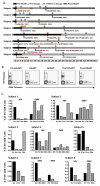
Figure 1. Virological and immunological characteristics of the study cohort
(A) Schematic representation of viremia and antigen sequence variation over time.A color code is assigned for each of the 3 main antigen load categories studied; high antigen load pre-HAART or pre-escape (black), low antigen load on HAART or after viral escape (diagonal lines) and antigen load rebound after cessation of HAART (grey).For subjects 1-4, the diagonal lines represent the administration ofHAART. For subjects 5-7, the diagonal lines represent epitope escape. For subject 8, the diagonal lines represent the combination of HAART and epitope escape.Month 0 indicates the estimated date of infection. Orange circles indicate the time pointsat which blood samples were analyzed for HIV-specific and CMV-specific CD8 T cell responses in each subject. Black arrows indicate the time points from which viral sequences were obtained. The targeted epitopes are depicted in each case; longitudinal epitope sequence variation is also displayed. (B) Representative tetramer co-staining showing the frequencies of HIV-specific and CMV-specific CD8 T cells longitudinally in subject 2. (C) Longitudinal frequencies of HIV-specific and CMV-specific CD8 T cells in all 8 subjects. Bars represent the following epitope-specific CD8 T cell populations: HLA-A*0201-FLGKIWPSHK (HIV Gag p2p7p1p6) and HLA-A*0201-NLVPMVATV (CMV pp65) in subject 1, HLA-A*0301-RLRPGGKKR (HIV Gag p17) and HLA-B*0702-TPRVTGGGAM (CMV pp65) in subject 2, HLA-B*0801-FLKEKGGL (HIV Nef) in subject 3, HLA-B*0702-TPGPGVRYPL (HIV Nef) and HLA-B*0702-TPRVTGGGAM (CMV pp65) in subject 4, HLA-B*0702-FPQGEAREL (HIV Pol) in subject 5, HLA-A*0301-RLRPGGKKK (HIV Gag p17) in subjects 6 and 7 (KKK and RKR represent wild type and variant epitopes in subject 7, respectively), and HLA-B*0801-GEIYKRWII (HIV Gag p24) and HLA-B*0702-TPRVTGGGAM (CMV pp65) in subject 8. The black, diagonal line and grey bars indicate pre-HAART or pre-escape, HAART or escape, and post-HAART time points, respectively.
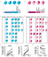
Figure 2. Phenotypic changes in HIV-specific CD8 T cell populations under conditions of reduced antigen load
(A&C) Maturation status of HIV-specific CD8 T cells at each time point, based on the expression of CCR7, CD27 and CD45RA. After the gates for the 3 markers were created on tetramer+cells, the Boolean gate platform was used to create 8 different combinations. The colored slices in each pie represent different phenotypic combinations;CD45RA+with other combinations (dark blue),CD45RA− with other positive combinations (medium blue), and CCR7−CD27−CD45RA− (light blue). **(B&D)**Exhaustion and survival capacity of HIV-specific CD8 T cells at each time point, based on the expression of CD28, CD127 and PD-1. The colored slices in each pie represent different phenotypic combinations; CD127+ with other combinations (purple), CD127−PD-1+ with other combinations (medium pink) and CD127−PD-1− with other combinations (light pink). In (A&B), which depict representative analyses for subject 1, the bars on the x-axis demonstrate the frequencies of cells belonging to a particular combination. The numbers under the pies indicatethe time points studied (m=month). Black, light grey and dark grey indicate pre-HAART or pre-escape, HAART or escape, and post-HAART time points, respectively. (E-G) Percentage of tetramer+ cells expressing CD45RA (E), CD127 (F) and PD-1 (G) at the first time point studied (high antigen load) and the second time point obtained after antigen decay (low antigen load). Ag=antigen.
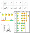
Figure 3. Functional changes in HIV-specific CD8 T cell populations under conditions of reduced antigen load
(A)Representative example of simultaneous multi-functional assessment of HIV-specific and CMV-specific CD8 T cells by multi-parametric flow cytometry at a given time point. Cells were stimulated for 6 hours with the corresponding cognate peptide before intracellular staining; αCD107a was present thoughout the assay to capture degranulating cells as described in the Materials and Methods. Percentages of function+tetramer+ cells are shown. Plots are gated on CD3+CD8+ cells. Dead cells were excluded from the analysis using a viability dye.(B) Multi-functional assessment of HIV-specific CD8 T cell responses by multi-parametric flow cytometry performed longitudinally for subject 1. After the gates for 4 functions were created (CD107a, IFN-γ, TNF and IL-2), the Boolean gate platform was used to create an array of 15 different positive combinations. The pies represent the functional profiles of HIV-specific CD8 T cell populations. The slices within each pie represent different functional combinations; 4+ (red), 3+ (orange), 2+ (yellow) and 1+ (green). The bars on the x-axis represent the response frequency for each combination. Numbers under the pies indicate the time points studied (m=month). Black, light grey and dark grey indicate pre-HAART or pre-escape, HAART or escape, and post-HAART time points, respectively.(C) Longitudinal flow cytometric analysis of HIV-specific CD8 T cell function for all 8 subjects. (D) Percentage of tetramer+ cellsexpressing CD107a, IFN-γ and TNF under conditions of high and low antigen load; the first time point before antigen decay and one time point after antigen decay, respectively, are shown. Ag=antigen.
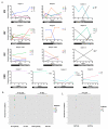
Figure 4. Evolution of the TCR repertoire in the context of antigen decay
(A) Longitudinal clonotype frequencies for HIV-specific and CMV-specific CD8 T cell responses. The major persistent clonotypes are represented for each subject, color-coded to match those shown in Supplementary Table 1. Under the x-axis, the black, diagonal line and grey bars indicate pre-HAART or pre-escape, HAART or escape, and post-HAART time points, respectively. (B) Morisita-Horn coefficients for HIV-specific and CMV-specific CD8 T cell populations before and after antigen decay (Ag and NoAg, respectively). The reference similarity for HIV-specific and CMV-specific CD8 T cell repertoireswas generated under the null hypothesis that the Ag and NoAg sets were randomly assigned from the same TCR population (HIV Ref and CMV Ref respectively). The similarity between Ag and NoAg time points was significantly lower than the reference similarities for HIV-specific but not for CMV-specific CD8 T cell repertoires.**(C)**Morisita-Horn coefficients for HIV-specific CD8 T cell responses comparing the time point before antigen decay (Ag) to either time points with low antigen load (NoAg) or time points after antigen rebound due to cessation of HAART (ReAg) for subjects 1, 2, 3 and 4. The similarity between time points before antigen decay and antigen rebound (Ag/ReAg) was significantly lower than between time points before and after antigen load decrease (Ag/NoAg).
Figure 5. Gain of function and persistence of individual HIV-specific CD8 T cell clonotypes under conditions of limited antigenic stimulation
**(A)**Phenotypic and multi-functional assessment of HLA-B*0801-restricted FL8-specific CD8 T cells from subject 3 before (1 month) and during (8 months) HAART. The dominant TRBV25-1/CASSVLRAAF/TRBJ1-1 clonotype showed changes in phenotype and gained functionality after antigendecay. **(B)Simultaneousphenotypic and multi-functional assessment of bulk and TCRVβ6-2+ HLA-A*0201-restricted FK10-specific CD8 T cells before and during HAART in subject 1.Tetramer+ cells comprising all clonotypes did not display an altered functional profile after antigendecay, whereas epitope-specific TCRVβ6-2+ CD8 T cells gained functionality within the same time frame.The experiment shown in this figure was performed at a different timethan the experiment presented in Figure 3 without co-stimulationto compare total tetramer+ cells and tetramer+TCRVβ6-2+ cells. No differences in phenotype were observed between tetramer+TCRVβ6-2+and tetramer+TCRVβ6-2− CD8 T cells at 17 months under HAART.(C)**Clonotypic composition of functional subsets within the HLA-B*0701-restricted FL9-specific CD8 T cell response at the 21 month pre-escape time point for subject 5, as determined by DNA-based sequence analysis. Pie colors represent the different functional combinations that were sorted: 3+ (orange), 2+ (yellow) and 1+ (green). The boxes show the CDR3 amino acid sequence, TRBV and TRBJ usage of the different functional cells.At 21 months, the tetramer+ CD8 T cell population was composed of one dominant clonotype (blue) and one subdominant clonotype(orange), both expressing TRBV18. All functional cells correspond to the subdominant clonotype at 21 months, which became dominant after antigen decay. No phenotypic differences were observed between functional and non-functional epitope-specific CD8 T cells at 21 months.
Similar articles
- Programmed death-1 expression on HIV-1-specific CD8+ T cells is shaped by epitope specificity, T-cell receptor clonotype usage and antigen load.
Kløverpris HN, McGregor R, McLaren JE, Ladell K, Stryhn A, Koofhethile C, Brener J, Chen F, Riddell L, Graziano L, Klenerman P, Leslie A, Buus S, Price DA, Goulder P. Kløverpris HN, et al. AIDS. 2014 Sep 10;28(14):2007-21. doi: 10.1097/QAD.0000000000000362. AIDS. 2014. PMID: 24906112 Free PMC article. - Fluctuations of functionally distinct CD8+ T-cell clonotypes demonstrate flexibility of the HIV-specific TCR repertoire.
Meyer-Olson D, Brady KW, Bartman MT, O'Sullivan KM, Simons BC, Conrad JA, Duncan CB, Lorey S, Siddique A, Draenert R, Addo M, Altfeld M, Rosenberg E, Allen TM, Walker BD, Kalams SA. Meyer-Olson D, et al. Blood. 2006 Mar 15;107(6):2373-83. doi: 10.1182/blood-2005-04-1636. Epub 2005 Dec 1. Blood. 2006. PMID: 16322475 Free PMC article. - Antigen load and viral sequence diversification determine the functional profile of HIV-1-specific CD8+ T cells.
Streeck H, Brumme ZL, Anastario M, Cohen KW, Jolin JS, Meier A, Brumme CJ, Rosenberg ES, Alter G, Allen TM, Walker BD, Altfeld M. Streeck H, et al. PLoS Med. 2008 May 6;5(5):e100. doi: 10.1371/journal.pmed.0050100. PLoS Med. 2008. PMID: 18462013 Free PMC article. - Abundant cytomegalovirus (CMV) reactive clonotypes in the CD8(+) T cell receptor alpha repertoire following allogeneic transplantation.
Link CS, Eugster A, Heidenreich F, Rücker-Braun E, Schmiedgen M, Oelschlägel U, Kühn D, Dietz S, Fuchs Y, Dahl A, Domingues AM, Klesse C, Schmitz M, Ehninger G, Bornhäuser M, Schetelig J, Bonifacio E. Link CS, et al. Clin Exp Immunol. 2016 Jun;184(3):389-402. doi: 10.1111/cei.12770. Epub 2016 Mar 8. Clin Exp Immunol. 2016. PMID: 26800118 Free PMC article. - Large TCR diversity of virus-specific CD8 T cells provides the mechanistic basis for massive TCR renewal after antigen exposure.
Miconnet I, Marrau A, Farina A, Taffé P, Vigano S, Harari A, Pantaleo G. Miconnet I, et al. J Immunol. 2011 Jun 15;186(12):7039-49. doi: 10.4049/jimmunol.1003309. Epub 2011 May 9. J Immunol. 2011. PMID: 21555537
Cited by
- Unravelling the mechanisms of durable control of HIV-1.
Walker BD, Yu XG. Walker BD, et al. Nat Rev Immunol. 2013 Jul;13(7):487-98. doi: 10.1038/nri3478. Nat Rev Immunol. 2013. PMID: 23797064 Review. - Programmed death-1 expression on HIV-1-specific CD8+ T cells is shaped by epitope specificity, T-cell receptor clonotype usage and antigen load.
Kløverpris HN, McGregor R, McLaren JE, Ladell K, Stryhn A, Koofhethile C, Brener J, Chen F, Riddell L, Graziano L, Klenerman P, Leslie A, Buus S, Price DA, Goulder P. Kløverpris HN, et al. AIDS. 2014 Sep 10;28(14):2007-21. doi: 10.1097/QAD.0000000000000362. AIDS. 2014. PMID: 24906112 Free PMC article. - Immunosequencing identifies signatures of cytomegalovirus exposure history and HLA-mediated effects on the T cell repertoire.
Emerson RO, DeWitt WS, Vignali M, Gravley J, Hu JK, Osborne EJ, Desmarais C, Klinger M, Carlson CS, Hansen JA, Rieder M, Robins HS. Emerson RO, et al. Nat Genet. 2017 May;49(5):659-665. doi: 10.1038/ng.3822. Epub 2017 Apr 3. Nat Genet. 2017. PMID: 28369038 - STING Ligand-Mediated Priming of Functional CD8+ T Cells Specific for HIV-1-Protective Epitopes from Naive T Cells.
Kuse N, Akahoshi T, Takiguchi M. Kuse N, et al. J Virol. 2021 Jul 26;95(16):e0069921. doi: 10.1128/JVI.00699-21. Epub 2021 Jul 26. J Virol. 2021. PMID: 34076478 Free PMC article. - Rapid perturbation in viremia levels drives increases in functional avidity of HIV-specific CD8 T cells.
Viganò S, Bellutti Enders F, Miconnet I, Cellerai C, Savoye AL, Rozot V, Perreau M, Faouzi M, Ohmiti K, Cavassini M, Bart PA, Pantaleo G, Harari A. Viganò S, et al. PLoS Pathog. 2013;9(7):e1003423. doi: 10.1371/journal.ppat.1003423. Epub 2013 Jul 4. PLoS Pathog. 2013. PMID: 23853580 Free PMC article.
References
- Jin X, Bauer DE, Tuttleton SE, Lewin S, Gettie A, Blanchard J, Irwin CE, Safrit JT, Mittler J, Weinberger L, Kostrikis LG, Zhang L, Perelson AS, Ho DD. Dramatic rise in plasma viremia after CD8(+) T cell depletion in simian immunodeficiency virus-infected macaques. J Exp Med. 1999;189:991–998. - PMC - PubMed
- Schmitz JE, Kuroda MJ, Santra S, Sasseville VG, Simon MA, Lifton MA, Racz P, Tenner-Racz K, Dalesandro M, Scallon BJ, Ghrayeb J, Forman MA, Montefiori DC, Rieber EP, Letvin NL, Reimann KA. Control of viremia in simian immunodeficiency virus infection by CD8+ lymphocytes. Science. 1999;283:857–860. - PubMed
Publication types
MeSH terms
Substances
Grants and funding
- CAPMC/ CIHR/Canada
- IDPIDA028871-01/PHS HHS/United States
- DP1 DA028871-01/DA/NIDA NIH HHS/United States
- DP1 DA028871/DA/NIDA NIH HHS/United States
- G0501963/MRC_/Medical Research Council/United Kingdom
LinkOut - more resources
Full Text Sources
Research Materials


