Natural amyloid-β oligomers acutely impair the formation of a contextual fear memory in mice - PubMed (original) (raw)
Natural amyloid-β oligomers acutely impair the formation of a contextual fear memory in mice
Kara A Kittelberger et al. PLoS One. 2012.
Abstract
Memory loss is one of the hallmark symptoms of Alzheimer's disease (AD). It has been proposed that soluble amyloid-beta (Abeta) oligomers acutely impair neuronal function and thereby memory. We here report that natural Abeta oligomers acutely impair contextual fear memory in mice. A natural Abeta oligomer solution containing Abeta monomers, dimers, trimers, and tetramers was derived from the conditioned medium of 7PA2 cells, a cell line that expresses human amyloid precursor protein containing the Val717Phe familial AD mutation. As a control we used 7PA2 conditioned medium from which Abeta oligomers were removed through immunodepletion. Separate groups of mice were injected with Abeta and control solutions through a cannula into the lateral brain ventricle, and subjected to fear conditioning using two tone-shock pairings. One day after fear conditioning, mice were tested for contextual fear memory and tone fear memory in separate retrieval trials. Three experiments were performed. For experiment 1, mice were injected three times: 1 hour before and 3 hours after fear conditioning, and 1 hour before context retrieval. For experiments 2 and 3, mice were injected a single time at 1 hour and 2 hours before fear conditioning respectively. In all three experiments there was no effect on tone fear memory. Injection of Abeta 1 hour before fear conditioning, but not 2 hours before fear conditioning, impaired the formation of a contextual fear memory. In future studies, the acute effect of natural Abeta oligomers on contextual fear memory can be used to identify potential mechanisms and treatments of AD associated memory loss.
Conflict of interest statement
Competing Interests: The authors have declared that no competing interests exist.
Figures
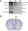
Figure 1. Natural Abeta oligomer solution and injection.
A) Blot image showing the presence of Abeta monomers, dimers, trimers, and tetramers in the Abeta solution. The 6E10 antibody was used for detection of Abeta oligomers that were removed from the Abeta solution using immunoprecipitation with A/G beads and 4G8 antibody (IP1, IP2, IP3: oligomers bound to beads used for first, second, and third immunoprecipitation). No oligomers were detected after three rounds of immunoprecipitation, which confirmed the absence of Abeta oligomers in the control solution (see “Materials and Methods” for a detailed description of how Abeta and control solutions were generated). B) Diagram showing the location of the guide cannula (green) and the injector cannula (red) in a Nissl-stained coronal section of the mouse brain . The tip of the guide cannula stopped just above the corpus callosum. The tip of the injection cannula extended into the lateral ventricle.
Figure 2. Experiment 1: repeated Abeta injection.
Top) Diagram showing the design of experiment 1. Separate groups of mice were injected three times with either control or Abeta solution. Bottom) Graphs showing average freezing scores during fear conditioning on day 1 and the two retrieval trials on day 2 (see “Materials and Methods: Analysis of freezing behavior” for explanation of intervals on the X axis). Mice injected with the Abeta solution (n = 5) had significantly lower freezing scores during the context fear retrieval trial as compared with mice injected with the control solution (n = 6). Error bars are standard errors of means. * P<0.05.
Figure 3. Experiment 2: single Abeta injection 1 hour before fear conditioning.
Top) Diagram showing the design of experiment 2. Separate groups of mice were injected one time with either control or Abeta solution 1 hour before fear conditioning. Bottom) Graphs showing average freezing scores during fear conditioning on day 1 and the two retrieval trials on day 2. Mice injected with the Abeta solution (n = 8) had significantly lower freezing scores during the context fear retrieval trial as compared with mice injected with the control solution (n = 10). Error bars are standard errors of means. * P<0.05.
Figure 4. Experiment 3: single Abeta injection 2 hours before fear conditioning.
Top) Diagram showing the design of experiment 3. Separate groups of mice were injected one time with either control or Abeta solution 2 hours before fear conditioning. Bottom) Graphs showing average freezing scores during fear conditioning on day 1 and the two retrieval trials on day 2. There was no difference during any of the intervals analyzed between mice injected with the Abeta solution (n = 10) and mice injected with the control solution (n = 8). Error bars are standard errors of means.
Similar articles
- Aβ oligomers inhibit synapse remodelling necessary for memory consolidation.
Freir DB, Fedriani R, Scully D, Smith IM, Selkoe DJ, Walsh DM, Regan CM. Freir DB, et al. Neurobiol Aging. 2011 Dec;32(12):2211-8. doi: 10.1016/j.neurobiolaging.2010.01.001. Epub 2010 Jan 25. Neurobiol Aging. 2011. PMID: 20097446 Free PMC article. - Secreted amyloid β-proteins in a cell culture model include N-terminally extended peptides that impair synaptic plasticity.
Welzel AT, Maggio JE, Shankar GM, Walker DE, Ostaszewski BL, Li S, Klyubin I, Rowan MJ, Seubert P, Walsh DM, Selkoe DJ. Welzel AT, et al. Biochemistry. 2014 Jun 24;53(24):3908-21. doi: 10.1021/bi5003053. Biochemistry. 2014. PMID: 24840308 Free PMC article. - In vivo application of beta amyloid oligomers: a simple tool to evaluate mechanisms of action and new therapeutic approaches.
Balducci C, Forloni G. Balducci C, et al. Curr Pharm Des. 2014;20(15):2491-505. doi: 10.2174/13816128113199990497. Curr Pharm Des. 2014. PMID: 23859553 Review. - The role of cell-derived oligomers of Abeta in Alzheimer's disease and avenues for therapeutic intervention.
Walsh DM, Klyubin I, Shankar GM, Townsend M, Fadeeva JV, Betts V, Podlisny MB, Cleary JP, Ashe KH, Rowan MJ, Selkoe DJ. Walsh DM, et al. Biochem Soc Trans. 2005 Nov;33(Pt 5):1087-90. doi: 10.1042/BST20051087. Biochem Soc Trans. 2005. PMID: 16246051 Review.
Cited by
- Hippocampal corticotropin-releasing hormone neurons support recognition memory and modulate hippocampal excitability.
Hooper A, Fuller PM, Maguire J. Hooper A, et al. PLoS One. 2018 Jan 18;13(1):e0191363. doi: 10.1371/journal.pone.0191363. eCollection 2018. PLoS One. 2018. PMID: 29346425 Free PMC article. - Interactions between Aβ oligomers and presynaptic cholinergic signaling: age-dependent effects on attentional capacities.
Parikh V, Bernard CS, Naughton SX, Yegla B. Parikh V, et al. Behav Brain Res. 2014 Nov 1;274:30-42. doi: 10.1016/j.bbr.2014.07.046. Epub 2014 Aug 4. Behav Brain Res. 2014. PMID: 25101540 Free PMC article. - TREM2 promotes Aβ phagocytosis by upregulating C/EBPα-dependent CD36 expression in microglia.
Kim SM, Mun BR, Lee SJ, Joh Y, Lee HY, Ji KY, Choi HR, Lee EH, Kim EM, Jang JH, Song HW, Mook-Jung I, Choi WS, Kang HS. Kim SM, et al. Sci Rep. 2017 Sep 11;7(1):11118. doi: 10.1038/s41598-017-11634-x. Sci Rep. 2017. PMID: 28894284 Free PMC article. - Mechanisms of U87 astrocytoma cell uptake and trafficking of monomeric versus protofibril Alzheimer's disease amyloid-β proteins.
Li Y, Cheng D, Cheng R, Zhu X, Wan T, Liu J, Zhang R. Li Y, et al. PLoS One. 2014 Jun 18;9(6):e99939. doi: 10.1371/journal.pone.0099939. eCollection 2014. PLoS One. 2014. PMID: 24941200 Free PMC article. - Ionic Environment Affects Biomolecular Interactions of Amyloid-β: SPR Biosensor Study.
Hemmerová E, Špringer T, Krištofiková Z, Homola J. Hemmerová E, et al. Int J Mol Sci. 2020 Dec 20;21(24):9727. doi: 10.3390/ijms21249727. Int J Mol Sci. 2020. PMID: 33419257 Free PMC article.
References
- Krafft GA, Klein WL. ADDLs and the signaling web that leads to Alzheimer's disease. Neuropharmacology. 2010;59:230–242. - PubMed
- Terry RD, Masliah E, Salmon DP, Butters N, DeTeresa R, et al. Physical basis of cognitive alterations in Alzheimer's disease: synapse loss is the major correlate of cognitive impairment. Ann Neurol. 1991;30:572–580. - PubMed
- McLean CA, Cherny RA, Fraser FW, Fuller SJ, Smith MJ, et al. Soluble pool of Abeta amyloid as a determinant of severity of neurodegeneration in Alzheimer's disease. Ann Neurol. 1999;46:860–866. - PubMed
- Van Dam D, D'Hooge R, Staufenbiel M, Van Ginneken C, Van Meir F, et al. Age-dependent cognitive decline in the APP23 model precedes amyloid deposition. European Journal of Neuroscience. 2003;17:388–396. - PubMed
Publication types
MeSH terms
Substances
LinkOut - more resources
Full Text Sources
Other Literature Sources
Medical
Research Materials


