NKG2D ligand tumor expression and association with clinical outcome in early breast cancer patients: an observational study - PubMed (original) (raw)
NKG2D ligand tumor expression and association with clinical outcome in early breast cancer patients: an observational study
Esther M de Kruijf et al. BMC Cancer. 2012.
Abstract
Background: Cell surface NKG2D ligands (NKG2DL) bind to the activating NKG2D receptor present on NK cells and subsets of T cells, thus playing a role in initiating an immune response. We examined tumor expression and prognostic effect of NKG2DL in breast cancer patients.
Methods: Our study population (n = 677) consisted of all breast cancer patients primarily treated with surgery in our center between 1985 and 1994. Formalin-fixed paraffin-embedded tumor tissue was immunohistochemically stained with antibodies directed against MIC-A/MIC-B (MIC-AB), ULBP-1, ULBP-2, ULBP-3, ULBP-4, and ULBP-5.
Results: NKG2DL were frequently expressed by tumors (MIC-AB, 50% of the cases; ULBP-1, 90%; ULBP-2, 99%; ULBP-3, 100%; ULBP-4, 26%; ULBP-5, 90%) and often showed co-expression: MIC-AB and ULBP-4 (p = 0.043), ULBP-1 and ULBP-5 (p = 0.006), ULBP-4 and ULBP-5 (p < 0.001). MIC-AB (p = 0.001) and ULBP-2 (p = 0.006) expression resulted in a statistically significant longer relapse free period (RFP). Combined expression of these ligands showed to be an independent prognostic parameter for RFP (p < 0.001, HR 0.41). Combined expression of all ligands showed no associations with clinical outcome.
Conclusions: We demonstrated for the first time that NKG2DL are frequently expressed and often co-expressed in breast cancer. Expression of MIC-AB and ULBP-2 resulted in a statistically significant beneficial outcome concerning RFP with high discriminative power. Combination of all NKG2DL showed no additive or interactive effect of ligands on each other, suggesting that similar and co-operative functioning of all NKG2DL can not be assumed. Our observations suggest that among driving forces in breast cancer outcome are immune activation on one site and tumor immune escape on the other site.
Figures
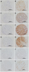
Figure 1
Representative examples of immunohistochemical stainings of primary breast cancer tissues for respectively no expression and high expression of MIC-AB (A: intensity 0 (negative); B: intensity 2 (intermediate)), ULBP-1 (C: intensity 0 (negative); D: intensity 2 (intermediate)), ULBP-2 (E: intensity 0 (negative); F: intensity 3 (strong)), ULBP-3 (G: intensity 0 (negative); H: intensity 3 (strong)), ULBP-4 (I: intensity 0 (negative); J: intensity 1 (weak)), and ULBP-5 (K: intensity 0 (negative); L: intensity 3 (strong)) in breast cancer. Immunohistochemistry was performed according to standard protocols as described in Materials and Methods.
Figure 2
Relapses over time related with expression of MIC-AB (A), ULBP-1 (B), ULBP-2 (C), ULBP-3 (D), ULBP-4 (E), and ULBP-5 (F). X-axis represents patient follow-up in years; Y-axis represents cumulative relapses in %. Log-rank p-values are shown in each graph. Only expression of MIC-AB and ULBP-2 resulted in statistically significantly favorable relapse-free period (RFP).
Figure 3
Relapses over time related with combined expression of MIC-AB and ULBP-2. X-axis represents patient follow-up in years; Y-axis represents cumulative relapses in %. Log-rank p-values are shown in the graph. Combined low expression of MIC-AB and ULBP-2 resulted in the worst outcome of patients concerning relapse-free period (RFP); while combined high expression of both ligands resulted in the most favorable outcome of patients.
Figure 4
Relapses over time related with combined number of NKG2D ligands with high expression (A) and amount of expression of NKG2D ligands (B). (A) legends in graph show total number of NKG2D ligands with high expression; (B) legends in graph show total intensity score of all NKG2D ligand expression. X-axis represents patient follow-up in years; Y-axis represents cumulative relapses in %. Log-rank p-values are shown in the graph. No associations were found with outcome concerning RFP for either combined number of expressed (A) or combined amount of expression (B) of ligands.
Similar articles
- Comparative genomic analysis of mammalian NKG2D ligand family genes provides insights into their origin and evolution.
Kondo M, Maruoka T, Otsuka N, Kasamatsu J, Fugo K, Hanzawa N, Kasahara M. Kondo M, et al. Immunogenetics. 2010 Jul;62(7):441-50. doi: 10.1007/s00251-010-0438-z. Epub 2010 Apr 8. Immunogenetics. 2010. PMID: 20376438 - Interferon-gamma down-regulates NKG2D ligand expression and impairs the NKG2D-mediated cytolysis of MHC class I-deficient melanoma by natural killer cells.
Schwinn N, Vokhminova D, Sucker A, Textor S, Striegel S, Moll I, Nausch N, Tuettenberg J, Steinle A, Cerwenka A, Schadendorf D, Paschen A. Schwinn N, et al. Int J Cancer. 2009 Apr 1;124(7):1594-604. doi: 10.1002/ijc.24098. Int J Cancer. 2009. PMID: 19089914 - Soluble ligands for the NKG2D receptor are released during HIV-1 infection and impair NKG2D expression and cytotoxicity of NK cells.
Matusali G, Tchidjou HK, Pontrelli G, Bernardi S, D'Ettorre G, Vullo V, Buonomini AR, Andreoni M, Santoni A, Cerboni C, Doria M. Matusali G, et al. FASEB J. 2013 Jun;27(6):2440-50. doi: 10.1096/fj.12-223057. Epub 2013 Feb 8. FASEB J. 2013. PMID: 23395909 - Natural killer group 2D receptor and its ligands in cancer immune escape.
Duan S, Guo W, Xu Z, He Y, Liang C, Mo Y, Wang Y, Xiong F, Guo C, Li Y, Li X, Li G, Zeng Z, Xiong W, Wang F. Duan S, et al. Mol Cancer. 2019 Feb 27;18(1):29. doi: 10.1186/s12943-019-0956-8. Mol Cancer. 2019. PMID: 30813924 Free PMC article. Review. - Cutting an NKG2D Ligand Short: Cellular Processing of the Peculiar Human NKG2D Ligand ULBP4.
Zöller T, Wittenbrink M, Hoffmeister M, Steinle A. Zöller T, et al. Front Immunol. 2018 Mar 29;9:620. doi: 10.3389/fimmu.2018.00620. eCollection 2018. Front Immunol. 2018. PMID: 29651291 Free PMC article. Review.
Cited by
- Altered Expression of Natural Cytotoxicity Receptors and NKG2D on Peripheral Blood NK Cell Subsets in Breast Cancer Patients.
Nieto-Velázquez NG, Torres-Ramos YD, Muñoz-Sánchez JL, Espinosa-Godoy L, Gómez-Cortés S, Moreno J, Moreno-Eutimio MA. Nieto-Velázquez NG, et al. Transl Oncol. 2016 Oct;9(5):384-391. doi: 10.1016/j.tranon.2016.07.003. Epub 2016 Sep 12. Transl Oncol. 2016. PMID: 27641642 Free PMC article. - Biological role of NK cells and immunotherapeutic approaches in breast cancer.
Roberti MP, Mordoh J, Levy EM. Roberti MP, et al. Front Immunol. 2012 Dec 12;3:375. doi: 10.3389/fimmu.2012.00375. eCollection 2012. Front Immunol. 2012. PMID: 23248625 Free PMC article. - Perturbation of NK cell peripheral homeostasis accelerates prostate carcinoma metastasis.
Liu G, Lu S, Wang X, Page ST, Higano CS, Plymate SR, Greenberg NM, Sun S, Li Z, Wu JD. Liu G, et al. J Clin Invest. 2013 Oct;123(10):4410-22. doi: 10.1172/JCI69369. Epub 2013 Sep 9. J Clin Invest. 2013. PMID: 24018560 Free PMC article. - Control of triple-negative breast cancer using ex vivo self-enriched, costimulated NKG2D CAR T cells.
Han Y, Xie W, Song DG, Powell DJ Jr. Han Y, et al. J Hematol Oncol. 2018 Jul 6;11(1):92. doi: 10.1186/s13045-018-0635-z. J Hematol Oncol. 2018. PMID: 29980239 Free PMC article. - Implication of combined PD-L1/PD-1 blockade with cytokine-induced killer cells as a synergistic immunotherapy for gastrointestinal cancer.
Dai C, Lin F, Geng R, Ge X, Tang W, Chang J, Wu Z, Liu X, Lin Y, Zhang Z, Li J. Dai C, et al. Oncotarget. 2016 Mar 1;7(9):10332-44. doi: 10.18632/oncotarget.7243. Oncotarget. 2016. PMID: 26871284 Free PMC article.
References
- Early breast cancer Trialists' Collaborative Group. Effects of chemotherapy and hormonal therapy for early breast cancer on recurrence and 15-year survival: an overview of the randomised trials. Lancet. 2005;365:1687–1717. - PubMed
Publication types
MeSH terms
Substances
LinkOut - more resources
Full Text Sources
Other Literature Sources
Medical


