Integrated genome and transcriptome sequencing identifies a novel form of hybrid and aggressive prostate cancer - PubMed (original) (raw)
doi: 10.1002/path.3987. Epub 2012 Mar 21.
Alexander W Wyatt, Anna V Lapuk, Andrew McPherson, Brian J McConeghy, Robert H Bell, Shawn Anderson, Anne Haegert, Sonal Brahmbhatt, Robert Shukin, Fan Mo, Estelle Li, Ladan Fazli, Antonio Hurtado-Coll, Edward C Jones, Yaron S Butterfield, Faraz Hach, Fereydoun Hormozdiari, Iman Hajirasouliha, Paul C Boutros, Robert G Bristow, Steven Jm Jones, Martin Hirst, Marco A Marra, Christopher A Maher, Arul M Chinnaiyan, S Cenk Sahinalp, Martin E Gleave, Stanislav V Volik, Colin C Collins
Affiliations
- PMID: 22294438
- PMCID: PMC3768138
- DOI: 10.1002/path.3987
Integrated genome and transcriptome sequencing identifies a novel form of hybrid and aggressive prostate cancer
Chunxiao Wu et al. J Pathol. 2012 May.
Abstract
Next-generation sequencing is making sequence-based molecular pathology and personalized oncology viable. We selected an individual initially diagnosed with conventional but aggressive prostate adenocarcinoma and sequenced the genome and transcriptome from primary and metastatic tissues collected prior to hormone therapy. The histology-pathology and copy number profiles were remarkably homogeneous, yet it was possible to propose the quadrant of the prostate tumour that likely seeded the metastatic diaspora. Despite a homogeneous cell type, our transcriptome analysis revealed signatures of both luminal and neuroendocrine cell types. Remarkably, the repertoire of expressed but apparently private gene fusions, including C15orf21:MYC, recapitulated this biology. We hypothesize that the amplification and over-expression of the stem cell gene MSI2 may have contributed to the stable hybrid cellular identity. This hybrid luminal-neuroendocrine tumour appears to represent a novel and highly aggressive case of prostate cancer with unique biological features and, conceivably, a propensity for rapid progression to castrate-resistance. Overall, this work highlights the importance of integrated analyses of genome, exome and transcriptome sequences for basic tumour biology, sequence-based molecular pathology and personalized oncology.
Copyright © 2012 Pathological Society of Great Britain and Ireland. Published by John Wiley & Sons, Ltd.
Conflict of interest statement
No conflicts of interest were declared.
Figures
Figure 1
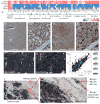
Histopathology and copy number analysis. Haematoxylin and eosin stains showing uniform adenocarcinoma with a Gleason score of 10 in patient 963's radical prostatectomy specimen (A, ×0.5; B, ×40) and a lymph node metastasis (C, ×20). No normal prostate structure can be discerned in (A, B). Lymphocytes can be seen in (C). Scale bars = 1 mm (A); 20 μm (B); 100 μm (C). (D) Differences between the samples, i.e. RP differs in signal intensity from LNmet at 2.9% of aCGH probe loci. The most and least similar pairs are emphasized. (E) Frequency plot showing the copy number (CN) aberrations in the primary tumour quadrants and the LNmet (green, gain; red, loss). A frequency of 100% indicates a CN aberration detected in all five samples; the majority of CN aberrations are detected in all samples. CN aberrant genes previously associated with PCa are annotated. Blue, fusion genes (the genomic breakpoints of 12 genes involved in fusion events coincided with aCGH segment breaks); pink, readthrough events. Note: Chromosomes X and Y demonstrated no aberrations and are not shown. (F, G) Chr11 CN profile in LT (F) and RP (G), indicating the marginal differences. (H, I) Chr17 CN profile in LP (H) and LNmet (I).
Figure 2
Fusion genes validated in the primary tumour and lymph node metastasis. The top three fusions involve either an androgen-related gene, a tumour suppressor or an oncogene. The bottom five fusions involve genes with a neuroendocrine function. Note that despite absence of RNA-Seq reads (ie expression) for some fusion genes, the underlying genomic breakpoints for each fusion were detected in both primary and metastatic tumours (see Supporting information, Figure S4).
Figure 3
Analysis of gene expression levels. (A) Heat map demonstrating expression of genes with a neural/endocrine function in the primary tumour (LP) and the lymph node metastatic tumour (LNmet) of patient 963 compared to adenocarcinoma cell lines and benign prostate. FP, filament protein; TF, transcription factor. (B–E) Antibody stains showing strong expression of AR (B, ×2; C, ×40) and CHGA (D, ×2; E, ×40) in the primary tumour. Bands of stromal cells are unstained. (F, G, I, J) Dual antibody stains of AR and CHGA (F, ×8; G, ×40; I, ×10; J, ×40) confirming co-expression in 100% of tumour cells. Note that in (I, J) benign prostate glands are visible, demonstrating that normal luminal cells are AR-positive only, while normal NE cells are CHGA-positive only. Scale bars = 1 mm (B, D); 200 μm (I); 100 μm (C, E, F); 20 μm (G, J). (H) Hexbin plot illustrating correlation of gene expression levels between LP and LNmet (log2 scale). Red and green lines indicate a four- and 16-fold expression difference, respectively, between LP and LNmet.
Similar articles
- From sequence to molecular pathology, and a mechanism driving the neuroendocrine phenotype in prostate cancer.
Lapuk AV, Wu C, Wyatt AW, McPherson A, McConeghy BJ, Brahmbhatt S, Mo F, Zoubeidi A, Anderson S, Bell RH, Haegert A, Shukin R, Wang Y, Fazli L, Hurtado-Coll A, Jones EC, Hach F, Hormozdiari F, Hajirasouliha I, Boutros PC, Bristow RG, Zhao Y, Marra MA, Fanjul A, Maher CA, Chinnaiyan AM, Rubin MA, Beltran H, Sahinalp SC, Gleave ME, Volik SV, Collins CC. Lapuk AV, et al. J Pathol. 2012 Jul;227(3):286-97. doi: 10.1002/path.4047. J Pathol. 2012. PMID: 22553170 Free PMC article. - Integration of copy number and transcriptomics provides risk stratification in prostate cancer: A discovery and validation cohort study.
Ross-Adams H, Lamb AD, Dunning MJ, Halim S, Lindberg J, Massie CM, Egevad LA, Russell R, Ramos-Montoya A, Vowler SL, Sharma NL, Kay J, Whitaker H, Clark J, Hurst R, Gnanapragasam VJ, Shah NC, Warren AY, Cooper CS, Lynch AG, Stark R, Mills IG, Grönberg H, Neal DE; CamCaP Study Group. Ross-Adams H, et al. EBioMedicine. 2015 Jul 29;2(9):1133-44. doi: 10.1016/j.ebiom.2015.07.017. eCollection 2015 Sep. EBioMedicine. 2015. PMID: 26501111 Free PMC article. - Intratumoral and Intertumoral Genomic Heterogeneity of Multifocal Localized Prostate Cancer Impacts Molecular Classifications and Genomic Prognosticators.
Wei L, Wang J, Lampert E, Schlanger S, DePriest AD, Hu Q, Gomez EC, Murakam M, Glenn ST, Conroy J, Morrison C, Azabdaftari G, Mohler JL, Liu S, Heemers HV. Wei L, et al. Eur Urol. 2017 Feb;71(2):183-192. doi: 10.1016/j.eururo.2016.07.008. Epub 2016 Jul 21. Eur Urol. 2017. PMID: 27451135 Free PMC article. - Molecular pathology of prostate cancer revealed by next-generation sequencing: opportunities for genome-based personalized therapy.
Huang J, Wang JK, Sun Y. Huang J, et al. Curr Opin Urol. 2013 May;23(3):189-93. doi: 10.1097/MOU.0b013e32835e9ef4. Curr Opin Urol. 2013. PMID: 23385974 Free PMC article. Review. - Neuroendocrine cells of prostate cancer: biologic functions and molecular mechanisms.
Huang YH, Zhang YQ, Huang JT. Huang YH, et al. Asian J Androl. 2019 May-Jun;21(3):291-295. doi: 10.4103/aja.aja_128_18. Asian J Androl. 2019. PMID: 30924452 Free PMC article.
Cited by
- Cancer genome-sequencing study design.
Mwenifumbo JC, Marra MA. Mwenifumbo JC, et al. Nat Rev Genet. 2013 May;14(5):321-32. doi: 10.1038/nrg3445. Nat Rev Genet. 2013. PMID: 23594910 Review. - Decoding the heterogeneous landscape in the development prostate cancer.
Segura-Moreno YY, Sanabria-Salas MC, Varela R, Mesa JA, Serrano ML. Segura-Moreno YY, et al. Oncol Lett. 2021 May;21(5):376. doi: 10.3892/ol.2021.12637. Epub 2021 Mar 15. Oncol Lett. 2021. PMID: 33777200 Free PMC article. Review. - Systematic identification and characterization of RNA editing in prostate tumors.
Mo F, Wyatt AW, Sun Y, Brahmbhatt S, McConeghy BJ, Wu C, Wang Y, Gleave ME, Volik SV, Collins CC. Mo F, et al. PLoS One. 2014 Jul 18;9(7):e101431. doi: 10.1371/journal.pone.0101431. eCollection 2014. PLoS One. 2014. PMID: 25036877 Free PMC article. - A 1536-well fluorescence polarization assay to screen for modulators of the MUSASHI family of RNA-binding proteins.
Minuesa G, Antczak C, Shum D, Radu C, Bhinder B, Li Y, Djaballah H, Kharas MG. Minuesa G, et al. Comb Chem High Throughput Screen. 2014;17(7):596-609. doi: 10.2174/1386207317666140609122714. Comb Chem High Throughput Screen. 2014. PMID: 24912481 Free PMC article. - Gene Fusion in Malignant Glioma: An Emerging Target for Next-Generation Personalized Treatment.
Xu T, Wang H, Huang X, Li W, Huang Q, Yan Y, Chen J. Xu T, et al. Transl Oncol. 2018 Jun;11(3):609-618. doi: 10.1016/j.tranon.2018.02.020. Epub 2018 Mar 20. Transl Oncol. 2018. PMID: 29571074 Free PMC article. Review.
References
- Stratton MR. Exploring the genomes of cancer cells: progress and promise. Science. 2011;331:1553–1558. - PubMed
- Hudson DL. Epithelial stem cells in human prostate growth and disease. Prostate Cancer Prostat Dis. 2004;7:188–194. - PubMed
- Long RM, Morrissey C, Fitzpatrick JM, et al. Prostate epithelial cell differentiation and its relevance to the understanding of prostate cancer therapies. Clin Sci (Lond) 2005;108:1–11. - PubMed
Publication types
MeSH terms
Substances
LinkOut - more resources
Full Text Sources
Medical
Molecular Biology Databases


