Microglia function in Alzheimer's disease - PubMed (original) (raw)
Microglia function in Alzheimer's disease
Egle Solito et al. Front Pharmacol. 2012.
Abstract
Contrary to early views, we now know that systemic inflammatory/immune responses transmit to the brain. The microglia, the resident "macrophages" of the brain's innate immune system, are most responsive, and increasing evidence suggests that they enter a hyper-reactive state in neurodegenerative conditions and aging. As sustained over-production of microglial pro-inflammatory mediators is neurotoxic, this raises great concern that systemic inflammation (that also escalates with aging) exacerbates or possibly triggers, neurological diseases (Alzheimer's, prion, motoneuron disease). It is known that inflammation has an essential role in the progression of Alzheimer's disease (AD), since amyloid-β (Aβ) is able to activate microglia, initiating an inflammatory response, which could have different consequences for neuronal survival. On one hand, microglia may delay the progression of AD by contributing to the clearance of Aβ, since they phagocyte Aβ and release enzymes responsible for Aβ degradation. Microglia also secrete growth factors and anti-inflammatory cytokines, which are neuroprotective. In addition, microglia removal of damaged cells is a very important step in the restoration of the normal brain environment, as if left such cells can become potent inflammatory stimuli, resulting in yet further tissue damage. On the other hand, as we age microglia become steadily less efficient at these processes, tending to become over-activated in response to stimulation and instigating too potent a reaction, which may cause neuronal damage in its own right. Therefore, it is critical to understand the state of activation of microglia in different AD stages to be able to determine the effect of potential anti-inflammatory therapies. We discuss here recent evidence supporting both the beneficial or detrimental performance of microglia in AD, and the attempt to find molecules/biomarkers for early diagnosis or therapeutic interventions.
Keywords: Alzheimer’s disease; NSAIDs; amyloid-β; annexin A1; immunity; inflammation; microglia.
Figures
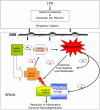
Figure 1
Peripheral infection/inflammation causes the release of pro-inflammatory mediators, including cytokines (TNF-α, IL-1β, IL-6), arachidonic acid (AA), prostaglandins (PGs), and nitric oxide (NO) synthesis. The brain also mounts an inflammatory response to systemic inflammation, as well as to local injury (neurodegeneration, trauma, stroke), with the microglial cells responding soonest and with production of the greatest amounts of pro-inflammatory mediators. However, the central response appears to be under tighter control than the peripheral response in that it is delayed and more modest, probably in order to avoid the dire consequences of a full-blown inflammatory response within the confines of the skull. Modified after Solito et al. (2008).
Figure 2
Different effects of NSAIDs on microglia. The response to NSAIDs may differ depending on whether they are used in early stages of disease, in which microglia present an alternatively activated phenotype compared with late stages which is associated with a classical microglia phenotype. (Adapted from Sastre and Gentleman, 2010). Abbreviations: ROS, reactive oxygen species; NOS2, same as iNOS; IDE, insulin degrading enzyme; SC, scavenger receptors.
Figure 3
Hypothetical annexin A1- FPRLI mechanism of action in microglia. Annexin A1 binds to FPRL-1 triggering an SOS signaling limiting microglia response to Aβ and preventing a constant uptake and apoptosis, altering microglia pro-inflammatory phenotype.
Similar articles
- Microglial Activation and Priming in Alzheimer's Disease: State of the Art and Future Perspectives.
Bivona G, Iemmolo M, Agnello L, Lo Sasso B, Gambino CM, Giglio RV, Scazzone C, Ghersi G, Ciaccio M. Bivona G, et al. Int J Mol Sci. 2023 Jan 3;24(1):884. doi: 10.3390/ijms24010884. Int J Mol Sci. 2023. PMID: 36614325 Free PMC article. Review. - Fibrillar Aβ triggers microglial proteome alterations and dysfunction in Alzheimer mouse models.
Sebastian Monasor L, Müller SA, Colombo AV, Tanrioever G, König J, Roth S, Liesz A, Berghofer A, Piechotta A, Prestel M, Saito T, Saido TC, Herms J, Willem M, Haass C, Lichtenthaler SF, Tahirovic S. Sebastian Monasor L, et al. Elife. 2020 Jun 8;9:e54083. doi: 10.7554/eLife.54083. Elife. 2020. PMID: 32510331 Free PMC article. - Activation of microglia and astrocytes: a roadway to neuroinflammation and Alzheimer's disease.
Kaur D, Sharma V, Deshmukh R. Kaur D, et al. Inflammopharmacology. 2019 Aug;27(4):663-677. doi: 10.1007/s10787-019-00580-x. Epub 2019 Mar 14. Inflammopharmacology. 2019. PMID: 30874945 Review. - The anti-inflammatory Annexin A1 induces the clearance and degradation of the amyloid-β peptide.
Ries M, Loiola R, Shah UN, Gentleman SM, Solito E, Sastre M. Ries M, et al. J Neuroinflammation. 2016 Sep 2;13(1):234. doi: 10.1186/s12974-016-0692-6. J Neuroinflammation. 2016. PMID: 27590054 Free PMC article. - Environmental Enrichment Potently Prevents Microglia-Mediated Neuroinflammation by Human Amyloid β-Protein Oligomers.
Xu H, Gelyana E, Rajsombath M, Yang T, Li S, Selkoe D. Xu H, et al. J Neurosci. 2016 Aug 31;36(35):9041-56. doi: 10.1523/JNEUROSCI.1023-16.2016. J Neurosci. 2016. PMID: 27581448 Free PMC article.
Cited by
- Fibrinogen and altered hemostasis in Alzheimer's disease.
Cortes-Canteli M, Zamolodchikov D, Ahn HJ, Strickland S, Norris EH. Cortes-Canteli M, et al. J Alzheimers Dis. 2012;32(3):599-608. doi: 10.3233/JAD-2012-120820. J Alzheimers Dis. 2012. PMID: 22869464 Free PMC article. Review. - Amyloid-β immunization enhances neurogenesis and cognitive ability in neonatal mice.
Chen H, Wang M, Jiao A, Tang G, Zhu W, Zou P, Li T, Cui G, Gong P. Chen H, et al. Int J Clin Exp Med. 2015 Apr 15;8(4):5340-50. eCollection 2015. Int J Clin Exp Med. 2015. PMID: 26131110 Free PMC article. - The restorative role of annexin A1 at the blood-brain barrier.
McArthur S, Loiola RA, Maggioli E, Errede M, Virgintino D, Solito E. McArthur S, et al. Fluids Barriers CNS. 2016 Sep 21;13(1):17. doi: 10.1186/s12987-016-0043-0. Fluids Barriers CNS. 2016. PMID: 27655189 Free PMC article. Review. - Microglia either promote or restrain TRAIL-mediated excitotoxicity caused by Aβ1-42 oligomers.
Zou J, McNair E, DeCastro S, Lyons SP, Mordant A, Herring LE, Vetreno RP, Coleman LG Jr. Zou J, et al. J Neuroinflammation. 2024 Sep 1;21(1):215. doi: 10.1186/s12974-024-03208-2. J Neuroinflammation. 2024. PMID: 39218898 Free PMC article. - Whole-body vibration ameliorates glial pathological changes in the hippocampus of hAPP transgenic mice, but does not affect plaque load.
Oroszi T, Geerts E, Rajadhyaksha R, Nyakas C, van Heuvelen MJG, van der Zee EA. Oroszi T, et al. Behav Brain Funct. 2023 Mar 20;19(1):5. doi: 10.1186/s12993-023-00208-9. Behav Brain Funct. 2023. PMID: 36941713 Free PMC article.
References
- Alafuzoff I., Overmyer M., Helisalmi S., Soininen H. (2000). Lower counts of astroglia and activated microglia in patients with Alzheimer’s disease with regular use of nonsteroidal anti-inflammatory drugs. J. Alzheimers Dis. 2, 37–46 - PubMed
- Arancio O., Zhang H. P., Chen X., Lin C., Trinchese F., Puzzo D., Liu S., Hegde A., Yan S. F., Stern A., Luddy J. S., Lue L. F., Walker D. G., Roher A., Buttini M., Mucke L., Li W., Schmidt A. M., Kindy M., Hyslop P. A., Stern D. M., Du Yan S. S. (2004). RAGE potentiates Abeta-induced perturbation of neuronal function in transgenic mice. EMBO J. 23, 4096–410510.1038/sj.emboj.7600415 - DOI - PMC - PubMed
LinkOut - more resources
Full Text Sources
Other Literature Sources
Research Materials


