A novel pathogenic role of the ER chaperone GRP78/BiP in rheumatoid arthritis - PubMed (original) (raw)
. 2012 Apr 9;209(4):871-86.
doi: 10.1084/jem.20111783. Epub 2012 Mar 19.
Sungyong You, Hyung-Ju Yoon, Dong-Ho Kim, Hyun-Sook Kim, Kyungho Lee, Jin Hee Ahn, Daehee Hwang, Amy S Lee, Ki-Jo Kim, Yune-Jung Park, Chul-Soo Cho, Wan-Uk Kim
Affiliations
- PMID: 22430489
- PMCID: PMC3328363
- DOI: 10.1084/jem.20111783
A novel pathogenic role of the ER chaperone GRP78/BiP in rheumatoid arthritis
Seung-Ah Yoo et al. J Exp Med. 2012.
Abstract
An accumulation of misfolded proteins can trigger a cellular survival response in the endoplasmic reticulum (ER). In this study, we found that ER stress-associated gene signatures were highly expressed in rheumatoid arthritis (RA) synoviums and synovial cells. Proinflammatory cytokines, such as TNF and IL-1β, increased the expression of GRP78/BiP, a representative ER chaperone, in RA synoviocytes. RA synoviocytes expressed higher levels of GRP78 than osteoarthritis (OA) synoviocytes when stimulated by thapsigargin or proinflammatory cytokines. Down-regulation of Grp78 transcripts increased the apoptosis of RA synoviocytes while abolishing TNF- or TGF-β-induced synoviocyte proliferation and cyclin D1 up-regulation. Conversely, overexpression of the Grp78 gene prevented synoviocyte apoptosis. Moreover, Grp78 small interfering RNA inhibited VEGF(165)-induced angiogenesis in vitro and also significantly impeded synoviocyte proliferation and angiogenesis in Matrigel implants engrafted into immunodeficient mice. Additionally, repeated intraarticular injections of BiP-inducible factor X, a selective GRP78 inducer, increased synoviocyte proliferation and angiogenesis in the joints of mice with experimental OA. In contrast, mice with Grp78 haploinsufficiency exhibited the suppression of experimentally induced arthritis and developed a limited degree of synovial proliferation and angiogenesis. In summary, this study shows that the ER chaperone GRP78 is crucial for synoviocyte proliferation and angiogenesis, the pathological hallmark of RA.
Figures
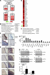
Figure 1.
ER stress response is increased in RA synovia and synovial cells. (A) Illustration of protein processing in the ER pathway with up-regulated genes in RA synovial tissue. Node colors represent fold change in RA synovial tissues as compared with normal synovial tissues (red, up-regulated; and green, down-regulated). (B) Functional enrichment analysis of up-regulated genes in OA tissues compared with normal. (C and D) Representative immunohistochemical stainings for ER stress–associated molecules in synovium sections obtained from RA and OA patients. (C) Tissues stained with anti-GRP78 antibody are representative of three RA and three OA patients. Intense staining in the synovium was observed in the lining layer (arrowheads). (D) Immunohistochemical staining of RA and OA synovia using anti-DDIT3 (CHOP), anti–p-IRE1, anti-ATF6, and anti-XBP1 antibody. Bars, 120 µm. (E) Heat map displaying 32 ER stress–associated up-regulated genes in RA macrophages. (F) Cellular processes enriched by DEGs in RA macrophages. Functional enrichment analysis of up-regulated DEGs was performed using DAVID software. (G) Quantitative real-time PCR assays of a subset of DEGs up-regulated in RA macrophages (n = 5) and proximal UPR genes. Four independent macrophage samples of healthy subjects and nine OA macrophages isolated from OA synovial tissues were used as controls. Fold inductions were calculated using the 2−ΔΔCt method. (H) Basal expression levels of ER stress–associated molecules in macrophages of healthy controls and RA patients, as determined by Western blot analysis using anti-GRP78 (left) and p-eIF2α (right) antibody. (I) Comparison of the optical density ratio ([GRP78 or p-eIF2α]/β-actin) between RA macrophages and normal (control) macrophages. *, P < 0.05 versus normal macrophages. (G and I) Data show mean ± SD.
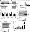
Figure 2.
Induction of GRP78 in synoviocytes by proinflammatory cytokines. (A and B) Regulation of GRP78 expression in RA FLSs by proinflammatory cytokines. Cells were stimulated with 10 ng/ml TNF, 1 ng/ml IL-1β, 10 ng/ml IL-10, or 100 µM CoCl2 for the indicated times. 20 µg/ml tunicamycin was used as a positive control. GRP78 levels in FLSs were determined by Western blot analysis. The data shown are representative of more than four experiments with similar results. (C and D) Quantitative real-time PCR assays of ER stress response genes induced by TNF (C) or IL-1β (D) in RA FLSs. Cells were stimulated with 10 ng/ml TNF or 1 ng/ml IL-1β for 48 h. Fold inductions were calculated using the 2−ΔΔCt method. Data show mean ± SD. (E and F) RA (n = 4) and OA (n = 4) FLSs were treated with 10 µM thapsigargin (E) or 20 µg/ml tunicamycin (F) for the indicated times. GRP78 expression was determined by Western blot analysis. In the lower panel, the optical density ratio of GRP78/β-actin (β-act) expression is presented as the mean values of four separate experiments. (G) Comparison of Grp78 mRNA expression in OA (n = 6) versus RA (n = 6) FLSs, as determined by real-time PCR. Cells were stimulated with 1 ng/ml IL-1β, 10 ng/ml TNF, 10 ng/ml TGF-β, 100 µM CoCl2, or 5 µM thapsigargin (Tg) for 8 h. Bars show mean and SEM. *, P < 0.05 versus OA FLSs.
Figure 3.
Role of GRP78 in synoviocyte survival. (A) Changes in ER sensor proteins in RA synoviocytes treated with 10 µM thapsigargin. The expressions of GRP78, p-eIF2α, ATF-6, BCL-2, and caspase-12 were determined by Western blot analysis. (B) The effect of Grp78 knockdown on synoviocyte survival. 2 d after transfection with siRNA for Grp78, the mRNA and protein expression levels of GRP78 in RA FLSs were determined by RT-PCR and Western blot analysis, respectively (left). The apoptosis of RA FLSs was induced by treating cells with 1 mM SNP for 12 h 2 d after Grp78 siRNA transfection. Degree of cell death was assessed by MTT assay (right). Results are the mean ± SD of more than four independent experiments performed in duplicate. *, P < 0.05 versus control siRNA–transfected cells. (C) RA FLSs were treated with BIX for 12 h, and then GRP78 expression was determined by Western blot analysis. Synoviocyte apoptosis was induced by treating RA FLSs with 10 µM thapsigargin in the presence or absence of BIX. *, P < 0.05 versus thapsigargin-treated cells in the absence of BIX. (D) SV40-immortalized RA FLSs were transfected with either the pFLAG-hGrp78 gene or pFLAG vector only. The protein expression levels for GRP78 were determined by Western blotting. (E and F) RA FLSs transfected with either the pFLAG-hGrp78 gene or pFLAG vector were treated with 5 µM thapsigargin (Tg) for 1 h, 10 µg/ml tunicamycin (Tm) for 12 h, or 5 mM SNP for 24 h. Cell viability was determined by MTT assay. *, P < 0.05 versus vector-transfected cells. (G) The apoptosis of RA FLSs harboring the pFLAG-hGrp78 gene or pFLAG vector was induced by treating cells with 5 µM thapsigargin or 10 µg/ml tunicamycin for 3 h. Degrees of apoptosis were assessed by APOPercentage apoptosis assay, a colorimetric method. Apoptotic cells appeared bright pink. Fold increase in apoptosis levels was expressed as pixel numbers. *, P < 0.05 versus vector-transfected cells. Bars, 100 µm. (C and E–G) Data show mean ± SD.
Figure 4.
Effects of GRP78 on TGF-β– or TNF-induced synoviocyte proliferation. (A) Synoviocyte proliferative responses to TGF-β and TNF. 24 h after transfection with Grp78 siRNA, RA FLSs (n = 4; 1 × 10⁴ cells) were treated with 10 ng/ml TGF-β or 10 ng/ml TNF for 72 h. FLS proliferation rate was determined by [3H]thymidine incorporation assay. Results are the means ± SD of four independent experiments performed in triplicate. *, P < 0.05 versus nontransfected cells. (B) 4 d after stimulating RA FLSs with 10 ng/ml TGF-β or 10 ng/ml TNF, manual cell counts were performed by trypan blue exclusion to identify viable cells. The results shown are the mean ± SD of three independent experiments performed in triplicate. *, P < 0.05 versus nontransfected cells. (C) Cyclin D1 expression in RA FLSs stimulated with 10 ng/ml TNF or 10 ng/ml TGF-β, as determined by Western blot analysis. A representative of three independent experiments is shown. (D) Effect of Grp78 knockdown on TNF- or TGF-β–induced increases in cyclin D1 expression in RA synoviocytes. Data are representative of three independent experiments. (E) Proliferation of FLSs obtained from Grp78+/− and Grp78+/+ mice. Cell number was determined by trypan blue exclusion 5 d after stimulating mouse FLSs with 10 ng/ml TNF or 10 ng/ml TGF-β. Data are presented as the mean ± SD of four mice per group. *, P < 0.05 versus FLSs from Grp78+/+ littermates.
Figure 5.
Grp78 siRNA inhibits proliferation, tube formation, migration, and chemotaxis of endothelial cells. (A) GRP78 expression levels in HUVECs were determined by Western blot analysis. (B) Effect of Grp78 siRNA on HUVEC proliferation. HUVECs were incubated for 24 h in DME supplemented with 1% FCS and then treated with Grp78 siRNA for 72 h in the presence of 20 ng/ml VEGF165. HUVEC proliferation was determined by [3H]thymidine incorporation assay. Results are mean ± SD and are representative of three independent experiments. *, P < 0.05. (C) HUVEC tube formation with Grp78 siRNA. 12 h after transfection with Grp78 siRNA or control siRNA, HUVECs were plated on Matrigel matrices with 20 ng/ml VEGF165 for 12 h. The total length of the tube network was calculated using Image-Pro Plus software. Results are mean ± SD and are representative of three independent experiments. *, P < 0.05. (D) Grp78 siRNA effect on HUVEC wound migration induced by VEGF165. After 12 h of the transfection with Grp78 siRNA or control siRNA, confluent HUVECs were incubated in M199 containing 1% FCS and 20 ng/ml VEGF165 for 12 h. Cells migrating beyond the reference line were photographed and counted. Data show mean ± SD. *, P < 0.05. (E) HUVEC chemotaxis in a Boyden chamber, as determined at 24 h after Grp78 siRNA transfection. Migrated HUVECs were stained violet using Diff-Quik kit. The mean ± SD of three independent experiments is presented on the right. *, P < 0.05. Bars, 100 µm.
Figure 6.
Inhibition of RA synoviocyte proliferation and angiogenesis by Grp78 siRNA in immunodeficient mice. Matrigel containing RA FLSs was subcutaneously injected into the back skin of immunodeficient mice. (A) 7 d after transfecting with the Grp78 siRNA, GRP78 expression levels in RA FLSs were determined by Western blot analysis. (B) Hematoxylin and eosin staining of Matrigels containing RA FLSs from immunodeficient mice. (C) Effect of Grp78 siRNA on RA FLS proliferation and HUVEC infiltration. RA FLSs were implanted in Matrigels for 7 d in the absence or presence of 50 ng/gel TGF-β. RA FLSs in Matrigel were identified by immunofluorescence labeling for HLA class I antigen. Infiltrating mouse endothelial cells were stained using mouse anti-vWF antibody. The cells positive for HLA class I are shown in green in the top and the middle panels. Representative photographs of endothelial cells are shown in red in the bottom panel. Bars: (B) 300 µm; (C) 100 µm. (D) Numbers of RA FLSs and endothelial cells in Matrigels treated with vehicle or TGF-β. Cells were manually counted under a magnification of ×200. Values are the mean ± SD of eight mice per group. *, P < 0.05 versus control siRNA–transfected cells.
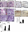
Figure 7.
Increase in synovial hyperplasia and bone erosion in mice treated with BIX, a selective BiP/GRP78 inducer. (A) Hematoxylin and eosin staining of the joints of mice administered periarticularly on alternate days for 2 or 4 wk with BIX. Black arrows and arrowheads in the top panel indicate intact cartilages and minimal synovial proliferation, respectively. Pink arrows in the middle panel indicate the enhanced proliferation of synoviocytes, and black arrows in the bottom panel represent bone erosion. The rectangular area in the middle left image is magnified in the middle right image. (B) The histological scores for degrees of synovial hyperplasia in mice injected with BIX alone, collagenase (COL) alone, and collagenase plus BIX (n = 6 per group). *, P < 0.05 versus collagenase (only)-treated mice without injecting BIX. (C) vWF staining of the synovia of mice treated with collagenase plus BIX versus collagenase alone. Positive cells are shown in brown (black arrows). (D) Evaluation of synovial proliferation by PCNA staining. Positive staining in the synoviums is indicated by brown nuclei. Ratios of positive cells (the number of positive synoviocytes/total synoviocytes counted) are presented in the bottom panel. *, P < 0.001. (B and D) Data show mean ± SD. Bars: (A, top and middle left) 300 µm; (A, middle right and bottom) 120 µm; (C and D) 60 µm.
Figure 8.
Arthritis induction in Grp78+/+ and Grp78+/− mice. (A) Western blot analysis of GRP78 in the joint tissues. Lysates obtained from the joints of Grp78+/− (n = 2) and wild-type littermates (Grp78+/+; n = 2) were subjected to Western blot analysis using anti-GRP78 and anti–β-actin antibodies. (B) The severity of anti–type II collagen antibody–induced arthritis in Grp78+/− (n = 7) and wild-type littermates (Grp78+/+; n = 7). Values are mean and SD. *, P < 0.05; **, P < 0.005 versus Grp78+/+ mice. (C) Effect of Grp78 deficiency on paw edema in mice with antibody-induced arthritis, as determined by the paw thickness index calculated at the indicated time points. Results are paw thickness index of forefoot (FF) and hind foot (HF). Values are mean. *, P < 0.05; **, P < 0.005 versus Grp78+/+ mice. (D) Hematoxylin and eosin staining of ankle joint sections obtained from Grp78+/− and Grp78+/+ mice 10 d after arthritis induction. The rectangular areas in the top images are magnified in the bottom images. (E) Mean histological scores of inflammatory cell infiltration (IFLM), synovial proliferation (SP), and joint destruction (JD) in Grp78+/− (n = 4) versus Grp78+/+ mice (n = 4) as determined on day 10 after antibody administration. Values are the mean and SD of four mice per group. *, P < 0.001 versus Grp78+/+ mice. (F) Immunohistochemical staining for vWF. Positive cells are shown in brown. Representative photographs are shown. Bars: (D, top) 300 µm; (D, bottom) 120 µm; (F) 60 µm.
Similar articles
- Role of endoplasmic reticulum stress in rheumatoid arthritis pathogenesis.
Park YJ, Yoo SA, Kim WU. Park YJ, et al. J Korean Med Sci. 2014 Jan;29(1):2-11. doi: 10.3346/jkms.2014.29.1.2. Epub 2013 Dec 26. J Korean Med Sci. 2014. PMID: 24431899 Free PMC article. Review. - Synovial-fluid-derived microparticles express vimentin and GRP78 in their surface and exhibit an in vitro stimulatory effect on fibroblast-like synoviocytes in rheumatoid arthritis.
Michael BNR, Mariaselvam CM, Kavadichanda CG, Negi VS. Michael BNR, et al. Int J Rheum Dis. 2023 Nov;26(11):2183-2194. doi: 10.1111/1756-185X.14912. Epub 2023 Sep 11. Int J Rheum Dis. 2023. PMID: 37695005 - Anti-neuropilin-1 peptide inhibition of synoviocyte survival, angiogenesis, and experimental arthritis.
Kong JS, Yoo SA, Kim JW, Yang SP, Chae CB, Tarallo V, De Falco S, Ryu SH, Cho CS, Kim WU. Kong JS, et al. Arthritis Rheum. 2010 Jan;62(1):179-90. doi: 10.1002/art.27243. Arthritis Rheum. 2010. PMID: 20039409 - NF-AT5 is a critical regulator of inflammatory arthritis.
Yoon HJ, You S, Yoo SA, Kim NH, Kwon HM, Yoon CH, Cho CS, Hwang D, Kim WU. Yoon HJ, et al. Arthritis Rheum. 2011 Jul;63(7):1843-52. doi: 10.1002/art.30229. Arthritis Rheum. 2011. PMID: 21717420 Free PMC article. - Immunoglobulin heavy-chain-binding protein (BiP): a stress protein that has the potential to be a novel therapy for rheumatoid arthritis.
Panayi GS, Corrigall VM. Panayi GS, et al. Biochem Soc Trans. 2014 Dec;42(6):1752-5. doi: 10.1042/BST20140230. Biochem Soc Trans. 2014. PMID: 25399601 Review.
Cited by
- Anti-Citrullinated Protein Antibodies Induce Macrophage Subset Disequilibrium in RA Patients.
Zhu W, Li X, Fang S, Zhang X, Wang Y, Zhang T, Li Z, Xu Y, Qu S, Liu C, Gao F, Pan H, Wang G, Li H, Sun B. Zhu W, et al. Inflammation. 2015 Dec;38(6):2067-75. doi: 10.1007/s10753-015-0188-z. Inflammation. 2015. PMID: 26063186 - Unveiling the dark side of glucose-regulated protein 78 (GRP78) in cancers and other human pathology: a systematic review.
Akinyemi AO, Simpson KE, Oyelere SF, Nur M, Ngule CM, Owoyemi BCD, Ayarick VA, Oyelami FF, Obaleye O, Esoe DP, Liu X, Li Z. Akinyemi AO, et al. Mol Med. 2023 Aug 21;29(1):112. doi: 10.1186/s10020-023-00706-6. Mol Med. 2023. PMID: 37605113 Free PMC article. Review. - Glucose-regulated protein 78 may play a crucial role in promoting the pulmonary microvascular remodeling in a rat model of hepatopulmonary syndrome.
Zhang H, Lv M, Zhao Z, Jia J, Zhang L, Xiao P, Wang L, Li C, Ji J, Tian X, Li X, Fan Y, Lai L, Liu Y, Li B, Zhang C, Liu M, Guo J, Han D, Ji C. Zhang H, et al. Gene. 2014 Jul 15;545(1):156-62. doi: 10.1016/j.gene.2014.04.041. Epub 2014 Apr 21. Gene. 2014. PMID: 24768185 Free PMC article. - Mechanisms Underlying Bone Loss Associated with Gut Inflammation.
Ke K, Arra M, Abu-Amer Y. Ke K, et al. Int J Mol Sci. 2019 Dec 15;20(24):6323. doi: 10.3390/ijms20246323. Int J Mol Sci. 2019. PMID: 31847438 Free PMC article. Review. - Association of HSP90B1 genetic polymorphisms with efficacy of glucocorticoids and improvement of HRQoL in systemic lupus erythematosus patients from Anhui Province.
Sun XX, Li SS, Zhang M, Xie QM, Xu JH, Liu SX, Gu YY, Pan FM, Tao JH, Xu SQ, Liu S, Cai J, Wang DG, Qian L, Wang CH, Lian L, Xiao H, Chen PL, Liang CM, Fang YB, Zhou Q, Huang HL, Su H, Pan HF, Ye DQ, Zou YF. Sun XX, et al. Am J Clin Exp Immunol. 2018 Apr 5;7(2):27-39. eCollection 2018. Am J Clin Exp Immunol. 2018. PMID: 29755855 Free PMC article.
References
- Arnett F.C., Edworthy S.M., Bloch D.A., McShane D.J., Fries J.F., Cooper N.S., Healey L.A., Kaplan S.R., Liang M.H., Luthra H.S., et al. 1988. The American Rheumatism Association 1987 revised criteria for the classification of rheumatoid arthritis. Arthritis Rheum. 31:315–324 10.1002/art.1780310302 - DOI - PubMed
- Bläss S., Union A., Raymackers J., Schumann F., Ungethüm U., Müller-Steinbach S., De Keyser F., Engel J.M., Burmester G.R. 2001. The stress protein BiP is overexpressed and is a major B and T cell target in rheumatoid arthritis. Arthritis Rheum. 44:761–771 10.1002/1529-0131(200104)44:4<761::AID-ANR132>3.0.CO;2-S - DOI - PubMed
- Corrigall V.M., Bodman-Smith M.D., Fife M.S., Canas B., Myers L.K., Wooley P., Soh C., Staines N.A., Pappin D.J., Berlo S.E., et al. 2001. The human endoplasmic reticulum molecular chaperone BiP is an autoantigen for rheumatoid arthritis and prevents the induction of experimental arthritis. J. Immunol. 166:1492–1498 - PubMed
Publication types
MeSH terms
Substances
LinkOut - more resources
Full Text Sources
Other Literature Sources
Medical
Molecular Biology Databases
Research Materials
Miscellaneous


