Molecular basis for the anchoring of proto-oncoprotein Nup98 to the cytoplasmic face of the nuclear pore complex - PubMed (original) (raw)
Molecular basis for the anchoring of proto-oncoprotein Nup98 to the cytoplasmic face of the nuclear pore complex
Tobias Stuwe et al. J Mol Biol. 2012.
Abstract
The cytoplasmic filament nucleoporins of the nuclear pore complex (NPC) are critically involved in nuclear export and remodeling of mRNA ribonucleoprotein particles and are associated with various human malignancies. Here, we report the crystal structure of the Nup98 C-terminal autoproteolytic domain, frequently missing from leukemogenic forms of the protein, in complex with the N-terminal domain of Nup82 and the C-terminal tail fragment of Nup159. The Nup82 β propeller serves as a noncooperative binding platform for both binding partners. Interaction of Nup98 with Nup82 occurs through a reciprocal exchange of loop structures. Strikingly, the same Nup98 groove promiscuously interacts with Nup82 and Nup96 in a mutually excusive fashion. Simultaneous disruption of both Nup82 interactions in yeast causes severe defects in mRNA export, while the severing of a single interaction is tolerated. Thus, the cytoplasmic filament network of the NPC is robust, consistent with its essential function in nucleocytoplasmic transport.
Published by Elsevier Ltd.
Conflict of interest statement
Conflict of interest
The authors declare that they have no conflict of interest.
Figures
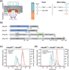
Fig. 1. Biochemical analysis of mammalian and chimeric Nup98 assemblies
(a) Schematic architecture of the NPC (left panel). The cylindrical symmetric core (orange) is decorated with cytoplasmic filaments (cyan) and a nuclear basket (magenta) and anchored in the nuclear envelope by integral pore membrane proteins (POMs, green). Natively unfolded FG repeats of a number of nups make up the transport barrier in the central channel and are indicated by a transparent plug. Schematic diagram of the cytoplasmic filament interaction network of the yeast and human NPC (right panel) as discussed in the text. (b) Domain organization of human Nup88 and the yeast homolog Nup82, mouse Nup98, and yeast Nup159. For hNup88 and yNup82, the N-terminal domain (NTD, blue) and the C-terminal α-helical domain (dark gray) are indicated. For mNup98, GLFG repeat regions (gray), the Gle2-binding sequence (GLEBS, dark gray), the unstructured region (gray), the autoproteolytic and NPC-targeting domain (APD, green), and the C-terminal extension (termed 6kDa fragment in the human protein) (gray) are indicated. For Nup159, the NTD (dark gray), the FG repeat region (gray), the unstructured region (gray), the dynein light chain interacting domain (DID, dark gray), the C-terminal α-helical region (dark gray), and the tail region (T, red) are indicated. The blue bar represents the domain boundaries of the human Nup88 N-terminal domain used for interaction studies. The black bars above the domain structures denote the crystallized fragments. (c) Multi-angle light scattering (MALS) analysis of the mammalian mNup98APD•hNup88NTD heterodimer. The differential refractive indices of mNup98APD (red), hNup88NTD (blue), and mNup98APD•hNup88NTD (green) are plotted against the elution volumes from a Superdex 200 10/300 GL gel filtration column (GE Healthcare) and are overlaid with the determined molecular masses for the selected peaks. (d) MALS analysis of the chimeric mNup98APD•yNup82NTD•yNup159T heterotrimer. The differential refractive indices of mNup98APD (red), yNup82NTD•yNup159T (blue), and mNup98APD•yNup82NTD•yNup159T (green) are plotted against the elution volumes from a Superdex 200 10/300 GL gel filtration column (GE Healthcare) and are overlaid with the determined molecular masses for the selected peaks.
Fig. 2. Structural overview of the heterotrimer
(a) Cartoon representation of the mNup98APD•yNup82NTD•yNup159T complex, showing the yNup82NTD in blue with various non-canonical insertions highlighted in yellow (3D4A or FGL loop), gray (4CD), and orange (6CD). The mNup98APD is displayed in green with the β6-αB connector (K loop) colored in magenta. yNup159T is shown in red. A 180° rotated view is shown on the right. (b) Cartoon representation of mNup98APD• yNup82NTD•yNup159T, rotated by 90° with respect to panel A, left panel. The seven blades of the β propeller are numbered. (c) Schematic representation of the architectures of the three domains in the mNup98APD•yNup82NTD•yNup159T heterotrimer and their interaction. Prominent insertions and secondary structure elements are labeled. The asterisk denotes the N-terminal region that links blades 1 and 7.
Fig. 3. Structural comparison of Nup98 complexes
Superposition of mNup98APD•yNup82NTD and hNup98APD•hNup986kDa complexes. For clarity, only the FGL loop of yNup82NTD is shown (stick representation). The FGL loop and the corresponding YGL segment of hNup986kDa bind to overlapping sites on Nup98APD as shown on the right.
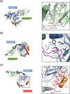
Fig. 4. Interfaces in the heterotrimer
(a) Detailed view of the interaction of the extended yNup82NTD FGL loop (yellow) that binds to the hydrophobic catalytic groove of mNup98APD between helix αB and strand β5. (b) Close-up view of the interaction between the tip of the mNup98APD K loop and its association with the yNup82NTD D pocket. A key salt-bridge is formed between the K loop K831 and the D pocket D204. (c) Close-up view of the interaction between yNup159T and yNup82NTD. The Nup159T binds to a hydrophobic groove in yNup82NTD β propeller that is formed by the non-canonical 4CD and 6CD insertions and blade 5. As a reference, ribbon representations of the heterotrimer are shown in the left panels. The black insets correspond to the magnified regions seen in the right panels.
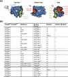
Fig. 5. Biochemical analysis of the interactions in the heterotrimer
(a) Surface rendition of yNup82NTD. The mNup98APD and Nup159T binding sites are colored in green and red, respectively. The positions of the DFY and the ΔFGL loop mutations that disrupt mNup98APD binding, and of the LILLF mutation that abolishes the yNup159T interaction are indicated in yellow and orange, respectively. As a reference, the ribbon representation of the heterotrimer is shown on the left. (b) Interaction table summarizing the results of the mutational analysis. Binding is scored as wild-type (+), intermediate (+/−), or no binding (−). Representative gel filtration profiles for the three scoring classes are illustrated in Fig. S5.
Fig. 6. Evolutionary conservation of the binding promiscuity
(a) hNup98APD interacts with hNup986kDa fragment and hNup88NTD in a mutually exclusive manner. Purified hNup88NTD was mixed at approximately equimolar ratio with the hNup98APD•hNup986kDa nucleoporin pair and analyzed by size exclusion chromatography. The inset shows the displaced hNup986kDa fragment upon binding of hNup88NTD. Note that the extinction coefficient of hNup986kDa is very low, resulting in weak absorbance and a small peak. (b) yNup145NAPD interacts with ySec13•yNup145C and yNup82NTD•yNup159T in a mutually exclusive manner. Nucleoporin complexes were mixed at approximate equimolar ratios and analyzed by size exclusion chromatography. Note that the ySec13•yNup145C pair was used for this experiment, as yNup145C is insoluble in the absence of ySec13. Grey bars and colored lines designate the analyzed fractions. Molecular weight standards and the positions of the proteins are indicated.
Fig. 7. The three GLFG nucleoporins of S. cerevisiae are functionally divergent
(a–c) Size exclusion chromatography interaction analysis of yNup145C•ySec13 with yNup145NAPD (a), with yNup116CTD (b) and with yNup100CTD (c). The analyzed proteins and complexes are indicated in each gel filtration profile. For analysis of complex formation, the ySec13•yNup145C heterodimer was mixed at approximately two-fold molar excess of yNup145NAPD, yNup116CTD, and yNup100CTD and injected onto a Superdex 200 10/300 GL gel filtration column. Grey bars and colored lines designate the analyzed fractions. Molecular weight standards and the positions of the proteins are indicated.
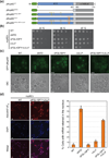
Fig. 8. In vivo analysis of Nup82 mutants in S. cerevisiae
(a) Domain structures of the N-terminally GFP-labeled yNup82 constructs. The positions of the ΔFGL, DFY and LILLF mutations are indicated by lines and colored according to Fig. 5a. (b) Yeast growth analysis using a nup82Δ strain transformed with the indicated GFP-yNup82 constructs. 10-fold serial dilutions were spotted on SD-Leu plates and grown for 2–3 days at the indicated temperatures. (c) In vivo localization of GFP-yNup82 variants at 37 °C visualized by fluorescence and differential interference contrast microscopy (DIC). (d) mRNA export assay of GFP-yNup82 variants. The detection of poly(A) mRNA and DNA was carried out using an Alexa-647-labeled oligo dT50 FISH probe and DAPI stain, respectively. Representative images of wild-type GFP-yNup82 (top) and GFP-yNup82ΔFGL+DFY+LILLF (bottom) complemented nup82Δ cells grown at 37 °C are shown (left panel). Quantitation of nuclear poly(A) mRNA retention is shown on the right. The percentages refer to the fraction of cells displaying a marked nuclear FISH staining. The error bars correspond to the standard deviations that are derived from four independent images. Each image contained approximately 1000 cells. The scale bar represents 5 µm.
Fig. 9. Model for the cytoplasmic filament interaction network of the NPC
Schematic representation of the cytoplasmic filament network of the yeast (left panel) and human (right panel) NPC. Asterisks indicate nucleoporins that are involved in human malignancies (red) and acute necrotizing encephalopathy (black).
Similar articles
- Architecture of the cytoplasmic face of the nuclear pore.
Bley CJ, Nie S, Mobbs GW, Petrovic S, Gres AT, Liu X, Mukherjee S, Harvey S, Huber FM, Lin DH, Brown B, Tang AW, Rundlet EJ, Correia AR, Chen S, Regmi SG, Stevens TA, Jette CA, Dasso M, Patke A, Palazzo AF, Kossiakoff AA, Hoelz A. Bley CJ, et al. Science. 2022 Jun 10;376(6598):eabm9129. doi: 10.1126/science.abm9129. Epub 2022 Jun 10. Science. 2022. PMID: 35679405 Free PMC article. - Structural and functional analysis of an essential nucleoporin heterotrimer on the cytoplasmic face of the nuclear pore complex.
Yoshida K, Seo HS, Debler EW, Blobel G, Hoelz A. Yoshida K, et al. Proc Natl Acad Sci U S A. 2011 Oct 4;108(40):16571-6. doi: 10.1073/pnas.1112846108. Epub 2011 Sep 19. Proc Natl Acad Sci U S A. 2011. PMID: 21930948 Free PMC article. - The N-terminal domain of Nup159 forms a beta-propeller that functions in mRNA export by tethering the helicase Dbp5 to the nuclear pore.
Weirich CS, Erzberger JP, Berger JM, Weis K. Weirich CS, et al. Mol Cell. 2004 Dec 3;16(5):749-60. doi: 10.1016/j.molcel.2004.10.032. Mol Cell. 2004. PMID: 15574330 - Into the basket and beyond: the journey of mRNA through the nuclear pore complex.
Ashkenazy-Titelman A, Shav-Tal Y, Kehlenbach RH. Ashkenazy-Titelman A, et al. Biochem J. 2020 Jan 17;477(1):23-44. doi: 10.1042/BCJ20190132. Biochem J. 2020. PMID: 31913454 Review. - Membrane-coating lattice scaffolds in the nuclear pore and vesicle coats: commonalities, differences, challenges.
Leksa NC, Schwartz TU. Leksa NC, et al. Nucleus. 2010 Jul-Aug;1(4):314-8. doi: 10.4161/nucl.1.4.11798. Epub 2010 Mar 12. Nucleus. 2010. PMID: 21327078 Free PMC article. Review.
Cited by
- Architecture of the fungal nuclear pore inner ring complex.
Stuwe T, Bley CJ, Thierbach K, Petrovic S, Schilbach S, Mayo DJ, Perriches T, Rundlet EJ, Jeon YE, Collins LN, Huber FM, Lin DH, Paduch M, Koide A, Lu V, Fischer J, Hurt E, Koide S, Kossiakoff AA, Hoelz A. Stuwe T, et al. Science. 2015 Oct 2;350(6256):56-64. doi: 10.1126/science.aac9176. Epub 2015 Aug 27. Science. 2015. PMID: 26316600 Free PMC article. - Proteomic elucidation of the targets and primary functions of the picornavirus 2A protease.
Serganov AA, Udi Y, Stein ME, Patel V, Fridy PC, Rice CM, Saeed M, Jacobs EY, Chait BT, Rout MP. Serganov AA, et al. J Biol Chem. 2022 Jun;298(6):101882. doi: 10.1016/j.jbc.2022.101882. Epub 2022 Mar 31. J Biol Chem. 2022. PMID: 35367208 Free PMC article. - Nanobodies: site-specific labeling for super-resolution imaging, rapid epitope-mapping and native protein complex isolation.
Pleiner T, Bates M, Trakhanov S, Lee CT, Schliep JE, Chug H, Böhning M, Stark H, Urlaub H, Görlich D. Pleiner T, et al. Elife. 2015 Dec 3;4:e11349. doi: 10.7554/eLife.11349. Elife. 2015. PMID: 26633879 Free PMC article. - Architecture of the cytoplasmic face of the nuclear pore.
Bley CJ, Nie S, Mobbs GW, Petrovic S, Gres AT, Liu X, Mukherjee S, Harvey S, Huber FM, Lin DH, Brown B, Tang AW, Rundlet EJ, Correia AR, Chen S, Regmi SG, Stevens TA, Jette CA, Dasso M, Patke A, Palazzo AF, Kossiakoff AA, Hoelz A. Bley CJ, et al. Science. 2022 Jun 10;376(6598):eabm9129. doi: 10.1126/science.abm9129. Epub 2022 Jun 10. Science. 2022. PMID: 35679405 Free PMC article. - The Nuclear Pore Complex as a Flexible and Dynamic Gate.
Knockenhauer KE, Schwartz TU. Knockenhauer KE, et al. Cell. 2016 Mar 10;164(6):1162-1171. doi: 10.1016/j.cell.2016.01.034. Cell. 2016. PMID: 26967283 Free PMC article. Review.
References
- Cook A, Bono F, Jinek M, Conti E. Structural biology of nucleocytoplasmic transport. Annu. Rev. Biochem. 2007;76:647–671. - PubMed
- Debler EW, Blobel G, Hoelz A. Nuclear transport comes full circle. Nat. Struct. Mol. Biol. 2009;16:457–459. - PubMed
- Hoelz A, Blobel G. Cell biology: popping out of the nucleus. Nature. 2004;432:815–816. - PubMed
- Hoelz A, Debler EW, Blobel G. The structure of the nuclear pore complex. Annu. Rev. Biochem. 2011;80:613–643. - PubMed
Publication types
MeSH terms
Substances
LinkOut - more resources
Full Text Sources
Molecular Biology Databases


