Master regulatory GATA transcription factors: mechanistic principles and emerging links to hematologic malignancies - PubMed (original) (raw)
Review
. 2012 Jul;40(13):5819-31.
doi: 10.1093/nar/gks281. Epub 2012 Apr 5.
Affiliations
- PMID: 22492510
- PMCID: PMC3401466
- DOI: 10.1093/nar/gks281
Review
Master regulatory GATA transcription factors: mechanistic principles and emerging links to hematologic malignancies
Emery H Bresnick et al. Nucleic Acids Res. 2012 Jul.
Abstract
Numerous examples exist of how disrupting the actions of physiological regulators of blood cell development yields hematologic malignancies. The master regulator of hematopoietic stem/progenitor cells GATA-2 was cloned almost 20 years ago, and elegant genetic analyses demonstrated its essential function to promote hematopoiesis. While certain GATA-2 target genes are implicated in leukemogenesis, only recently have definitive insights emerged linking GATA-2 to human hematologic pathophysiologies. These pathophysiologies include myelodysplastic syndrome, acute myeloid leukemia and an immunodeficiency syndrome with complex phenotypes including leukemia. As GATA-2 has a pivotal role in the etiology of human cancer, it is instructive to consider mechanisms underlying normal GATA factor function/regulation and how dissecting such mechanisms may reveal unique opportunities for thwarting GATA-2-dependent processes in a therapeutic context. This article highlights GATA factor mechanistic principles, with a heavy emphasis on GATA-1 and GATA-2 functions in the hematopoietic system, and new links between GATA-2 dysregulation and human pathophysiologies.
Figures
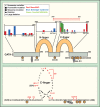
Figure 1.
GATA factor mechanistic principles. The models depict mechanistic principles derived from studies of GATA-1 and GATA-2. While the fundamental nature of these principles is likely to be shared by other GATA factors, additional GATA factor-specific mechanistic permutations are expected. Principle 1: GATA factors occupy a very small percent of the WGATAA motifs in a genome (<1%), suggesting that crucial mechanisms exist that control the discrimination among these highly abundant motifs. However, such mechanisms are not firmly established. The model depicts the occlusion of select GATA motifs, thus creating an obligate requirement for chromatin remodeling/modification reactions to increase access of the WGATAA residues required for GATA factor binding and/or to selectively occlude the vast majority of sites. At certain sites, FOG-1 (56,57) and GATA-1 acetylation (95) enhance chromatin access. Presumably, a host of regulatory factors mediate the essential process of establishing/maintaining accessible and occluded sites. Principle 2: GATA factors activate and repress target genes via multiple mechanisms, including with or without FOG-1 (36). Presumably, this mechanistic diversity reflects the specific chromatin architecture at a genetic locus, the subnuclear environment in which the locus resides and the regulatory mileau characteristic of the specific environment. Principle 3: GATA-1 and GATA-2 commonly co-localize with Scl/TAL1, another master regulator of hematopoiesis (96), at chromatin sites. The model illustrates GATA factor and Scl/TAL1 occupancy of a composite element consisting of an E-box and a WGATAA motif, which was originally described by Wadman et al. (76). Similar to the description above, only a very small percentage of composite elements are occupied by GATA factors in cells (53,58). As co-localization does not require the E-box (72), there is much to be learned about the biochemical nature of the GATA factor and Scl/TAL1 interaction. However, the co-localization measured by ChIP often correlates with transcriptional activity (54,58,72). Principle 4: GATA switches are defined as a molecular transition in which one GATA factor replaces another from a chromatin site, which is often associated with an altered transcriptional output. The GATA switch depicted reflects that occurring at the Gata2 locus during erythropoiesis, in which GATA-1 displaces GATA-2 from chromatin, which rapidly instigates repression (87). Context-dependent GATA switches may either activate or repress transcription and, in certain cases, may sustain the original transcriptional output (36).
Figure 2.
GATA-2 mutations in human hematologic disorders (143–146). Specific GATA-2 mutations in human patients are indicated, with details denoted in the legend. Each symbol represents a single patient with the particular mutation. The diagram of GATA-2 protein organization illustrates the N- and C-fingers, acetylation sites (124), the serine 401 phosphorylation site and two sumoylation consensus motifs. The diagram at the bottom illustrates the amino acid sequence composition of the C-finger and neighboring regions, with the positions of disease mutations highlighted. Stop, mutation that creates stop codon; frs, frameshift mutation; del, deletion.
Similar articles
- The role of the GATA2 transcription factor in normal and malignant hematopoiesis.
Vicente C, Conchillo A, García-Sánchez MA, Odero MD. Vicente C, et al. Crit Rev Oncol Hematol. 2012 Apr;82(1):1-17. doi: 10.1016/j.critrevonc.2011.04.007. Epub 2011 May 24. Crit Rev Oncol Hematol. 2012. PMID: 21605981 Review. - [GATA factor-related hematopoietic neoplasms].
Shimizu R. Shimizu R. Rinsho Ketsueki. 2024;65(9):902-910. doi: 10.11406/rinketsu.65.902. Rinsho Ketsueki. 2024. PMID: 39358289 Review. Japanese. - GATA-related hematologic disorders.
Shimizu R, Yamamoto M. Shimizu R, et al. Exp Hematol. 2016 Aug;44(8):696-705. doi: 10.1016/j.exphem.2016.05.010. Epub 2016 May 25. Exp Hematol. 2016. PMID: 27235756 Review. - The role of GATA family transcriptional factors in haematological malignancies: A review.
Abunimye DA, Okafor IM, Okorowo H, Obeagu EI. Abunimye DA, et al. Medicine (Baltimore). 2024 Mar 22;103(12):e37487. doi: 10.1097/MD.0000000000037487. Medicine (Baltimore). 2024. PMID: 38518015 Free PMC article. Retracted. Review. - The GATA factor revolution in hematology.
Katsumura KR, Bresnick EH; GATA Factor Mechanisms Group. Katsumura KR, et al. Blood. 2017 Apr 13;129(15):2092-2102. doi: 10.1182/blood-2016-09-687871. Epub 2017 Feb 8. Blood. 2017. PMID: 28179282 Free PMC article. Review.
Cited by
- Endogenous small molecule effectors in GATA transcription factor mechanisms governing biological and pathological processes.
Liao R, Bresnick EH. Liao R, et al. Exp Hematol. 2024 Sep;137:104252. doi: 10.1016/j.exphem.2024.104252. Epub 2024 Jun 12. Exp Hematol. 2024. PMID: 38876253 Review. - The PRC2 complex epigenetically silences GATA4 to suppress cellular senescence and promote the progression of breast cancer.
Yu W, Lin X, Leng S, Hou Y, Dang Z, Xue S, Li N, Zhang F. Yu W, et al. Transl Oncol. 2024 Aug;46:102014. doi: 10.1016/j.tranon.2024.102014. Epub 2024 Jun 5. Transl Oncol. 2024. PMID: 38843657 Free PMC article. - Integrating circulating T follicular memory cells and autoantibody repertoires for characterization of autoimmune disorders.
Harris EM, Chamseddine S, Chu A, Senkpeil L, Nikiciuk M, Al-Musa A, Woods B, Ozdogan E, Saker S, van Konijnenburg DPH, Yee CSK, Nelson R, Lee P, Halyabar O, Hale RC, Day-Lewis M, Henderson LA, Nguyen AA, Elkins M, Ohsumi TK, Gutierrez-Arcelus M, Peyper JM, Platt CD, Grace RF, LaBere B, Chou J. Harris EM, et al. medRxiv [Preprint]. 2024 Mar 7:2024.02.25.24303331. doi: 10.1101/2024.02.25.24303331. medRxiv. 2024. PMID: 38464255 Free PMC article. Preprint. - Long noncoding RNA GATA2AS influences human erythropoiesis by transcription factor and chromatin landscape modulation.
Liu G, Kim J, Nguyen N, Zhou L, Dean A. Liu G, et al. Blood. 2024 May 30;143(22):2300-2313. doi: 10.1182/blood.2023021287. Blood. 2024. PMID: 38447046 - Pathogenic GATA2 genetic variants utilize an obligate enhancer mechanism to distort a multilineage differentiation program.
Katsumura KR, Liu P, Kim JA, Mehta C, Bresnick EH. Katsumura KR, et al. Proc Natl Acad Sci U S A. 2024 Mar 5;121(10):e2317147121. doi: 10.1073/pnas.2317147121. Epub 2024 Feb 29. Proc Natl Acad Sci U S A. 2024. PMID: 38422019 Free PMC article.
References
- Yamamoto M, Ko LJ, Leonard MW, Beug H, Orkin SH, Engel JD. Activity and tissue-specific expression of the transcription factor NF-E1 multigene family. Genes Dev. 1990;4:1650–1662. - PubMed
- Dorfman DM, Wilson DB, Bruns GA, Orkin SH. Human transcription factor GATA-2. Evidence for regulation of preproendothelin-1 gene expression in endothelial cells. J. Biol. Chem. 1992;267:1279–1285. - PubMed
Publication types
MeSH terms
Substances
Grants and funding
- DK50107/DK/NIDDK NIH HHS/United States
- R01 DK068634/DK/NIDDK NIH HHS/United States
- CA120313/CA/NCI NIH HHS/United States
- R37 DK050107/DK/NIDDK NIH HHS/United States
- DK68034/DK/NIDDK NIH HHS/United States
LinkOut - more resources
Full Text Sources
Other Literature Sources
Research Materials

