The extra domain A of fibronectin increases VEGF-C expression in colorectal carcinoma involving the PI3K/AKT signaling pathway - PubMed (original) (raw)
The extra domain A of fibronectin increases VEGF-C expression in colorectal carcinoma involving the PI3K/AKT signaling pathway
Lisha Xiang et al. PLoS One. 2012.
Abstract
The extra domain A (EDA)-containing fibronectin (EDA-FN), an alternatively spliced form of the extracellular matrix protein fibronectin, is predominantly expressed in various malignancies but not in normal tissues. In the present study, we investigated the potential pro-lymphangiogenesis effects of extra domain A (EDA)-mediated vascular endothelial growth factor-C (VEGF-C) secretion in colorectal carcinoma (CRC). We detected the expressions of EDA and VEGF-C in 52 human colorectal tumor tissues and their surrounding mucosae by immunohistochemical analysis, and further tested the correlation between the expressions of these two proteins in aforementioned CRC tissues. Both EDA and VEGF-C were abundantly expressed in the specimens of human CRC tissues. And VEGF-C was associated with increased expression of EDA in human CRC according to linear regression analysis. Besides, EDA expression was significantly correlated with lymph node metastasis, tumor differentiation and clinical stage by clinicopathological analysis of tissue microarrays containing tumor tissues of 115 CRC patients. Then, human CRC cell SW480 was transfected with lentivectors to elicit expression of shRNA against EDA (shRNA-EDA), and SW620 was transfected with a lentiviral vector to overexpress EDA (pGC-FU-EDA), respectively. We confirmed that VEGF-C was upregulated in EDA-overexpressed cells, and downregulated in shRNA-EDA cells. Moreover, a PI3K-dependent signaling pathway was found to be involved in EDA-mediated VEGF-C secretion. The in vivo result demonstrated that EDA could promote tumor growth and tumor-induced lymphangiogenesis in mouse xenograft models. Our findings provide evidence that EDA could play a role in tumor-induced lymphangiogenesis via upregulating autocrine secretion of VEGF-C in colorectal cancer, which is associated with the PI3K/Akt-dependent pathway.
Conflict of interest statement
Competing Interests: The authors have declared that no competing interests exist.
Figures
Figure 1. Immunohistochemical staining for EDA and VEGF-C in human colorectal carcinoma.
Representative images of EDA expression in human colorectal carcinoma tissues (B) and in normal colorectal mucosae (A) are shown. Representative images of VEGF-C expression in colorectal carcinoma tissues (E) and in normal mucosae (D) are shown. Inset shows higher magnification of EDA(C) and VEGF-C (F) staining (brown), distinct from the colorectal benign mucosa. (G) Linear regression of EDA and VEGF-C of 52 human colorectal cancer samples was performed. (H) Representative images of EDA expression in tissue microarrays containing tumor samples from 115 CRC patients are shown. The bottom panel shows higher magnification of EDA staining. The slides were counterstained with hematoxylin. Scale bar = 50 µm.
Figure 2. Expression of cellular and secreted VEGF-C protein in transfected cells and control cells.
(A) shRNA-EDA SW480, mock SW480, pGC-FU-EDA SW620 and mock SW620 were confirmed by fluorescent microscopy. (B) Expression of EDA and VEGF-C protein was determined by western blotting. GAPDH was a loading control. VEGF-C protein concentration in transfected SW480 group(C) and transfected SW620 group (D) was detected by ELISA. This experiment was repeated in duplicate more than three times. The error bars are the means ± SEM. and ** p < 0.01 is considered as statistically significant. Scale bar = 50 µm.
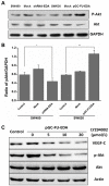
Figure 3. Activation of PI3K/Akt signaling pathway in transfected cells and control cells.
(A) p-Akt and Akt proteins of SW620 group and SW480 group were performed by western blotting analysis. (B) Quantitative analysis shows the ratio of p-Akt/GAPDH. The error bars are standard deviations of three independent experimental results. * p < 0.05 and ** p < 0.01 are considered statistically significant and highly significant, respectively. (C) VEGF-C, p-Akt and Akt proteins of EDA-overexpressed SW620 cells were performed by western blotting when the EDA-overexpressed cells were preincubated in medium containing 0–20 µM LY294002 for 24 h. Wild type SW620 was set up as the control group. The results shown are the average of three experiments.
Figure 4. BALB/c nude mice were subcutaneouly injected with transfected cells and negative control cells.
Xenografts were excised and sized 42 days later. (A) Effect of EDA on tumor proliferation. (B) Tumor volumes were measured when mice were sacrificed and the data are presented as mean determinants (±SEM). * p < 0.05. (C) Immunohistochemical staining of EDA and VEGF-C was performed in nude mouse xenografts. Scale bar = 50 µm.
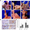
Figure 5. Orthotopic xenografts derived from transfected cells and control cells.
Seven to 8 wk old male BALB/c nude mice were orthotopically implanted with 6 groups of tumor xenografts. (A) Representative images of xenografts. Orthotopic images of xenograft tumors derived from 6 groups of transfected cells and control cells. Scale bar = 5 mm. (B) Expression of LYVE-1 was used to count LMVD in xenografts by immunohistochemical staining. The distribution of lymph vessels was stained brown under the light microscope. Scale bar = 50 µm. (C) Quantification of lymph microvessel density (LMVD) is shown. The results shown are the average of three experiments. ** p < 0.01, as compared with control group. The values are displayed as the mean ± SEM.
Similar articles
- TGFβ1-mediated PI3K/Akt and p38 MAP kinase dependent alternative splicing of fibronectin extra domain A in human podocyte culture.
Madne TH, Dockrell MEC. Madne TH, et al. Cell Mol Biol (Noisy-le-grand). 2018 Apr 30;64(5):127-135. Cell Mol Biol (Noisy-le-grand). 2018. PMID: 29729706 - Qingjie Fuzheng Granule suppresses lymphangiogenesis in colorectal cancer via the VEGF-C/VEGFR-3 dependent PI3K/AKT pathway.
Huang B, Lu Y, Gui M, Guan J, Lin M, Zhao J, Mao Q, Lin J. Huang B, et al. Biomed Pharmacother. 2021 May;137:111331. doi: 10.1016/j.biopha.2021.111331. Epub 2021 Feb 9. Biomed Pharmacother. 2021. PMID: 33578235 - Loss of Smad4 in colorectal cancer induces resistance to 5-fluorouracil through activating Akt pathway.
Zhang B, Zhang B, Chen X, Bae S, Singh K, Washington MK, Datta PK. Zhang B, et al. Br J Cancer. 2014 Feb 18;110(4):946-57. doi: 10.1038/bjc.2013.789. Epub 2014 Jan 2. Br J Cancer. 2014. PMID: 24384683 Free PMC article. - PI3K/Akt and MAPK/ERK1/2 signaling pathways are involved in IGF-1-induced VEGF-C upregulation in breast cancer.
Zhu C, Qi X, Chen Y, Sun B, Dai Y, Gu Y. Zhu C, et al. J Cancer Res Clin Oncol. 2011 Nov;137(11):1587-94. doi: 10.1007/s00432-011-1049-2. Epub 2011 Sep 9. J Cancer Res Clin Oncol. 2011. PMID: 21904903 Review. - Lymphangiogenesis and colorectal cancer.
Huang C, Chen Y. Huang C, et al. Saudi Med J. 2017 Mar;38(3):237-244. doi: 10.15537/smj.2017.3.16245. Saudi Med J. 2017. PMID: 28251217 Free PMC article. Review.
Cited by
- Construction of an Exudative Age-Related Macular Degeneration Diagnostic and Therapeutic Molecular Network Using Multi-Layer Network Analysis, a Fuzzy Logic Model, and Deep Learning Techniques: Are Retinal and Brain Neurodegenerative Disorders Related?
Latifi-Navid H, Barzegar Behrooz A, Jamehdor S, Davari M, Latifinavid M, Zolfaghari N, Piroozmand S, Taghizadeh S, Bourbour M, Shemshaki G, Latifi-Navid S, Arab SS, Soheili ZS, Ahmadieh H, Sheibani N. Latifi-Navid H, et al. Pharmaceuticals (Basel). 2023 Nov 2;16(11):1555. doi: 10.3390/ph16111555. Pharmaceuticals (Basel). 2023. PMID: 38004422 Free PMC article. - Endothelial VEGFR Coreceptors Neuropilin-1 and Neuropilin-2 Are Essential for Tumor Angiogenesis.
Benwell CJ, Johnson RT, Taylor JAGE, Price CA, Robinson SD. Benwell CJ, et al. Cancer Res Commun. 2022 Dec 14;2(12):1626-1640. doi: 10.1158/2767-9764.CRC-22-0250. eCollection 2022 Dec. Cancer Res Commun. 2022. PMID: 36970722 Free PMC article. - Continuous dynamic identification of key genes and molecular signaling pathways of periosteum in guided bone self-generation in swine model.
Yu BF, Wang Z, Chen XX, Zeng Q, Dai CC, Wei J. Yu BF, et al. J Orthop Surg Res. 2023 Jan 18;18(1):53. doi: 10.1186/s13018-023-03524-y. J Orthop Surg Res. 2023. PMID: 36653843 Free PMC article. - Elevated plasma EDA fibronectin in primary myelofibrosis is determined by high allele burden of _JAK2_V617F mutation and strongly predicts splenomegaly progression.
Malara A, Gruppi C, Massa M, Tira ME, Rosti V, Balduini A, Barosi G. Malara A, et al. Front Oncol. 2022 Sep 21;12:987643. doi: 10.3389/fonc.2022.987643. eCollection 2022. Front Oncol. 2022. PMID: 36212480 Free PMC article. - Targeting the extra domain A of fibronectin for cancer therapy with CAR-T cells.
Martín-Otal C, Lasarte-Cia A, Serrano D, Casares N, Conde E, Navarro F, Sánchez-Moreno I, Gorraiz M, Sarrión P, Calvo A, De Andrea CE, Echeveste J, Vilas A, Rodriguez-Madoz JR, San Miguel J, Prosper F, Hervas-Stubbs S, Lasarte JJ, Lozano T. Martín-Otal C, et al. J Immunother Cancer. 2022 Aug;10(8):e004479. doi: 10.1136/jitc-2021-004479. J Immunother Cancer. 2022. PMID: 35918123 Free PMC article.
References
- Sundlisaeter E, Dicko A, Sakariassen PØ, Sondenaa K, Enger PØ, et al. Lymphangiogenesis in colorectal cancer--prognostic and therapeutic aspects. Int J Cancer. 2007;121:1401–1409. - PubMed
- Saad RS, Lindner JL, Liu Y, Silverman JF. Lymphatic vessel density as prognostic marker in esophageal adenocarcinoma. Am J Clin Pathol. 2009;131:92–98. - PubMed
- Liu HT, Ma R, Yang QF, Du G, Zhang CJ. Lymphangiogenic characteristics of triple negativity in node-negative breast cancer. Int J Surg Pathol. 2009;17:426–431. - PubMed
- Clasper S, Royston D, Baban D, Cao Y, Ewers S, et al. A novel gene expression profile in lymphatics associated with tumor growth and nodal metastasis. Cancer Res. 2008;68:7293–7303. - PubMed
- Barresi V, Reggiani-Bonetti L, Di Gregorio C, De Leon MP, Barresi G. Lymphatic vessel density and its prognostic value in stage I colorectal carcinoma. J Clin Pathol. 2011;64:6–12. - PubMed
MeSH terms
Substances
LinkOut - more resources
Full Text Sources
Other Literature Sources
Medical
Miscellaneous


