Immunization with SARS coronavirus vaccines leads to pulmonary immunopathology on challenge with the SARS virus - PubMed (original) (raw)
Immunization with SARS coronavirus vaccines leads to pulmonary immunopathology on challenge with the SARS virus
Chien-Te Tseng et al. PLoS One. 2012.
Erratum in
- PLoS One. 2012;7(8). doi:10.1371/annotation/2965cfae-b77d-4014-8b7b-236e01a35492
Abstract
Background: Severe acute respiratory syndrome (SARS) emerged in China in 2002 and spread to other countries before brought under control. Because of a concern for reemergence or a deliberate release of the SARS coronavirus, vaccine development was initiated. Evaluations of an inactivated whole virus vaccine in ferrets and nonhuman primates and a virus-like-particle vaccine in mice induced protection against infection but challenged animals exhibited an immunopathologic-type lung disease.
Design: Four candidate vaccines for humans with or without alum adjuvant were evaluated in a mouse model of SARS, a VLP vaccine, the vaccine given to ferrets and NHP, another whole virus vaccine and an rDNA-produced S protein. Balb/c or C57BL/6 mice were vaccinated i.m. on day 0 and 28 and sacrificed for serum antibody measurements or challenged with live virus on day 56. On day 58, challenged mice were sacrificed and lungs obtained for virus and histopathology.
Results: All vaccines induced serum neutralizing antibody with increasing dosages and/or alum significantly increasing responses. Significant reductions of SARS-CoV two days after challenge was seen for all vaccines and prior live SARS-CoV. All mice exhibited histopathologic changes in lungs two days after challenge including all animals vaccinated (Balb/C and C57BL/6) or given live virus, influenza vaccine, or PBS suggesting infection occurred in all. Histopathology seen in animals given one of the SARS-CoV vaccines was uniformly a Th2-type immunopathology with prominent eosinophil infiltration, confirmed with special eosinophil stains. The pathologic changes seen in all control groups lacked the eosinophil prominence.
Conclusions: These SARS-CoV vaccines all induced antibody and protection against infection with SARS-CoV. However, challenge of mice given any of the vaccines led to occurrence of Th2-type immunopathology suggesting hypersensitivity to SARS-CoV components was induced. Caution in proceeding to application of a SARS-CoV vaccine in humans is indicated.
Conflict of interest statement
Competing Interests: The authors have declared that no competing interests exist.
Figures
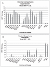
Figure 1. Vaccine Comparisons of Three SARS-CoV Vaccines, Experiment 1.
Serum neutralizing (neut) antibody and lung virus titers for each vaccine dosage group. A. Geometric mean serum antibody titer as log2 and standard error of the mean (S.E.) on day 56 for each vaccine dosage group. Seven to eight mice per group. Vaccines: double inactivated whole virus (DIV), recombinant S protein (SV), viral-like particle vaccine (VLP), with alum (+A). Five mice per group were given 0.1 ml of vaccine intramuscularly on days 0 and 28. B. Geometric mean virus titer (log10 TCID50/g) and standard error of the mean (S.E.) in lungs on day 58 (two days after SARS-CoV challenge) for each vaccine dosage group. Analyses: A. GMT with compared to without alum: DIV p>.05, VLP p>.05, SV p = .001. GMT for different vaccine dosage: DIV with alum p = .007, DIV without alum p>.05, SV with alum p = .028, SV without alum p = .01. Multiple regression: GMT increased for alum p = .012 and dosage p<.001, for SV alum only p = .001. B. GMT for all DIV groups not different p>.05, GMT for SV group without alum p .008 and with alum p .023. GMT for VLP group is not different p>.05.
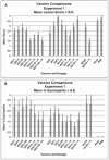
Figure 2. Vaccine Comparisons of Three SARS-CoV Vaccines, Experiment 1.
Mean lung cellular infiltration/lesion pathology and percent eosinophils in infiltrates for each vaccine dosage group two days after challenge with SARS-CoV. A. Mean lesion score and standard error of the mean (S.E.) for each vaccine dosage group. All mice exhibited lung histopathology. Scores are mean of scores for seven to eight mice per group. Scoring. 0 – no pathology, 1 and 2 – (1) minimal (2) moderate peribronchiole and perivascular cellular infiltration, 3 and 4 – 1 and/or 2 plus minimal (3) or moderate (4) epithelial cell necrosis of bronchioles with cell debris in the lumen. B. Mean percent eosinophils on histologic evaluation for seven to eight mice in each vaccine dosage group. Mean for each mouse is the mean percent eosinophils on five separate microscopy fields of lung sections. Analyses: A. Mean lesion scores were different p<.001. DIV without alum greater than with alum p = .001, VLP without alum greater than with alum p = .008. Posthoc comparisons: DIV lower than SV p = .001 and controls p<.001 but not VLP p>.05. SV lower than controls p .048. B. Mean percent eosinophils were different p<.001. Mean percent eosinophils lower for DIV with alum than without alum p = .049 and lower for SV with alum than without alum p = .001. Mean percent eosinophils lower for SV than DIV p = .002 or VLP. P = <.001. Mean percent eosinophils greater than controls for DIV, SV and VLP, all three vaccines p<.001.
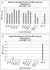
Figure 3. Higher Dosages of SV Vaccine plus DIV and BPV Vaccine Comparisons, Experiment 2.
Serum neutralizing (neut) antibody and lung virus titers for each vaccine dosage group. A. Geometric mean serum antibody titer and standard error of the mean (S.E.) on day 56 for each vaccine dosage group. Five mice per group given 0.1 ml of vaccine intramuscularly on days 0 and 28. B. Geometric mean virus titer (log10 TCID50/g) and standard error of the mean (S.E.) in lungs on day 58 (two days after SARS-CoV challenge) for each vaccine dosage group. Seven to eight mice per group. Vaccines: double inactivated whole virus (DIV), recombinant S protein (SV), β propiolactone inactivated whole virus (BPV) with alum (+A). Analyses: A. GMT with alum greater than without alum: SV p<.001, DIV p = .014. GMT for the two BPV groups are different p = .039. Multiple regression: DIV and SV increased with alum p≤.01, no dosage effect p>.05.
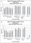
Figure 4. Higher Dosages of SV Vaccine plus DIV and BPV Vaccine Comparisons, Experiment 2.
Mean lung cellular infiltration/lesion pathology and mean percent eosinophils in infiltrates for each vaccine dosage group two days after challenge with SARS-CoV. A. Mean lesion score and standard error of the mean (S.E.) for each vaccine dosage group. Scores are mean of scores for seven to eight mice per group. Scoring - 0 - no definite pathology, 1 - mild peribronchiole and perivascular cellular infiltration, 2 - moderate peribronchiole and perivascular cellular infiltration, 3 - severe peribronchiolar and perivascular cellular infiltration with thickening of alveolar walls, alveolar infiltration and bronchiole epithelial cell necrosis and debris in the lumen. Ten to 20 microscopy fields were scored for each mouse lung. B. Mean score and standard error of the mean (S.E.) for eosinophils as percent of infiltrating cells for each vaccine dosage group. Scores are mean of scores for seven to eight mice per group. Scoring: 0 - <5% of cells, 1 - 5–10% of cells, 2 - 10–20% of cells, 3 - >20% of cells. Ten to 20 microscopy fields were scored for each mouse lung. Analyses: A. Mean lesion scores were different p<.001. Mean scores were lower for SV than DIV p<.001 and less than BPV p = .006. B. Mean eosinophil scores were lower for SV than DIV p<.001 and less than BPV p<.001. Eosinophil scores greater for SV than PBS or live virus p<.001.
Figure 5. Photographs of Lung Tissue.
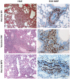
Representative photomicrographs of lung tissue two days after challenge of Balb/c mice with SARS-CoV that had previously been given a SARS-CoV vaccine. Lung sections were separately stained with hematoxylin and eosin (H&E) and an immunohistochemical protocol using an eosinophil-specific staining procedure with a monoclonal antibody to a major basic protein of eosinophils. DAB chromogen provided the brown eosinophil identity stain. The procedure and antibody were kindly provided by the Lee Laboratory, Mayo Clinic, Arizona. The H&E stain column is on the left and eosinophil-specific major basic protein (EOS MBP) stain column is on the right. Vaccines: double inactivated whole virus (DIV), β propiolactone inactivated whole virus vaccine (BPV). As shown in the images, eosinophils are prominent (brown DAB staining) in all sections examined. Exposure to SARS-CoV is associated with prominent inflammatory infiltrates characterized by a predominant eosinophilic component.
Figure 6. Photomicrographs of Lung Tissue.
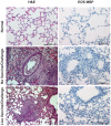
Representative photomicrographs of lung tissue from unvaccinated unchallenged mice (normal) and from Balb/c mice two days after challenge with SARS-CoV that had previously been given PBS only (no vaccine) or live virus. H&E and immunohistochemical stains for eosinophil major basic protein were performed as described for figure 5. The H&E column is on the left and the Eos MBP column is on the right. Shown are sections from normal mice (no vaccine or live virus) and mice given PBS (no vaccine) or live SARS-CoV and then challenged with SARS-CoV. As shown in the middle and bottom row images, although exposure to SARS-CoV elicits inflammatory infiltrates and accumulation of debris in the bronchial lumen, eosinophils in all groups remain within normal limits.
Figure 7. Mouse and Vaccine Specificity, Experiment 3.
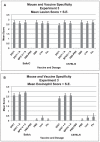
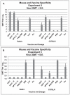
Serum neutralizing (neut) antibody and lung virus titers for each vaccine dosage group. A. Geometric mean serum antibody titer and standard error of the mean (S.E.) on day 56 for each vaccine dosage group for each mouse strain (Balb/c or C57BL/6). Five mice per group given 0.1 ml of vaccine intramuscularly on days 0 and 28. B. Geometric mean virus titer (log10 TCID50/g) and standard error of the mean (S.E.) in lungs on day 58 (two days after SARS-CoV challenge for each vaccine dosage group for each mouse strain. Seven to eight mice per group. Vaccines: Double inactivated whole virus, (DIV), β propiolactone inactivated whole virus (BPV), with alum (+A). Analyses: A. GMT for highest DIV dosage without alum greater for Balb/c than C57BL/6 p = .008 but not for alum p>.05. GMT for the BPV vaccine and live virus were not different for the two strains p>.05. B. GMT for PBS control mice were not different p>.05. GMT for DIV without alum and BPV with alum greater for C57BL/6 than Balb/c p = .004.
Figure 8. Mouse and Vaccine Specificity, Experiment 3.
Mean lung cellular infiltration/lesion pathology and percent eosinophils in infiltrates for each vaccine dosage group for each mouse strain (Balb/c or C57BL/6) two days after challenge with SARS-CoV. A. Mean lesion score and standard error of the mean (S.E.) for each vaccine dosage group. Scores are mean of scores for seven to eight mice per group. Scoring 0 - no definite pathology, 1 - mild peribronchiole and perivascular cellular infiltration, 2 - moderate peribronchiole and perivascular cellular infiltration, 3 - severe peribronchiole and perivascular cellular infiltration with thickening of alveolar walls, alveolar infiltration and bronchiole epithelial cell necrosis and debris in the lumen. Ten to 20 microscopy fields were scored for each mouse lung. B. Mean score and standard error of the mean (S.E.) for eosinophils as percent of infiltrating cells for each vaccine dosage group. Scores are mean of scores for seven to eight mice per group. Scoring: 0 - <5% of cells, 1 - 5–10% of cells, 2 - 10–20% of cells, 3 - >20% of cells. Ten to 20 microscopy fields were scored for each mouse lung. Analyses: A. Mean lesion scores were not different p>.05. B. Mean eosinophil scores were different p<.001. Mean scores for vaccine groups greater than non-vaccine groups for Balb/c and C57BL/6 p<.001 for all comparisons. Mean eosinophil scores for the same groups not different for Balb/c and C57BL/6 p>.05.
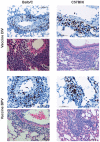
Figure 9. Photomicrographs of Lung Tissue.
Representative photomicrographs of lung tissue two days after challenge of Balb/c and C57BL/6 mice that had previously been given a SARS-CoV vaccine. Lung sections were separately stained with H&E (pink and blue micrographs) or the immunohistochemical stain for eosinophil major basic protein (blue and brown micrographs). Balb/c mice lung sections are in the left column and C57BL/6 are in the right column; doubly inactivated whole virus vaccine is in the upper four panels and those from mice given the β propiolactone inactivated whole virus vaccine are in the lower four panels. Pathologic changes observed (inflammatory infiltrates) are similar in Balb/c and C57BL/6 and eosinophils are prominent in both groups.
Similar articles
- Severe acute respiratory syndrome-associated coronavirus vaccines formulated with delta inulin adjuvants provide enhanced protection while ameliorating lung eosinophilic immunopathology.
Honda-Okubo Y, Barnard D, Ong CH, Peng BH, Tseng CT, Petrovsky N. Honda-Okubo Y, et al. J Virol. 2015 Mar;89(6):2995-3007. doi: 10.1128/JVI.02980-14. Epub 2014 Dec 17. J Virol. 2015. PMID: 25520500 Free PMC article. - Gold nanoparticle-adjuvanted S protein induces a strong antigen-specific IgG response against severe acute respiratory syndrome-related coronavirus infection, but fails to induce protective antibodies and limit eosinophilic infiltration in lungs.
Sekimukai H, Iwata-Yoshikawa N, Fukushi S, Tani H, Kataoka M, Suzuki T, Hasegawa H, Niikura K, Arai K, Nagata N. Sekimukai H, et al. Microbiol Immunol. 2020 Jan;64(1):33-51. doi: 10.1111/1348-0421.12754. Epub 2019 Nov 18. Microbiol Immunol. 2020. PMID: 31692019 Free PMC article. - Receptor-binding domain of SARS-CoV spike protein induces long-term protective immunity in an animal model.
Du L, Zhao G, He Y, Guo Y, Zheng BJ, Jiang S, Zhou Y. Du L, et al. Vaccine. 2007 Apr 12;25(15):2832-8. doi: 10.1016/j.vaccine.2006.10.031. Epub 2006 Oct 30. Vaccine. 2007. PMID: 17092615 Free PMC article. - Severe acute respiratory syndrome vaccine development: experiences of vaccination against avian infectious bronchitis coronavirus.
Cavanagh D. Cavanagh D. Avian Pathol. 2003 Dec;32(6):567-82. doi: 10.1080/03079450310001621198. Avian Pathol. 2003. PMID: 14676007 Free PMC article. Review. - Vaccine design for severe acute respiratory syndrome coronavirus.
He Y, Jiang S. He Y, et al. Viral Immunol. 2005;18(2):327-32. doi: 10.1089/vim.2005.18.327. Viral Immunol. 2005. PMID: 16035944 Review.
Cited by
- A Pathophysiological Perspective on COVID-19's Lethal Complication: From Viremia to Hypersensitivity Pneumonitis-like Immune Dysregulation.
Sanchez-Gonzalez MA, Moskowitz D, Issuree PD, Yatzkan G, Rizvi SAA, Day K. Sanchez-Gonzalez MA, et al. Infect Chemother. 2020 Sep;52(3):335-344. doi: 10.3947/ic.2020.52.3.335. Epub 2020 Jul 15. Infect Chemother. 2020. PMID: 32537960 Free PMC article. Review. - Vaccines targeting SARS-CoV-2 tested in humans.
Edwards KM. Edwards KM. Nat Med. 2020 Sep;26(9):1336-1338. doi: 10.1038/s41591-020-1048-4. Nat Med. 2020. PMID: 32839620 No abstract available. - Therapeutic Strategies in the Fight against COVID-19: From Bench to Bedside.
Abadi B, Aarabi Jeshvaghani AH, Fathalipour H, Dehghan L, Rahimi Sirjani K, Forootanfar H. Abadi B, et al. Iran J Med Sci. 2022 Nov;47(6):517-532. doi: 10.30476/IJMS.2021.92662.2396. Iran J Med Sci. 2022. PMID: 36380976 Free PMC article. Review. - Challenges in evaluating SARS-CoV-2 vaccines during the COVID-19 pandemic.
Abu-Raya B, Gantt S, Sadarangani M. Abu-Raya B, et al. CMAJ. 2020 Aug 24;192(34):E982-E985. doi: 10.1503/cmaj.201237. Epub 2020 Jul 9. CMAJ. 2020. PMID: 32646869 Free PMC article. No abstract available. - A roadmap for MERS-CoV research and product development: report from a World Health Organization consultation.
Modjarrad K, Moorthy VS, Ben Embarek P, Van Kerkhove M, Kim J, Kieny MP. Modjarrad K, et al. Nat Med. 2016 Jul 7;22(7):701-5. doi: 10.1038/nm.4131. Nat Med. 2016. PMID: 27387881 Free PMC article.
References
- World Health Organization website. 2003. Available: http://www.who.int/csr/media/sars_wha.pdf. Accessed 2012 Apr 2. Severe acute respiratory syndrome (SARS): Status of the outbreak and lessons for the immediate future; unmasking a new disease. CSR/WHO, Geneva. 20 May 2003.
- Tsang KW, Ho PL, Ooi CG, Yee WK, Wang T, et al. A cluster of cases of severe acute respiratory syndrome in Hong Kong. N Engl J Med. 2003;348:1953–66. - PubMed
- Poutanen SM, Low D, Henry B, Finkelstein S, Rose D, et al. Identification of severe acute respiratory syndrome in Canada. N Engl J Med. 2003;348:1953–66. - PubMed
- World Health Organization Website. Available: http://www.who.int/csr/sars/country/2003_04_04/en/index.html. Accessed 2004 April 21.
- Lee N, Hui D, Wu A, Chan P, Cameron P, et al. A major outbreak of severe acute respiratory syndrome in Hong Kong. N Engl J Med. 2003;348:1986–94. - PubMed
Publication types
MeSH terms
Substances
LinkOut - more resources
Full Text Sources
Other Literature Sources
Medical
Molecular Biology Databases
Miscellaneous
