Current status of thermal ablation treatments for lung malignancies - PubMed (original) (raw)
Current status of thermal ablation treatments for lung malignancies
Damian E Dupuy et al. Semin Intervent Radiol. 2010 Sep.
Abstract
About 75% of lung cancer patients are not surgical candidates, either due to advanced disease or medical comorbidities. Furthermore, conventional treatments that can be offered to these patients are beneficial only to a small percentage of them. Thermal ablation is a minimally invasive treatment that is commonly used in this group of patients, and which has shown promising results. Currently, the most widely used ablation techniques in the treatment of lung malignancies are radiofrequency ablation (RFA), microwave ablation, and cryoablation. Although the most studied technique is RFA, recent studies with microwave ablation and cryoablation have shown some advantages over RFA. This article reviews the application of thermal ablation in the thorax, including patient selection, basic aspects of procedure technique, imaging follow-up, treatment outcomes, and comparison of ablation techniques.
Keywords: Lung cancer; cryoablation; microwave ablation; radiofrequency ablation; thermal ablation.
Figures
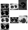
Figure 1
An 81-year-old man with right upper lobe nonsmall cell lung cancer felt to be poor candidate for lobectomy due to underlying heart disease. Axial positron emission tomography-computed tomography (PET-CT) image (A) shows intense avidity in right upper lobe mass (arrow) without evidence of regional or distant spread of disease. Axial CT (B) shows 2.2-cm mass (arrow) to have speculated margins and small pleural tail consistent with biopsy diagnosis of adenocarcinoma, stage T1b, N0, M0 (stage IA). Axial CT image (C) with patient prone during radiofrequency ablation (RFA) shows ground glass halo sign (arrows) after application of RF energy. Halo gives rough indication of energy penetration into aerated lung around mass. Axial contrast-enhanced CT images (D) in mediastinal (top) and lung (bottom) windows 3 months post-RFA shows lesion consolidation with peripheral and pleural reactive enhancement and central nonenhancement consistent with adequate treatment. Axial PET-CT image (E) shows intense uptake in mass (arrow), which is unusual given size and technical success of treatment. Therefore, repeat biopsy performed (F), which confirmed activity was due to underlying inflammatory reaction and not due to residual tumor. This is an example of false-positive PET examination due to reactionary inflammation, which is seen commonly along a pleural surface. Axial contrast-enhanced CT images (G) 9 months after RFA show contraction of mass (arrow) consistent with involution of thermal scar.
Figure 2
An 80-year-old man with cavitary squamous cell carcinoma. Axial computed tomography (CT) (A) one week after biopsy shows residual pneumothorax and cavitary mass in left upper lobe (arrow). (B) Three microwave antennae placed after instillation of fluid into air filled cavity. Note ground glass halo (arrows) after a single 10-minute treatment. Axial CT (C) 3 months after microwave ablation shows enlargement of mass with central cavitation.
Figure 3
A 75-year-old man with chest wall recurrence of nonsmall cell lung cancer after external beam radiotherapy presented with pain. Axial positron emission tomography-computed tomography (PET-CT) image (A) shows extensive metabolically active tumor invading chest wall. Axial CT image (B) after second freeze cycle from three cryoprobes shows expanding ice ball into chest wall (arrows). (C) 3-month follow-up (left) PET-CT shows cavitation and rim activity, which improved 6 months later (right).
Similar articles
- Clinical experiences with microwave thermal ablation of lung malignancies.
Sidoff L, Dupuy DE. Sidoff L, et al. Int J Hyperthermia. 2017 Feb;33(1):25-33. doi: 10.1080/02656736.2016.1204630. Epub 2016 Jul 24. Int J Hyperthermia. 2017. PMID: 27411731 Review. - Lung cancer ablation: technologies and techniques.
Alexander ES, Dupuy DE. Alexander ES, et al. Semin Intervent Radiol. 2013 Jun;30(2):141-50. doi: 10.1055/s-0033-1342955. Semin Intervent Radiol. 2013. PMID: 24436530 Free PMC article. Review. - Thermal Ablation of Lung Tumors: Focus on Microwave Ablation.
Vogl TJ, Nour-Eldin NA, Albrecht MH, Kaltenbach B, Hohenforst-Schmidt W, Lin H, Panahi B, Eichler K, Gruber-Rouh T, Roman A. Vogl TJ, et al. Rofo. 2017 Sep;189(9):828-843. doi: 10.1055/s-0043-109010. Epub 2017 May 16. Rofo. 2017. PMID: 28511267 Review. English. - Image guided thermal ablation in lung cancer treatment.
Lin M, Eiken P, Blackmon S. Lin M, et al. J Thorac Dis. 2020 Nov;12(11):7039-7047. doi: 10.21037/jtd-2019-cptn-08. J Thorac Dis. 2020. PMID: 33282409 Free PMC article. Review. - Radiofrequency ablation in lung cancer: promising results in safety and efficacy.
Suh R, Reckamp K, Zeidler M, Cameron R. Suh R, et al. Oncology (Williston Park). 2005 Oct;19(11 Suppl 4):12-21. Oncology (Williston Park). 2005. PMID: 16366374 Review.
Cited by
- TAK1 inhibition by natural cyclopeptide RA-V promotes apoptosis and inhibits protective autophagy in Kras-dependent non-small-cell lung carcinoma cells.
Yang J, Yang T, Yan W, Li D, Wang F, Wang Z, Guo Y, Bai P, Tan N, Chen L. Yang J, et al. RSC Adv. 2018 Jun 27;8(41):23451-23458. doi: 10.1039/c8ra04241a. eCollection 2018 Jun 21. RSC Adv. 2018. PMID: 35540129 Free PMC article. - Thermal ablation of thyroid nodules: are radiofrequency ablation, microwave ablation and high intensity focused ultrasound equally safe and effective methods?
Korkusuz Y, Gröner D, Raczynski N, Relin O, Kingeter Y, Grünwald F, Happel C. Korkusuz Y, et al. Eur Radiol. 2018 Mar;28(3):929-935. doi: 10.1007/s00330-017-5039-x. Epub 2017 Sep 11. Eur Radiol. 2018. PMID: 28894936 - Current therapy and development of therapeutic agents for lung cancer.
Wang Z, Kim J, Zhang P, Galvan Achi JM, Jiang Y, Rong L. Wang Z, et al. Cell Insight. 2022 Feb 9;1(2):100015. doi: 10.1016/j.cellin.2022.100015. eCollection 2022 Apr. Cell Insight. 2022. PMID: 37193130 Free PMC article. Review. - Alternative to surgery in early stage NSCLC-interventional radiologic approaches.
Lee KS, Pua BB. Lee KS, et al. Transl Lung Cancer Res. 2013 Oct;2(5):340-53. doi: 10.3978/j.issn.2218-6751.2013.10.02. Transl Lung Cancer Res. 2013. PMID: 25806253 Free PMC article. Review. - Percutaneous treatment of chest wall chondroid hamartomas: the experience of a single center.
Inserra A, Martucci C, Cassanelli G, Crocoli A, Paolantonio G, Gregori LM, Natali GL. Inserra A, et al. Pediatr Radiol. 2023 Feb;53(2):249-255. doi: 10.1007/s00247-022-05498-1. Epub 2022 Sep 5. Pediatr Radiol. 2023. PMID: 36058941 Free PMC article.
References
- American Cancer Society Cancer Facts and Figures 2008. Atlanta, GA: American Cancer Society; 2008.
- Robinson L A, Ruckdeschel J C, Wagner H, Jr, Stevens C W, American College of Chest Physicians Treatment of non-small cell lung cancer-stage IIIA: ACCP evidence-based clinical practice guidelines (2nd edition) Chest. 2007;132(3, Suppl):243S–265S. - PubMed
- Gandhi N S, Dupuy D E. Image-guided radiofrequency ablation as a new treatment option for patients with lung cancer. Semin Roentgenol. 2005;40(2):171–181. - PubMed
- McTaggart R A, Dupuy D E, Dipetrillo T. Image guided ablation in the thorax. In: In: Geschwind J-F H, Soulen MC, editor. Interventional Oncology: Principles and Practice. New York: Cambridge University Press; 2008. pp. 440–474.
- Jain S K, Dupuy D E, Cardarelli G A, Zheng Z, DiPetrillo T A. Percutaneous radiofrequency ablation of pulmonary malignancies: combined treatment with brachytherapy. AJR Am J Roentgenol. 2003;181(3):711–715. - PubMed
LinkOut - more resources
Full Text Sources
Other Literature Sources


