Mutant human myocilin induces strain specific differences in ocular hypertension and optic nerve damage in mice - PubMed (original) (raw)
Mutant human myocilin induces strain specific differences in ocular hypertension and optic nerve damage in mice
Colleen M McDowell et al. Exp Eye Res. 2012 Jul.
Abstract
Elevated intraocular pressure (IOP) is a causative risk factor for the development and progression of glaucoma. Glaucomatous mutations in myocilin (MYOC) damage the trabecular meshwork and elevate IOP in humans and in mice. Animal models of glaucoma are important to discover and better understand molecular pathogenic pathways and to test new glaucoma therapeutics. Although a number of different animal models of glaucoma have been developed and characterized, there are no true models of human primary open angle glaucoma (POAG). The overall goal of this work is to develop the first inducible mouse model of POAG using a human POAG relevant transgene (i.e. mutant MYOC) expression in mouse eyes to elevate IOP and cause pressure-induced damage to the optic nerve. Four mouse strains (A/J, BALB/cJ, C57BL/6J, and C3H/HeJ) were used in this study. Ad5.MYOC.Y437H (5 × 10(7) pfu) was injected intravitreally into one eye, with the uninjected contralateral eye serving as the control eye. Conscious IOP measurements were taken using a TonoLab rebound tonometer. Optic nerve damage was determined by scoring PPD stained optic nerve cross sections. Retinal ganglion cell and superior colliculus damage was assessed by Nissl stain cell counts. Intravitreal administration of viral vector Ad5.MYOC.Y437H caused a prolonged, reproducible, and statistically significant IOP elevation in BALB/cJ, A/J, and C57BL/6J mice. IOPs increased to approximately 25 mm Hg for 8 weeks (p < 0.0001). In contrast, the C3H/HeJ mouse strain was resistant to Ad5.MYOC.Y437H induced IOP elevation for the 8-week time period. IOPs were stable (12-15 mm Hg) in the uninjected control eyes. We also determined whether there were any strain differences in pressure-induced optic nerve damage. Even though IOP was similarly elevated in three of the strains tested (BALB/cJ, C57BL/6J, and A/J) only the A/J strain had considerable and significant optic nerve damage at the end of 8 weeks with optic nerve damage score of 2.64 ± 0.19 (n = 18, p < 0.001) in the injected eye. There was no statistical difference in retinal ganglion cell death or superior colliculus damage at the 8-week time point in any of the strains tested. These results demonstrate strain dependent responses to Ad5.MYOC.Y437H-induced ocular hypertension and pressure-induced optic nerve damage.
Copyright © 2012 Elsevier Ltd. All rights reserved.
Figures
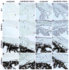
Figure 1. Myocilin is overexpressed in the trabecular meshwork after adenovirus transduction
All mice were injected in one eye with 5×10⁷ pfu Ad5.MYOC.Y437H. The contralateral uninjected eye served as a control. All samples were collected 7 days post-injection. The first two columns are labeled with anti-myocilin antibody and myocilin expression is represented by the brown labeling (A–B, E–F, I–J, M–N; n= 5 mice per strain). Myocilin expression in the TM is indicated by black arrows (B, F, J, N). The last two columns are sections from the same eye used in the first two columns and are stained with hematoxylin. The stars represent an open iridiocorneal angle (C–D, G–H, K–L, O–P; n=5 mice per strain).
Figure 2. Ad5.MYOC.Y437H induces strain dependent ocular hypertension in mice
All mice were injected in one eye with 5×10⁷ pfu Ad5.MYOC.Y437H and IOP was measured for 8 weeks post injection. The contralateral uninjected eye served as a control. (A) A/J mice had significant IOP elevation beginning at 5 days post-injection (p=0.0002). The IOP elevation remained significant for at least 56 days (p<0.0001, Days 7–56). (B) BALB/cJ mice had significant IOP elevation beginning at 5 days post-injection (p<0.0001). The IOP elevation remained significant for at least 56 days (p<0.0001, Days 7–56). (C) C57BL/6J mice had significant IOP elevation beginning at 5 days post-injection (p=0.0393). The IOP elevation remained significant for at least 56 days (p<0.0001, Days 7–56). (D) C3H/HeJ mice had significant IOP elevation at Day 5 (p=0.0018), Day 7 (p=0.0042) and Day 14 (p=0.0104). At all other time points there was no significant elevation in IOP. Ad5.MYOC.Y437H injected eyes represented in grey, uninjected control eye represented in black. Data reported as mean +/− SD.
Figure 3. Ad5.MYOC.WT has no effect on IOP
BALB/cJ mice were injected in one eye with 5×10⁷ pfu Ad5.MYOC.WT and IOP was measured for 8 weeks post injection. The contralateral uninjected eye served as a control. There was no statistical difference in IOP between the injected and uninjected eye at any time point throughout the 8-week time period. Ad5.MYOC.WT injected eyes represented in grey, uninjected control eye represented in black. Data reported as mean +/− SD.
Figure 4. Ad5.MYOC.Y437H damages the optic nerve in A/J mice at 8 weeks post injection
Optic nerve damage was assessed by PPD stain and quantified by using the optic nerve damage score (ONDS) system, which clinically grades optic nerve damage from 1 (no damage) to 5 (severe damage). At 8 weeks post-injection there was a significant difference in ONDS in A/J mice (injected eye ONDS=2.64 +/− 0.19, n=18; uninjected eye ONDS=1.40 +/− 0.14, n=18, p<0.001). There was no significant difference in ONDS in BALB/cJ, C57BL/6J or C3H/HeJ mice at 8 weeks. Data represented as mean +/− SEM.
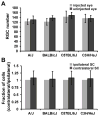
Figure 5. Ad5.MYOC.Y437H has no effect on RGC loss or SC damage at 8 weeks post injection
(A) Nissl stained retina flat mounts were used to quantify RGC loss 8 weeks after Ad5.MYOC.Y437H injection. At 8 weeks post-injection there was no significant difference in RGC number in A/J (n=8), BALB/cJ (n=10), C57BL/6J (n=10), or C3H/HeJ (n=10) mice. Two images (400X) were taken from each quadrant of the retina flat mount (8 images total) and four regions within each image were counted, representing about 2% of total RGC’s. (B) Nissl stained superior colliculus sections were used to quantitate damage 8 weeks after Ad5.MYOC.Y437H injection. At 8 weeks post-injection there was no significant difference in cell number in the superior colliculus in A/J (n=10), BALB/cJ (n=10), C57BL/6J (n=10), or C3H/HeJ (n=8) mice. Data represented as mean +/− SD.
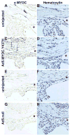
Figure 6. Mutant myocilin is expressed in the TM 8 weeks post-injection in A/J mice
All mice were injected in one eye with 5×10⁷ pfu Ad5.MYOC.Y437H. The contralateral uninjected eye served as a control. All samples were collected 8 weeks post-injection. The left column is labeled with anti-myocilin antibody and myocilin expression is represented by the brown labeling (A, C, E, G; n= 5 mice per strain). Myocilin expression in the TM is indicated by black arrows. The right column is sections from the same eye used in the left column and are stained with hematoxylin. The stars represent open iridiocorneal angles (B, D, F, H; n=5 mice per strain).
Similar articles
- Mutated myocilin and heterozygous Sod2 deficiency act synergistically in a mouse model of open-angle glaucoma.
Joe MK, Nakaya N, Abu-Asab M, Tomarev SI. Joe MK, et al. Hum Mol Genet. 2015 Jun 15;24(12):3322-34. doi: 10.1093/hmg/ddv082. Epub 2015 Mar 3. Hum Mol Genet. 2015. PMID: 25740847 Free PMC article. - Topical ocular sodium 4-phenylbutyrate rescues glaucoma in a myocilin mouse model of primary open-angle glaucoma.
Zode GS, Bugge KE, Mohan K, Grozdanic SD, Peters JC, Koehn DR, Anderson MG, Kardon RH, Stone EM, Sheffield VC. Zode GS, et al. Invest Ophthalmol Vis Sci. 2012 Mar 21;53(3):1557-65. doi: 10.1167/iovs.11-8837. Print 2012 Mar. Invest Ophthalmol Vis Sci. 2012. PMID: 22328638 Free PMC article. - Reduction of ER stress via a chemical chaperone prevents disease phenotypes in a mouse model of primary open angle glaucoma.
Zode GS, Kuehn MH, Nishimura DY, Searby CC, Mohan K, Grozdanic SD, Bugge K, Anderson MG, Clark AF, Stone EM, Sheffield VC. Zode GS, et al. J Clin Invest. 2011 Sep;121(9):3542-53. doi: 10.1172/JCI58183. Epub 2011 Aug 8. J Clin Invest. 2011. PMID: 21821918 Free PMC article. - Mendelian genes in primary open angle glaucoma.
Sears NC, Boese EA, Miller MA, Fingert JH. Sears NC, et al. Exp Eye Res. 2019 Sep;186:107702. doi: 10.1016/j.exer.2019.107702. Epub 2019 Jun 22. Exp Eye Res. 2019. PMID: 31238079 Free PMC article. Review. - Elevation of intraocular pressure in rodents using viral vectors targeting the trabecular meshwork.
Pang IH, Millar JC, Clark AF. Pang IH, et al. Exp Eye Res. 2015 Dec;141:33-41. doi: 10.1016/j.exer.2015.04.003. Epub 2015 May 27. Exp Eye Res. 2015. PMID: 26025608 Free PMC article. Review.
Cited by
- Lentiviral mediated delivery of CRISPR/Cas9 reduces intraocular pressure in a mouse model of myocilin glaucoma.
Patil SV, Kaipa BR, Ranshing S, Sundaresan Y, Millar JC, Nagarajan B, Kiehlbauch C, Zhang Q, Jain A, Searby CC, Scheetz TE, Clark AF, Sheffield VC, Zode GS. Patil SV, et al. Res Sq [Preprint]. 2023 Dec 19:rs.3.rs-3740880. doi: 10.21203/rs.3.rs-3740880/v1. Res Sq. 2023. PMID: 38196579 Free PMC article. Updated. Preprint. - A murine glaucoma model induced by rapid in vivo photopolymerization of hyaluronic acid glycidyl methacrylate.
Guo C, Qu X, Rangaswamy N, Leehy B, Xiang C, Rice D, Prasanna G. Guo C, et al. PLoS One. 2018 Jun 27;13(6):e0196529. doi: 10.1371/journal.pone.0196529. eCollection 2018. PLoS One. 2018. PMID: 29949582 Free PMC article. - Generating cell-derived matrices from human trabecular meshwork cell cultures for mechanistic studies.
Yemanyi F, Vranka J, Raghunathan V. Yemanyi F, et al. Methods Cell Biol. 2020;156:271-307. doi: 10.1016/bs.mcb.2019.10.008. Epub 2020 Jan 7. Methods Cell Biol. 2020. PMID: 32222223 Free PMC article. - Exploiting the interaction between Grp94 and aggregated myocilin to treat glaucoma.
Stothert AR, Suntharalingam A, Huard DJ, Fontaine SN, Crowley VM, Mishra S, Blagg BS, Lieberman RL, Dickey CA. Stothert AR, et al. Hum Mol Genet. 2014 Dec 15;23(24):6470-80. doi: 10.1093/hmg/ddu367. Epub 2014 Jul 15. Hum Mol Genet. 2014. PMID: 25027323 Free PMC article. - Lentiviral mediated delivery of CRISPR/Cas9 reduces intraocular pressure in a mouse model of myocilin glaucoma.
Patil SV, Kaipa BR, Ranshing S, Sundaresan Y, Millar JC, Nagarajan B, Kiehlbauch C, Zhang Q, Jain A, Searby CC, Scheetz TE, Clark AF, Sheffield VC, Zode GS. Patil SV, et al. Sci Rep. 2024 Mar 23;14(1):6958. doi: 10.1038/s41598-024-57286-6. Sci Rep. 2024. PMID: 38521856 Free PMC article.
References
- Ackert-Bicknell CL, Demissie S, Marin de Evsikova C, Hsu YH, DeMambro VE, Karasik D, Cupples LA, Ordovas JM, Tucker KL, Cho K, Canalis E, Paigen B, Churchill GA, Forejt J, Beamer WG, Ferrari S, Bouxsein ML, Kiel DP, Rosen CJ. PPARG by dietary fat interaction influences bone mass in mice and humans. J Bone Miner Res. 2008;23:1398–1408. - PMC - PubMed
- Alward WL, Fingert JH, Coote MA, Johnson AT, Lerner SF, Junqua D, Durcan FJ, McCartney PJ, Mackey DA, Sheffield VC, Stone EM. Clinical features associated with mutations in the chromosome 1 open-angle glaucoma gene (GLC1A) N Engl J Med. 1998;338:1022–1027. - PubMed
- Butler EG, England PJ, Williams GM. Genetic differences in enzymes associated with peroxisome proliferation and hydrogen peroxide metabolism in inbred mouse strains. Carcinogenesis. 1988;9:1459–1463. - PubMed
Publication types
MeSH terms
Substances
Grants and funding
- R01 EY011721/EY/NEI NIH HHS/United States
- R29 EY011721/EY/NEI NIH HHS/United States
- HHMI/Howard Hughes Medical Institute/United States
- EY11721/EY/NEI NIH HHS/United States
- R21 EY019977/EY/NEI NIH HHS/United States
LinkOut - more resources
Full Text Sources
Medical
Molecular Biology Databases


