Comparison of different methods for the calculation of the microvascular flow index - PubMed (original) (raw)
Comparison of different methods for the calculation of the microvascular flow index
Mario O Pozo et al. Crit Care Res Pract. 2012.
Abstract
The microvascular flow index (MFI) is commonly used to semiquantitatively characterize the velocity of microcirculatory perfusion as absent (0), intermittent (1), sluggish (2), or normal (3). There are three approaches to compute MFI: (1) the average of the predominant flow in each of the four quadrants (MFI(by quadrants)), (2) the direct assessment during the bedside video acquisition (MFI(point of care)), and (3) the mean value of the MFIs determined in each individual vessel (MFI(vessel by vessel)). We hypothesized that the agreement between the MFIs is poor and that the MFI(vessel by vessel) better reflects the microvascular perfusion. For this purpose, we analyzed 100 videos from septic patients. In 25 of them, red blood cell (RBC) velocity was also measured. There were wide 95% limits of agreement between MFI(by quadrants) and MFI(point of care) (1.46), between MFI(by quadrants) and MFI(vessel by vessel) (2.85), and between MFI(by point of care) and MFI(vessel by vessel) (2.56). The MFIs significantly correlated with the RBC velocity and with the fraction of perfused small vessels, but MFI(vessel by vessel) showed the best R(2). Although the different methods for the calculation of MFI reflect microvascular perfusion, they are not interchangeable and MFI(vessel by vessel) might be better.
Figures
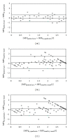
Figure 1
Bland and Altman analysis for the different methods used for the calculation of microvascular flow index (MFI). Panel (a): bedside point of care MFI (MFIpoint of care) and MFI determined by quadrants (MFIby quadrants). Panel (b): MFIpoint of care) and MFI determined by vessel by vessel analysis (MFIvessel by vessel). Panel (c): (MFIby quadrants) and (MFIvessel by vessel). Lines are bias and 95% limits of agreement.
Figure 2
Correlations of the red blood cell velocity with the microvascular flow index determined by vessel by vessel analysis (MFIvessel by vessel) Panel (a), the microvascular flow index determined by quadrants (MFIby quadrants) Panel (b), and the bedside point-of-care microvascular flow index (MFIpoint of care) Panel (c).
Figure 3
Correlations of the proportion of perfused small vessels with the microvascular flow index determined by vessel by vessel analysis (MFIvessel by vessel) Panel (a), the microvascular flow index determined by quadrants (MFIby quadrants) Panel (b), and the bedside point-of-care microvascular flow index (MFIpoint of care) Panel (c).
Similar articles
- Ability and efficiency of an automatic analysis software to measure microvascular parameters.
Carsetti A, Aya HD, Pierantozzi S, Bazurro S, Donati A, Rhodes A, Cecconi M. Carsetti A, et al. J Clin Monit Comput. 2017 Aug;31(4):669-676. doi: 10.1007/s10877-016-9928-3. Epub 2016 Sep 1. J Clin Monit Comput. 2017. PMID: 27586243 - Real-time point of care microcirculatory assessment of shock: design, rationale and application of the point of care microcirculation (POEM) tool.
Naumann DN, Mellis C, Husheer SL, Hopkins P, Bishop J, Midwinter MJ, Hutchings SD. Naumann DN, et al. Crit Care. 2016 Sep 30;20(1):310. doi: 10.1186/s13054-016-1492-1. Crit Care. 2016. PMID: 27716373 Free PMC article. - Point-of-care assessment of microvascular blood flow in critically ill patients.
Arnold RC, Parrillo JE, Phillip Dellinger R, Chansky ME, Shapiro NI, Lundy DJ, Trzeciak S, Hollenberg SM. Arnold RC, et al. Intensive Care Med. 2009 Oct;35(10):1761-6. doi: 10.1007/s00134-009-1517-1. Epub 2009 Jun 24. Intensive Care Med. 2009. PMID: 19554307 - Assessment of microcirculatory perfusion in healthy anesthetized cats undergoing ovariohysterectomy using sidestream dark field microscopy.
Goodnight ME, Cooper ES, Butler AL. Goodnight ME, et al. J Vet Emerg Crit Care (San Antonio). 2015 May-Jun;25(3):349-57. doi: 10.1111/vec.12296. Epub 2015 Mar 4. J Vet Emerg Crit Care (San Antonio). 2015. PMID: 25736201 - Septic shock and chemotherapy-induced cytopenia: effects on microcirculation.
Karvunidis T, Chvojka J, Lysak D, Sykora R, Krouzecky A, Radej J, Novak I, Matejovic M. Karvunidis T, et al. Intensive Care Med. 2012 Aug;38(8):1336-44. doi: 10.1007/s00134-012-2582-4. Epub 2012 May 15. Intensive Care Med. 2012. PMID: 22584795
Cited by
- MicroTools enables automated quantification of capillary density and red blood cell velocity in handheld vital microscopy.
Hilty MP, Guerci P, Ince Y, Toraman F, Ince C. Hilty MP, et al. Commun Biol. 2019 Jun 19;2:217. doi: 10.1038/s42003-019-0473-8. eCollection 2019. Commun Biol. 2019. PMID: 31240255 Free PMC article. - Effects of norepinephrine on tissue perfusion in a sheep model of intra-abdominal hypertension.
Ferrara G, Kanoore Edul VS, Caminos Eguillor JF, Martins E, Canullán C, Canales HS, Ince C, Estenssoro E, Dubin A. Ferrara G, et al. Intensive Care Med Exp. 2015 Dec;3(1):46. doi: 10.1186/s40635-015-0046-1. Epub 2015 Mar 31. Intensive Care Med Exp. 2015. PMID: 26215810 Free PMC article. - Sublingual microcirculatory blood flow and vessel density in Sherpas at high altitude.
Gilbert-Kawai E, Coppel J, Court J, van der Kaaij J, Vercueil A, Feelisch M, Levett D, Mythen M, Grocott MP, Martin D; Xtreme Everest 2 Research Group. Gilbert-Kawai E, et al. J Appl Physiol (1985). 2017 Apr 1;122(4):1011-1018. doi: 10.1152/japplphysiol.00970.2016. Epub 2017 Jan 26. J Appl Physiol (1985). 2017. PMID: 28126908 Free PMC article. - A guide to human in vivo microcirculatory flow image analysis.
Massey MJ, Shapiro NI. Massey MJ, et al. Crit Care. 2016 Feb 10;20:35. doi: 10.1186/s13054-016-1213-9. Crit Care. 2016. PMID: 26861691 Free PMC article. - Systemic and microcirculatory effects of blood transfusion in experimental hemorrhagic shock.
Ferrara G, Edul VSK, Canales HS, Martins E, Canullán C, Murias G, Pozo MO, Caminos Eguillor JF, Buscetti MG, Ince C, Dubin A. Ferrara G, et al. Intensive Care Med Exp. 2017 Dec;5(1):24. doi: 10.1186/s40635-017-0136-3. Epub 2017 Apr 21. Intensive Care Med Exp. 2017. PMID: 28432665 Free PMC article.
References
- Kanoore Edul VS, Enrico C, Laviolle B, Risso Vazquez A, Ince C, Dubin A. Quantitative assessment of the microcirculation in healthy volunteers and in septic shock patients. Critical Care Medicine. 2012;40(5):1443–1448. - PubMed
- Groner W, Winkelman JW, Harris AG, et al. Orthogonal polarization spectral imaging: a new method for study of the microcirculation. Nature Medicine. 1999;5(10):1209–1213. - PubMed
- Goedhart PT, Khalilzada M, Bezemer R, Merza J, Ince C. Sidestream Dark Field (SDF) imaging: a novel stroboscopic LED ring-based imaging modality for clinical assessment of the microcirculation. Optics Express. 2007;15(23):15101–15114. - PubMed
- De Backer D, Creteur J, Preiser JC, Dubois MJ, Vincent JL. Microvascular blood flow is altered in patients with sepsis. American Journal of Respiratory and Critical Care Medicine. 2002;166(1):98–104. - PubMed
- Trzeciak S, Dellinger RP, Parrillo JE, et al. Early microcirculatory perfusion derangements in patients with severe sepsis and septic shock: relationship to hemodynamics, oxygen transport, and survival. Annals of Emergency Medicine. 2007;49(1):88–98. - PubMed
LinkOut - more resources
Full Text Sources


