Serotonin receptors in hippocampus - PubMed (original) (raw)
Review
Serotonin receptors in hippocampus
Laura Cristina Berumen et al. ScientificWorldJournal. 2012.
Abstract
Serotonin is an ancient molecular signal and a recognized neurotransmitter brainwide distributed with particular presence in hippocampus. Almost all serotonin receptor subtypes are expressed in hippocampus, which implicates an intricate modulating system, considering that they can be localized as autosynaptic, presynaptic, and postsynaptic receptors, even colocalized within the same cell and being target of homo- and heterodimerization. Neurons and glia, including immune cells, integrate a functional network that uses several serotonin receptors to regulate their roles in this particular part of the limbic system.
Figures
Figure 1
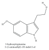
Serotonin (5-HT). Modified image from NCBI PubChem Substance Database CID 5202.
Figure 2
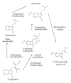
Serotonin metabolism. Tryptophan is the precursor for serotonin synthesis, with different enzymatic reactions in plant and animals [6]; hydroxylation is the rate limiting step (enzyme mediated by tryptophan hydroxylase in animals or tryptamine hydroxylase in plants), while decarboxylation is a rapid conversion by the aromatic amino acid decarboxylase (tryptophan decarboxylase). The catabolic metabolite of serotonin is 5-hydroxyindoleacetic acid, via 5-hydroxyindole acetaldehyde enzymatically converted by the membrane-bound mitochondrial flavoprotein monoamino oxidase. Modified images from NCBI PubChem Substance Database.
Figure 3
Serotonin main signaling pathways. 5-HT or agonists/antagonists for each receptor (•) interact in the extracellular side and the conformational changes of 5-HTRs modify the activity of specific intracellular enzymes, which in time modify other targets state to provoke different cellular responses [43]. G-protein βγ pathways are not represented in the figure. All of the serotonin receptor subtypes are represented for a hippocampal pyramidal cell, as reported, but subpopulations of these neurons might differentially express 5-HT receptors. AC, adenylate cyclase; PLC, phospholipase C. The 7TMD images of each subtype receptor are represented with the defined number of exons that code for the mature protein [44]; putative intron location in correspondent pre-mRNA is marked by a lightning symbol (↯), and alternative splicing sites are marked with stars (⋆⋆⋆).
Figure 4
cAMP signaling pathways. Serotonin receptors 5-HT1 and 5-HT5 interact with α i/0 G-protein inhibiting the formation of cyclic adenylate monophosphate (cAMP) by adenylate cyclase (AC), while 5-HT4, 5-HT6, and 5-HT7 activate AC by means of α S G-protein. βγ subunits of G-protein may interact in other signaling pathways, for example, modulating GIRK or calcium voltage gated channels. Representation of _β_-arrestin (_β_-arr) is made to indicate other signaling pathways. Traditional ligands to study different subtype receptors are written in the extracellular zone; note that 5-HT7 may bind the traditional ligand for 5-HT1A as well as LSD (5-HT2 ligand).
Figure 5
5-HT2 receptors signaling. Main pathways of intracellular signaling for these serotonine receptors subtype involve rupture of membrane phospholipids, particularly with phospholipase C (PLC) producing diacylglycerol (DAG) and inositol 1,4,5-trisphosphate (IP3) from phosphatidylinositol 4,5-bisphosphate (PIP2). These second messengers activate protein kinase C (PKC) which in time may activate the extracellular signal-regulated kinases 1 and 2 (ERK1/2) [50]. Phospholipase A2 is eventually activated producing arachidonic acid (AA) from phosphatidylcholine (PC), or phosphatidic acid (PA) by means of phospholipase D (PLD) [44, 50]. SERT is included in the diagram, coexisting in astrocytes for example, to emphasize the intracellular participation of serotonin itself [14]. Other pathways including (Rho-GEF) and (PI3K) are shown [51]. MEK, mitogen-activated protein kinase; PH, phosphohydrolase enzyme; PKA, protein kinase A-relation to cAMP pathways; SERT, serotonin transporter.
Figure 6
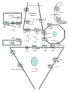
Serotonin receptors in hippocampus. The functional glia-neuron-vascular cells network uses several serotonin receptors (5-HTRs). The 7TMD images of each subtype receptor are represented with the defined number of exons that code for the mature protein (Bockaert et al., 2006) [44]; putative intron location in correspondent pre-mRNA is marked by a lightning symbol (↯); alternative splicing sites are marked with stars (⋆⋆⋆). Neuron metabotropic 5-HTRs are mainly somatodendritic volume receptors although there is an association with synaptic specializations for some of them. 5-HT3 with the five 4TMD subunits of a ligand activated ion channel is shown as synaptic receptor although this fact remains to be determined in hippocampus. Microglia is also included in the network for its relevance in pathophysiological responses, with 5-HT2B receptor expression (Capone et al., 2007) [90]. The 12TMD image of the serotonin transporter (SERT; 5-HTT) and vesicular monoamine transporter (VMAT) are represented in the serotonergic neuron and only SERT in the astrocyte.
Similar articles
- Functional effects of chronic electroconvulsive shock on serotonergic 5-HT(1A) and 5-HT(1B) receptor activity in rat hippocampus and hypothalamus.
Gur E, Dremencov E, Garcia F, Van de Kar LD, Lerer B, Newman ME. Gur E, et al. Brain Res. 2002 Oct 11;952(1):52-60. doi: 10.1016/s0006-8993(02)03193-1. Brain Res. 2002. PMID: 12363404 - Differential subcellular localization of the 5-HT3-As receptor subunit in the rat central nervous system.
Miquel MC, Emerit MB, Nosjean A, Simon A, Rumajogee P, Brisorgueil MJ, Doucet E, Hamon M, Vergé D. Miquel MC, et al. Eur J Neurosci. 2002 Feb;15(3):449-57. doi: 10.1046/j.0953-816x.2001.01872.x. Eur J Neurosci. 2002. PMID: 11876772 - Distribution of serotonin 5-hydroxytriptamine 1B (5-HT(1B)) receptors in the normal rat hypothalamus.
Makarenko IG, Meguid MM, Ugrumov MV. Makarenko IG, et al. Neurosci Lett. 2002 Aug 9;328(2):155-9. doi: 10.1016/s0304-3940(02)00345-2. Neurosci Lett. 2002. PMID: 12133578 - [Serotonergic control of prefrontal cortex].
Puig MV, Celada P, Artigas F. Puig MV, et al. Rev Neurol. 2004 Sep 16-30;39(6):539-47. Rev Neurol. 2004. PMID: 15467993 Review. Spanish. - Growing Evidence for Heterogeneous Synaptic Localization of 5-HT2A Receptors.
Bécamel C, Berthoux C, Barre A, Marin P. Bécamel C, et al. ACS Chem Neurosci. 2017 May 17;8(5):897-899. doi: 10.1021/acschemneuro.6b00409. Epub 2017 May 1. ACS Chem Neurosci. 2017. PMID: 28459524 Review.
Cited by
- Mechanistic study on vasodilatory and antihypertensive effects of hesperetin: ex vivo and in vivo approaches.
Tew WY, Tan CS, Yan CS, Loh HW, Wang X, Wen X, Wei X, Yam MF. Tew WY, et al. Hypertens Res. 2024 Sep;47(9):2416-2434. doi: 10.1038/s41440-024-01652-4. Epub 2024 Jun 24. Hypertens Res. 2024. PMID: 38914702 - Prenatal Hypoxia Triggers a Glucocorticoid-Associated Depressive-like Phenotype in Adult Rats, Accompanied by Reduced Anxiety in Response to Stress.
Stratilov V, Potapova S, Safarova D, Tyulkova E, Vetrovoy O. Stratilov V, et al. Int J Mol Sci. 2024 May 28;25(11):5902. doi: 10.3390/ijms25115902. Int J Mol Sci. 2024. PMID: 38892090 Free PMC article. - Sex-specific expression of distinct serotonin receptors mediates stress vulnerability of adult hippocampal neural stem cells in mice.
Luo YJ, Bao H, Crowther A, Li YD, Chen ZK, Tart DS, Asrican B, Zhang L, Song J. Luo YJ, et al. Cell Rep. 2024 May 28;43(5):114140. doi: 10.1016/j.celrep.2024.114140. Epub 2024 Apr 23. Cell Rep. 2024. PMID: 38656873 Free PMC article. - Linking alterations in estrogen receptor expression to memory deficits and depressive behavior in an ovariectomy mouse model.
Baek DC, Kang JY, Lee JS, Lee EJ, Son CG. Baek DC, et al. Sci Rep. 2024 Mar 21;14(1):6854. doi: 10.1038/s41598-024-57611-z. Sci Rep. 2024. PMID: 38514828 Free PMC article. - Pharmacological Evaluation of Signals of Disproportionality Reporting Related to Adverse Reactions to Antiepileptic Cannabidiol in VigiBase.
Calapai F, Mannucci C, McQuain L, Salvo F. Calapai F, et al. Pharmaceuticals (Basel). 2023 Oct 5;16(10):1420. doi: 10.3390/ph16101420. Pharmaceuticals (Basel). 2023. PMID: 37895891 Free PMC article.
References
- Rapport MM, Green AA, Page IH. Serum vasoconstrictor (serotonin). IV. Isolation and characterization. The Journal of Biological Chemistry. 1948;176:1243–1251. - PubMed
- Erspamer V, Asero B. Identification of enteramine, the specific hormone of the enterochromaffin cell system, as 5-hydroxytryptamine. Nature. 1952;169(4306):800–801. - PubMed
- Turlejski K. Evolutionary ancient roles of serotonin: long-lasting regulation of activity and development. Acta Neurobiologiae Experimentalis. 1996;56(2):619–636. - PubMed
- Nichols DE, Nichols CD. Serotonin receptors. Chemical Reviews. 2008;108(5):1614–1641. - PubMed
Publication types
MeSH terms
Substances
LinkOut - more resources
Full Text Sources


