Neural correlates of socioeconomic status in the developing human brain - PubMed (original) (raw)
Neural correlates of socioeconomic status in the developing human brain
Kimberly G Noble et al. Dev Sci. 2012 Jul.
Abstract
Socioeconomic disparities in childhood are associated with remarkable differences in cognitive and socio-emotional development during a time when dramatic changes are occurring in the brain. Yet, the neurobiological pathways through which socioeconomic status (SES) shapes development remain poorly understood. Behavioral evidence suggests that language, memory, social-emotional processing, and cognitive control exhibit relatively large differences across SES. Here we investigated whether volumetric differences could be observed across SES in several neural regions that support these skills. In a sample of 60 socioeconomically diverse children, highly significant SES differences in regional brain volume were observed in the hippocampus and the amygdala. In addition, SES × age interactions were observed in the left superior temporal gyrus and left inferior frontal gyrus, suggesting increasing SES differences with age in these regions. These results were not explained by differences in gender, race or IQ. Likely mechanisms include differences in the home linguistic environment and exposure to stress, which may serve as targets for intervention at a time of high neural plasticity.
© 2012 Blackwell Publishing Ltd.
Figures
Figure 1
Hypothesized mechanisms by which SES operates to influence cognitive development. See text for details.
Figure 2
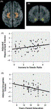
Family SES predicts child hippocampus and amygdala volumes. (A) Hippocampus ROI defined in orange. (B) Amygdala ROI defined in yellow. (C) Family income-to-needs ratio is positively correlated with child hippocampal volume, adjusted for child age, gender, and total cortical volume (Beta = 0.22; p < .032). (D) Number of years of parent education is negatively correlated with child amygdala volume, adjusted for child age, gender and total cortical volume (Beta = −0.29; p < .002). In all plots, regional volume is portrayed as the standardized residual, in standard deviations, after adjusting for covariates.
Figure 3
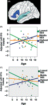
SES × Age interactions in left superior temporal gyrus and left inferior frontal gyrus. A priori ROIs defined in FreeSurfer were chosen that related to language processing. Among the 44 children whose parent education ranged from 11 to 17.5 years, SES × child age interactions were observed in (A) left inferior frontal gyrus (IFG; dark blue) and left superior temporal gyrus (STG; light blue). This is portrayed graphically in (B) left IFG (Beta = 2.769; p < .014), and (C) left STG (Beta = 2.516; p < .012), suggesting that SES differences in regional brain volume are not uniform at different ages. In all plots, regional volume is portrayed as the standardized residual, in standard deviations, after adjusting for total cortical volume. All analyses were performed using continuous variables for child age, parent education, and ROI volume, but are displayed with parental education represented in terciles, with 11–13.5 years of parental education in green; 13.5–15 years of parental education in orange; and 15–17.5 years of parental education in blue.
Similar articles
- Age-Related Differences in Cortical Thickness Vary by Socioeconomic Status.
Piccolo LR, Merz EC, He X, Sowell ER, Noble KG; Pediatric Imaging, Neurocognition, Genetics Study. Piccolo LR, et al. PLoS One. 2016 Sep 19;11(9):e0162511. doi: 10.1371/journal.pone.0162511. eCollection 2016. PLoS One. 2016. PMID: 27644039 Free PMC article. - Longitudinally Mapping Childhood Socioeconomic Status Associations with Cortical and Subcortical Morphology.
McDermott CL, Seidlitz J, Nadig A, Liu S, Clasen LS, Blumenthal JD, Reardon PK, Lalonde F, Greenstein D, Patel R, Chakravarty MM, Lerch JP, Raznahan A. McDermott CL, et al. J Neurosci. 2019 Feb 20;39(8):1365-1373. doi: 10.1523/JNEUROSCI.1808-18.2018. Epub 2018 Dec 26. J Neurosci. 2019. PMID: 30587541 Free PMC article. - Prenatal socioeconomic status and social support are associated with neonatal brain morphology, toddler language and psychiatric symptoms.
Spann MN, Bansal R, Hao X, Rosen TS, Peterson BS. Spann MN, et al. Child Neuropsychol. 2020 Feb;26(2):170-188. doi: 10.1080/09297049.2019.1648641. Epub 2019 Aug 6. Child Neuropsychol. 2020. PMID: 31385559 Free PMC article. - Socioeconomic status and the developing brain.
Hackman DA, Farah MJ. Hackman DA, et al. Trends Cogn Sci. 2009 Feb;13(2):65-73. doi: 10.1016/j.tics.2008.11.003. Epub 2009 Jan 8. Trends Cogn Sci. 2009. PMID: 19135405 Free PMC article. Review. - Neurocognitive development in socioeconomic context: Multiple mechanisms and implications for measuring socioeconomic status.
Ursache A, Noble KG. Ursache A, et al. Psychophysiology. 2016 Jan;53(1):71-82. doi: 10.1111/psyp.12547. Psychophysiology. 2016. PMID: 26681619 Free PMC article. Review.
Cited by
- Age-Related Differences in Cortical Thickness Vary by Socioeconomic Status.
Piccolo LR, Merz EC, He X, Sowell ER, Noble KG; Pediatric Imaging, Neurocognition, Genetics Study. Piccolo LR, et al. PLoS One. 2016 Sep 19;11(9):e0162511. doi: 10.1371/journal.pone.0162511. eCollection 2016. PLoS One. 2016. PMID: 27644039 Free PMC article. - Childhood Poverty Predicts Adult Amygdala and Frontal Activity and Connectivity in Response to Emotional Faces.
Javanbakht A, King AP, Evans GW, Swain JE, Angstadt M, Phan KL, Liberzon I. Javanbakht A, et al. Front Behav Neurosci. 2015 Jun 12;9:154. doi: 10.3389/fnbeh.2015.00154. eCollection 2015. Front Behav Neurosci. 2015. PMID: 26124712 Free PMC article. - Neurodevelopmental Optimization after Early-Life Adversity: Cross-Species Studies to Elucidate Sensitive Periods and Brain Mechanisms to Inform Early Intervention.
Luby JL, Baram TZ, Rogers CE, Barch DM. Luby JL, et al. Trends Neurosci. 2020 Oct;43(10):744-751. doi: 10.1016/j.tins.2020.08.001. Epub 2020 Aug 27. Trends Neurosci. 2020. PMID: 32863044 Free PMC article. Review. - Regional Brain Growth Trajectories in Fetuses with Congenital Heart Disease.
Rollins CK, Ortinau CM, Stopp C, Friedman KG, Tworetzky W, Gagoski B, Velasco-Annis C, Afacan O, Vasung L, Beaute JI, Rofeberg V, Estroff JA, Grant PE, Soul JS, Yang E, Wypij D, Gholipour A, Warfield SK, Newburger JW. Rollins CK, et al. Ann Neurol. 2021 Jan;89(1):143-157. doi: 10.1002/ana.25940. Epub 2020 Nov 4. Ann Neurol. 2021. PMID: 33084086 Free PMC article. - Individual differences in infancy research: Letting the baby stand out from the crowd.
Pérez-Edgar K, Vallorani A, Buss KA, LoBue V. Pérez-Edgar K, et al. Infancy. 2020 Jul;25(4):438-457. doi: 10.1111/infa.12338. Epub 2020 May 4. Infancy. 2020. PMID: 32744796 Free PMC article.
References
- Adams MJ (1990). Beginning to read: Thinking and learning about print. Cambridge, MA: MIT Press.
- Blair C, Granger D, & Peters Razza R (2005). Cortisol reactivity is positively related to executive function in preschool children attending Head Start. Child Development, 76 (3), 554–567. - PubMed
- Bradley RH, Corwyn RF, Burchinal M, McAdoo HP, & Garcia Coll C (2001). The home environments of children in the United States part II: Relations with behavioral development through age thirteen. Child Development, 72 (6), 1844–1867. - PubMed
- Bremner JD, Randall P, Vermetten E, Staib L, Bronen RA, Mazure C, Capelli S, McCarthy G, Innis RB, & Charney DS (1997). Magnetic resonance imaging-based measurement of hippocampal volume in posttraumatic stress disorder related to childhood physical and sexual abuse - a preliminary report. Biological Psychiatry, 41 (1), 23–32. - PMC - PubMed
- Brooks-Gunn J, & Duncan GJ (1997). The effects of poverty on children. Future of Children, 7 (2), 55–71. - PubMed
Publication types
MeSH terms
Grants and funding
- R01DA017831/DA/NIDA NIH HHS/United States
- R01MH087563/MH/NIMH NIH HHS/United States
- R01 HD053893-01/HD/NICHD NIH HHS/United States
- R01 DA017830/DA/NIDA NIH HHS/United States
- R01 HD053893/HD/NICHD NIH HHS/United States
- R01 MH087563/MH/NIMH NIH HHS/United States
LinkOut - more resources
Full Text Sources


