Polymorphic toxin systems: Comprehensive characterization of trafficking modes, processing, mechanisms of action, immunity and ecology using comparative genomics - PubMed (original) (raw)
Polymorphic toxin systems: Comprehensive characterization of trafficking modes, processing, mechanisms of action, immunity and ecology using comparative genomics
Dapeng Zhang et al. Biol Direct. 2012.
Abstract
Background: Proteinaceous toxins are observed across all levels of inter-organismal and intra-genomic conflicts. These include recently discovered prokaryotic polymorphic toxin systems implicated in intra-specific conflicts. They are characterized by a remarkable diversity of C-terminal toxin domains generated by recombination with standalone toxin-coding cassettes. Prior analysis revealed a striking diversity of nuclease and deaminase domains among the toxin modules. We systematically investigated polymorphic toxin systems using comparative genomics, sequence and structure analysis.
Results: Polymorphic toxin systems are distributed across all major bacterial lineages and are delivered by at least eight distinct secretory systems. In addition to type-II, these include type-V, VI, VII (ESX), and the poorly characterized "Photorhabdus virulence cassettes (PVC)", PrsW-dependent and MuF phage-capsid-like systems. We present evidence that trafficking of these toxins is often accompanied by autoproteolytic processing catalyzed by HINT, ZU5, PrsW, caspase-like, papain-like, and a novel metallopeptidase associated with the PVC system. We identified over 150 distinct toxin domains in these systems. These span an extraordinary catalytic spectrum to include 23 distinct clades of peptidases, numerous previously unrecognized versions of nucleases and deaminases, ADP-ribosyltransferases, ADP ribosyl cyclases, RelA/SpoT-like nucleotidyltransferases, glycosyltranferases and other enzymes predicted to modify lipids and carbohydrates, and a pore-forming toxin domain. Several of these toxin domains are shared with host-directed effectors of pathogenic bacteria. Over 90 families of immunity proteins might neutralize anywhere between a single to at least 27 distinct types of toxin domains. In some organisms multiple tandem immunity genes or immunity protein domains are organized into polyimmunity loci or polyimmunity proteins. Gene-neighborhood-analysis of polymorphic toxin systems predicts the presence of novel trafficking-related components, and also the organizational logic that allows toxin diversification through recombination. Domain architecture and protein-length analysis revealed that these toxins might be deployed as secreted factors, through directed injection, or via inter-cellular contact facilitated by filamentous structures formed by RHS/YD, filamentous hemagglutinin and other repeats. Phyletic pattern and life-style analysis indicate that polymorphic toxins and polyimmunity loci participate in cooperative behavior and facultative 'cheating' in several ecosystems such as the human oral cavity and soil. Multiple domains from these systems have also been repeatedly transferred to eukaryotes and their viruses, such as the nucleo-cytoplasmic large DNA viruses.
Conclusions: Along with a comprehensive inventory of toxins and immunity proteins, we present several testable predictions regarding active sites and catalytic mechanisms of toxins, their processing and trafficking and their role in intra-specific and inter-specific interactions between bacteria. These systems provide insights regarding the emergence of key systems at different points in eukaryotic evolution, such as ADP ribosylation, interaction of myosin VI with cargo proteins, mediation of apoptosis, hyphal heteroincompatibility, hedgehog signaling, arthropod toxins, cell-cell interaction molecules like teneurins and different signaling messengers.
Figures
Figure 1
(A) Workflow for identification and analysis of toxin and immunity domains in bacterial polymorphic toxin systems. (B) General domain architecture template for polymorphic toxins along with representative architectures seen in different secretory systems. Trafficking domains are colored grey, repeats light green, pre-toxin domains (PT-domain) yellow, releasing peptidases blue, and toxin domains pink. Newly identified domains are encircled in dashed lines in all figures in this paper. Proteins are not drawn to scale. Note, only repeats automatically detected by profiles are shown in all figures; the proteins usually have much longer repeat units than shown due to repeats being below the detection threshold. Toxins are grouped based on their secretion pathways that are defined by their canonical trafficking domains (Table 1). Proteins are denoted by their gene name, species abbreviations and GI (Genbank Index) numbers separated by underscores. (C) General gene-neighborhoods template for polymorphic toxin operons. Individual genes are represented as arrows pointing from the 5′ to the 3′-end of the coding frame. Genes are labeled by their domain architectures. The gene neighborhood is labeled by the gene name, species abbreviation and GI number of the SUKH gene marked with an asterisk. Toxins are colored pink, immunity proteins orange, and other trafficking related proteins grey. For species abbreviations refer to supplementary material.
Figure 2
Domain architectures of selected examples of polymorphic toxins containing distinct releasing peptidases: (A) HINT, (B) ZU5, (C) PrsW peptidase, (D) Caspase peptidase, (E) MCF1-SHE-like predicted peptidase. The alignment of MCF1-SHE domain is shown with predicted catalytic residues marked with blue asterisks. For all alignments in this study, proteins are denoted by their gene name, species abbreviations and GI (Genbank Index) numbers separated by underscores. Secondary structure assignments are shown above the alignment, where the blue arrow represents the β-strand and the red cylinder the α-helix. Poorly conserved inserts are excluded in the alignment and replaced by the length of the inserts. Columns in the alignment are colored based on their amino acid conservation at consensus shown below the alignment. The coloring scheme and consensus abbreviations are as follows: h, hydrophobic (ACFILMVWY), l, aliphatic (LIV) and a, aromatic (FWY) residues shaded yellow; b, big residues (LIYERFQKMW), shaded gray; s, small residues (AGSVCDN) and u, tiny residues (GAS), shaded green; p, polar residues (STEDKRNQHC) shaded blue; and c, charged residues (DEHKR) shaded magenta. Absolutely conserved residues are shaded red.
Figure 3
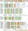
(A) Multiple sequence alignment, (B) predicted topology diagram, (C) domain architectures, and (D) gene neighborhoods for papain-like peptidase 1 (Tox-PL1) toxins. (E) Domain architectures of OTU papain-like peptidase toxins. The labeling scheme for domain architectures and alignments, and the coloring scheme and consensus abbreviations are as in Figure 3.
Figure 4
Features of PVC metallopeptidase toxins: (A) multiple sequence alignment, (B) predicted topology diagram, (C) representative domain architectures, and (D) conserved gene neighborhoods for PVC containing genes across different bacterial lineages and archaea. In (D), PVC toxins are shown in blue, the AAA + ATPase associated with the PVC system (PVC-AAA) in orange and phage-derived proteins in yellow. Gene neighborhoods are labeled with the corresponding information for the PVC-metallopeptidase containing genes marked with an asterisk. The labeling scheme for domain architectures and alignments, and the coloring scheme and consensus abbreviations are as in Figure 3.
Figure 5
Representative domain architectures for toxin proteins containing: (A) several distinct metallopeptidase toxin domains such as HopH1 peptidase and Tox-MPTases 1 – 5, (B) LD-peptidase, (C) Tox-HDC domain. (D) Sequence alignment and domain architectures of inactive transglutaminase-containing toxins. The labeling scheme for domain architectures and alignments, and the coloring scheme and consensus abbreviations are as in Figure 3.
Figure 6
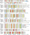
Sequence alignment and representative domain architectures of novel HNH nuclease families: (A) Tox-HHH, (B) Tox-EHHH, (C) Tox-SHH, (D) Tox-GHH2, and (E) Tox-GHH. ‘#’ indicates residues involved in metal ion-binding, ‘%’ indicates the conserved histidine which is required for activation of the water molecule for hydrolysis, and ‘*’ indicates polar residues (often asparagine) that are conserved in the HNH fold. The labeling scheme for domain architectures and alignments, and the coloring scheme and consensus abbreviations are as in 3.
Figure 7
Sequence alignment and representative domain architectures of novel restriction endonuclease families described in this study: (A) Tox-REase-2, (B) Tox-REase-3, (C) Tox-REase-4, (D) Tox-REase-5, (E) Tox-REase-6, (F) Tox-REase-7, (G) Tox-REase-8, (H) Tox-REase-9, (I) Tox-REase-10. The labeling scheme for domain architectures and alignments, and the coloring scheme and consensus abbreviations are as in Figure 3.
Figure 8
Representative domain architectures of several nucleic acid-targeting toxin domains: (A) two distinct families of URI nucleases (Tox-URI1 and Tox-URI2), (B) Tox-ComI nuclease, (C) two distinct ParB fold families (Tox-ParB, Tox-ParBL1), (D) two novel JAB families (Tox-JAB-1, Tox-JAB-2). (E) Multiple sequence alignment of the Het-C domain with Zinc-dependent phospholipase C and S1-P1 nuclease, showing their homologous relationship. Conserved catalytic residues are labeled with blue ‘#’. The labeling scheme for domain architectures and alignments, and the coloring scheme and consensus abbreviations are as in Figure 3.
Figure 9
(A) Shared common core of the BECR fold illustrated with representative structures from Barnase, RelE, ColE5, ColD, and EndoU families. PDB ids are shown in brackets. All structural cartoons are shown in an approximately similar orientation. The α-helices are colored red, β-sheets yellow and loops gray. The predicted and known active site residues are labeled. Representative domain architectures of polymorphic toxins containing (B) Tox-Barnase, (C) Tox-RelE, (D)Colicin E5 (Tox-ColE5), (E) Tox-EndoU, (F) Colicin D (Tox-ColD), and (G) several other novel toxin domains predicted to contain the BECR fold. The labeling scheme for domain architectures and alignments, and the coloring scheme and consensus abbreviations are as in Figure 3.
Figure 10
Domain architectures of polymorphic toxins containing (A) Two novel deaminase families reported in this study, (B) Cytotoxic necrotizing factor (Tox-CNF), (C) several families of ADP-ribosyltransferases (Tox-ART), (D) Phospholipase A2 toxin (Tox-PLA2) and toxin RelE (Tox-RelE), (E) three novel α/β hydrolase families, (F) Tox-W-TIP, (G) Ntox38, (H) novel Latrotoxin C-terminal domain (LatrotoxinCTD), and (I) MafBN secretion related domain. Also shown in (F) and (G) are the multiple sequence alignments of the Tox-W-TIP and Ntox38 domains respectively. The labeling scheme for domain architectures and alignments, and the coloring scheme and consensus abbreviations are as in Figure 3.
Figure 11
(A) Representative examples of poly-immunity gene loci. (B) Representative examples of poly-immunity proteins. (C) Domain architecture network of immunity domains in poly-immunity proteins. (D) Frequency of immunity protein families that neutralize a given number of toxin domains.
Figure 12
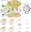
Network derived from the domain architectures of toxins. The central panel shows the network for all toxins in all species, whereas the lower panels show networks derived for major bacterial clades. The network is a directional graph with edges connecting neighboring domains in a polypeptide, in which the N-terminal domain is the source node, whereas the C-terminal domain is the target node. Edges are colored to match the source node color to illustrate the main direction of flow in the graph. Domains with similar properties are grouped together as shown.
Figure 13
Length distribution for predicted complete active toxins in different bacterial clades. Complete active toxins, as against cassettes, were identified based on characteristic marker domains for each of the distinct secretory systems associated with the toxin either in the same polypeptide or in gene neighborhoods (Table 1). The topmost row shows the combined statistics for all active toxins while other panels present the breakdown of these distributions based on secretory bacterial clades. The toxin length distribution is represented as beanplot[182] (e.g. left panel in the first row) and a raw histogram (top row, central panel) and clearly indicates the multimodal nature of toxin length. The barplot on the first row (rightmost panel) shows the frequencies of consecutive toxin and/or immunity gene pairs in theses genomes. Only pairs of gene encoded by the same strand where considered. The labels indicate whether an immunity protein (I) or a toxin (T) is encoded upstream or downstream of its neighbor in putative operons, e.g. TI corresponds to a pair where an immunity gene is preceded by a toxin gene. Note that the TI (toxin - > immunity) architecture is the most frequent pair observed in all graphs except for bacteroidetes/chlorobi and firmicutes, where the presence of polyimmunity loci inflates the II category. Dashed vertical lines correspond to the median protein length for the data on each panel, and the solid vertical lines over each beanplot correspond to the median length in that secretory system alone. The axes at the right of each panel contain the number of active toxins per secretory system.
Figure 14
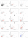
Scatterplots of the number of toxins versus number of immunity proteins per genome. In scatter plots, black or gray dots in the background represent all taxa, and red or blue dots correspond to taxa belonging to the clade or ecological properties described on each plot’s title. The dashed line corresponds to the diagonal (x = y) and the ellipses encircle taxa that are characterized by an excess of immunity proteins as discussed in the text.
Similar articles
- Polymorphic Toxins and Their Immunity Proteins: Diversity, Evolution, and Mechanisms of Delivery.
Ruhe ZC, Low DA, Hayes CS. Ruhe ZC, et al. Annu Rev Microbiol. 2020 Sep 8;74:497-520. doi: 10.1146/annurev-micro-020518-115638. Epub 2020 Jul 17. Annu Rev Microbiol. 2020. PMID: 32680451 Free PMC article. Review. - A novel immunity system for bacterial nucleic acid degrading toxins and its recruitment in various eukaryotic and DNA viral systems.
Zhang D, Iyer LM, Aravind L. Zhang D, et al. Nucleic Acids Res. 2011 Jun;39(11):4532-52. doi: 10.1093/nar/gkr036. Epub 2011 Feb 8. Nucleic Acids Res. 2011. PMID: 21306995 Free PMC article. - New players in the toxin field: polymorphic toxin systems in bacteria.
Jamet A, Nassif X. Jamet A, et al. mBio. 2015 May 5;6(3):e00285-15. doi: 10.1128/mBio.00285-15. mBio. 2015. PMID: 25944858 Free PMC article. Review. - Evolution of the deaminase fold and multiple origins of eukaryotic editing and mutagenic nucleic acid deaminases from bacterial toxin systems.
Iyer LM, Zhang D, Rogozin IB, Aravind L. Iyer LM, et al. Nucleic Acids Res. 2011 Dec;39(22):9473-97. doi: 10.1093/nar/gkr691. Epub 2011 Sep 3. Nucleic Acids Res. 2011. PMID: 21890906 Free PMC article. - Comprehensive analysis of the HEPN superfamily: identification of novel roles in intra-genomic conflicts, defense, pathogenesis and RNA processing.
Anantharaman V, Makarova KS, Burroughs AM, Koonin EV, Aravind L. Anantharaman V, et al. Biol Direct. 2013 Jun 15;8:15. doi: 10.1186/1745-6150-8-15. Biol Direct. 2013. PMID: 23768067 Free PMC article.
Cited by
- Novel transglutaminase-like peptidase and C2 domains elucidate the structure, biogenesis and evolution of the ciliary compartment.
Zhang D, Aravind L. Zhang D, et al. Cell Cycle. 2012 Oct 15;11(20):3861-75. doi: 10.4161/cc.22068. Epub 2012 Sep 14. Cell Cycle. 2012. PMID: 22983010 Free PMC article. - Analysis of two domains with novel RNA-processing activities throws light on the complex evolution of ribosomal RNA biogenesis.
Burroughs AM, Aravind L. Burroughs AM, et al. Front Genet. 2014 Dec 23;5:424. doi: 10.3389/fgene.2014.00424. eCollection 2014. Front Genet. 2014. PMID: 25566315 Free PMC article. - A new family of secreted toxins in pathogenic Neisseria species.
Jamet A, Jousset AB, Euphrasie D, Mukorako P, Boucharlat A, Ducousso A, Charbit A, Nassif X. Jamet A, et al. PLoS Pathog. 2015 Jan 8;11(1):e1004592. doi: 10.1371/journal.ppat.1004592. eCollection 2015 Jan. PLoS Pathog. 2015. PMID: 25569427 Free PMC article. - Identification of A Putative T6SS Immunity Islet in Salmonella Typhi.
Barretto LAF, Fowler CC. Barretto LAF, et al. Pathogens. 2020 Jul 11;9(7):559. doi: 10.3390/pathogens9070559. Pathogens. 2020. PMID: 32664482 Free PMC article. - Polymorphic Toxins and Their Immunity Proteins: Diversity, Evolution, and Mechanisms of Delivery.
Ruhe ZC, Low DA, Hayes CS. Ruhe ZC, et al. Annu Rev Microbiol. 2020 Sep 8;74:497-520. doi: 10.1146/annurev-micro-020518-115638. Epub 2020 Jul 17. Annu Rev Microbiol. 2020. PMID: 32680451 Free PMC article. Review.
References
- Rochat H, Martin-Eauclaire H. Animal toxins: facts and protocols. Basel Boston: Birkhauser Verlag; 2000.
- Keeler RF, Tu AT. Toxicology of plant and fungal compounds. New York: Dekker; 1991.
- Mackessy SP. Handbook of venoms and toxins of reptiles. Boca Raton: CRC Press; 2010.
- Alouf JE, Popoff MR. The comprehensive sourcebook of bacterial protein toxins. 3. Amsterdam; Boston: Elsevier Academic Press; 2006.
- Walsh C. Antibiotics: actions, origins, resistance. Washington, D.C.: ASM Press; 2003.
Publication types
MeSH terms
Substances
LinkOut - more resources
Full Text Sources
Other Literature Sources
Molecular Biology Databases


