Fiji: an open-source platform for biological-image analysis - PubMed (original) (raw)
. 2012 Jun 28;9(7):676-82.
doi: 10.1038/nmeth.2019.
Ignacio Arganda-Carreras, Erwin Frise, Verena Kaynig, Mark Longair, Tobias Pietzsch, Stephan Preibisch, Curtis Rueden, Stephan Saalfeld, Benjamin Schmid, Jean-Yves Tinevez, Daniel James White, Volker Hartenstein, Kevin Eliceiri, Pavel Tomancak, Albert Cardona
Affiliations
- PMID: 22743772
- PMCID: PMC3855844
- DOI: 10.1038/nmeth.2019
Fiji: an open-source platform for biological-image analysis
Johannes Schindelin et al. Nat Methods. 2012.
Abstract
Fiji is a distribution of the popular open-source software ImageJ focused on biological-image analysis. Fiji uses modern software engineering practices to combine powerful software libraries with a broad range of scripting languages to enable rapid prototyping of image-processing algorithms. Fiji facilitates the transformation of new algorithms into ImageJ plugins that can be shared with end users through an integrated update system. We propose Fiji as a platform for productive collaboration between computer science and biology research communities.
Figures
Figure 1. Fiji as a high-powered distribution of ImageJ
(a) The Fiji platform enables interaction with multidimentional image data in point-and-click interface identical to ImageJ. It simplifies transition from mathematical formulations coming from computer vision researchers to functional programs written by software engineers using version control and algorithmic libraries (schematic diagram of ImgLib design) and allows bioinformaticians to construct powerful image processing pipelines using scripting languages (Script Editor plugin screenshot). (b) The Fiji Updater provides a unique mechanism for releasing new algorithmic developments to the user community. Every time Fiji is launched it offers to download available updates from the main server in Dresden, Germany or from alternative update sites. New plugins approved by the Fiji developer community are instantaneously delivered to thousands of Fiji users worldwide or can be offered directly through user initiated update sites.
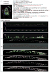
Figure 2. Scripting and ImgLib
(a) An example of a simple Jython script that achieves complex segmentation and 3d visualization drawing on the Fiji libraries steered by simple scripting commands. The task is to open a 3d RGB image (line 5) of a Drosophila first instar brain where cortex and neuropile glia are labeled in green by Nirvana-Gal4 and UASmcd8GFP, surface glial cells are labeled red with anti-repo antibody, and all nuclei are labeled blue with Sytox. The goal is to automatically count red surface glial cells using the Difference of Gaussian (DoG) detector (line 9) applying the constraints for cell size and labeling intensity (lines 3 and 4). These constraints are expressed as DoG sigma parameters (lines 7 and 8) by extracting image dimensions from metadata (line 6). The cell count is printed in the dialog box (line 10) and cells are subsequently displayed in the 4d viewer as red spheres of fixed diameter, overlaid with orthogonal view of the raw 3d images (lines 13–16). (b–j) Algorithms implemented using generic ImgLib constructs operate on images regardless of dimensionality. The figure shows the output of two ImgLib algorithms: Maximally Stable Extremal Regions (MSER) and again DoG. The 3d input image is a confocal stack of a C. elegans worm expressing nuclear marker. Scale bar 10 μm. (h). A slice from the stack is used as the 2d input image. Scale bar 10 μm. (e). A line segment from the slice is used as the 1d input image (b). The results of the DoG algorithm for 1d, 2d, and 3d are visualized in (c), (f), and (i). The results of the MSER algorithm for 1d, 2d, and 3d are shown in (d), (g), and (j). The algorithms are run on 1d (c,d), 2d (f,g), and 3d (i,j) input without changing a single line of code (see Supplementary Fig. 4). The nested MSER regions representing competing segmentation hypotheses for the nuclei are color coded (green, red, blue and magenta).
Figure 3. Fiji projects
(a–e) Stitching plugin for globally optimal registration of tiled 3d confocal images. The source data of Drosophila first instar larval nervous system (a) were registered using phase correlation with global optimization (b) and visualized in Fiji 4d Viewer (c). Four labeled neurons (colour coded in 4d Viewer (d)) were segmented using a manual segmentation plugin (Segmentation Editor) and their volumes were measured (e). (f–j) Globally optimal reconstruction of large ssTEM mosaics using TrakEM2 plugin. Schematic view of ssTEM mosaic, each square is an individual image tile and the independent sections are color coded (f). (g) Screenshot of a video visualizing the progress of global optimization for a single section. SIFT features are in cyan and the residual error signifying displacement of corresponding SIFT features at the current iteration is depicted by red lines. (h) Dual color overlay of two registered consecutive sections showing the entire hemisphere of the larval brain. Scale bar 10 μm. (i) Axonal profiles within a small part of a single section in the ssTEM dataset were manually segmented using TrakEM2. Each profile is labeled with a different color. Scale bar 0.5 μm. (j) Relationship between numbers of pre-synaptic partners and post-synaptic sites extracted manually with TrakEM2 from a micro-cube of the registered data. (k–o) Plugin suite for processing of multi-view SPIM data. (k) Schematic representation of multi-view SPIM imaging showing the 3d stacks of the same specimen acquired from different angles. (l) Progress of the global optimization of multi-view SPIM acquisition of Drosophila embryo. Corresponding geometric bead descriptors are colored according to their residual displacement at the current iteration of the optimizer. (m) The resulting reconstructed embryo at the 12th (top) and 13th (bottom) nuclear division cycle shown as 3d rendering in Fiji’s 3d viewer. Scale bar 50 μm. (n) Results of Difference of Gaussian segmentation of the nuclei marked with His-YFP; same stages as in (m). Each nucleus is marked with a different color. (o) Quantification of the nuclear counts at the 12th and 13th nuclear division in the embryo shown in (m) and (n).
Figure 4. Fiji usage
(a) A chart showing the number of unique visitors to Fiji wiki per month over the last three years. (b) World map overlaid with geolocations of computers that updated Fiji over a period of one week.
Similar articles
- The ImageJ ecosystem: Open-source software for image visualization, processing, and analysis.
Schroeder AB, Dobson ETA, Rueden CT, Tomancak P, Jug F, Eliceiri KW. Schroeder AB, et al. Protein Sci. 2021 Jan;30(1):234-249. doi: 10.1002/pro.3993. Epub 2020 Nov 20. Protein Sci. 2021. PMID: 33166005 Free PMC article. - Current challenges in open-source bioimage informatics.
Cardona A, Tomancak P. Cardona A, et al. Nat Methods. 2012 Jun 28;9(7):661-5. doi: 10.1038/nmeth.2082. Nat Methods. 2012. PMID: 22743770 - MorphoLibJ: integrated library and plugins for mathematical morphology with ImageJ.
Legland D, Arganda-Carreras I, Andrey P. Legland D, et al. Bioinformatics. 2016 Nov 15;32(22):3532-3534. doi: 10.1093/bioinformatics/btw413. Epub 2016 Jul 13. Bioinformatics. 2016. PMID: 27412086 - Quantitating the cell: turning images into numbers with ImageJ.
Arena ET, Rueden CT, Hiner MC, Wang S, Yuan M, Eliceiri KW. Arena ET, et al. Wiley Interdiscip Rev Dev Biol. 2017 Mar;6(2). doi: 10.1002/wdev.260. Epub 2016 Dec 2. Wiley Interdiscip Rev Dev Biol. 2017. PMID: 27911038 Review. - Open-source tools for immersive environmental visualization.
Sherman WR, Su S, McDonald PA, Mu Y, Harris F Jr. Sherman WR, et al. IEEE Comput Graph Appl. 2007 Mar-Apr;27(2):88-91. doi: 10.1109/mcg.2007.44. IEEE Comput Graph Appl. 2007. PMID: 17388206 Review. No abstract available.
Cited by
- Mlf mediates proteotoxic response via formation of cellular foci for protein folding and degradation in Giardia.
Vinopalová M, Arbonová L, Füssy Z, Dohnálek V, Samad A, Bílý T, Vancová M, Doležal P. Vinopalová M, et al. PLoS Pathog. 2024 Oct 21;20(10):e1012617. doi: 10.1371/journal.ppat.1012617. eCollection 2024 Oct. PLoS Pathog. 2024. PMID: 39432513 Free PMC article. - Kinetochores grip microtubules with directionally asymmetric strength.
Larson JD, Heitkamp NA, Murray LE, Popchock AR, Biggins S, Asbury CL. Larson JD, et al. J Cell Biol. 2025 Jan 6;224(1):e202405176. doi: 10.1083/jcb.202405176. Epub 2024 Nov 1. J Cell Biol. 2025. PMID: 39485274 Free PMC article. - Regeneration-specific promoter switching facilitates Mest expression in the mouse digit tip to modulate neutrophil response.
Jou V, Peña SM, Lehoczky JA. Jou V, et al. NPJ Regen Med. 2024 Oct 28;9(1):32. doi: 10.1038/s41536-024-00376-w. NPJ Regen Med. 2024. PMID: 39468052 Free PMC article. - Detailed cell-level analysis of sperm nuclear quality among the different hypo-osmotic swelling test (HOST) classes.
Bloch A, Rogers EJ, Nicolas C, Martin-Denavit T, Monteiro M, Thomas D, Morel H, Lévy R, Siffroi JP, Dupont C, Rouen A. Bloch A, et al. J Assist Reprod Genet. 2021 Sep;38(9):2491-2499. doi: 10.1007/s10815-021-02232-y. Epub 2021 Jun 2. J Assist Reprod Genet. 2021. PMID: 34076795 Free PMC article. - Optical Coherence Tomography Angiography-Based Quantitative Assessment of Morphologic Changes in Active Myopic Choroidal Neovascularization During Anti-vascular Endothelial Growth Factor Therapy.
Wang Y, Hu Z, Zhu T, Su Z, Fang X, Lin J, Chen Z, Su Z, Ye P, Ma J, Zhang L, Li J, Feng L, Sun CB, Zhang Z, Shentu X. Wang Y, et al. Front Med (Lausanne). 2021 May 7;8:657772. doi: 10.3389/fmed.2021.657772. eCollection 2021. Front Med (Lausanne). 2021. PMID: 34026789 Free PMC article.
References
- Turing AM. The chemical basis of morphogenesis. 1953. Bull Math Biol. 1990;52:153–197. discussion 119–152. - PubMed
- Altschul SF, Gish W, Miller W, Myers EW, Lipman DJ. Basic local alignment search tool. J Mol Biol. 1990;215:403–410. - PubMed
- Myers EW, et al. A whole-genome assembly of Drosophila. Science. 2000;287:2196–2204. - PubMed
- Collinet C, et al. Systems survey of endocytosis by multiparametric image analysis. Nature. 2010;464:243–249. - PubMed
Publication types
MeSH terms
LinkOut - more resources
Full Text Sources
Other Literature Sources
Molecular Biology Databases


