Withaferin A synergizes the therapeutic effect of doxorubicin through ROS-mediated autophagy in ovarian cancer - PubMed (original) (raw)
Withaferin A synergizes the therapeutic effect of doxorubicin through ROS-mediated autophagy in ovarian cancer
Miranda Y Fong et al. PLoS One. 2012.
Abstract
Application of doxorubicin (Dox) for the treatment of cancer is restricted due to its severe side effects. We used combination strategy by combining doxorubicin (Dox) with withaferin A (WFA) to minimize the ill effects of Dox. Treatment of various epithelial ovarian cancer cell lines (A2780, A2780/CP70 and CaOV3) with combination of WFA and Dox (WFA/DOX) showed a time- and dose-dependent synergistic effect on inhibition of cell proliferation and induction of cell death, thus reducing the dosage requirement of Dox. Combination treatment resulted in a significant enhancement of ROS production resulting in immense DNA damage, induction of autophagy analyzed by transmission electron microscope and increase in expression of autophagy marker LC3B, and culminated in cell death analyzed by cleaved caspase 3. We validated combination therapy on tumor growth using an in vitro 3Dimension (3D) tumor model and the more classic in vivo xenograft model of ovarian cancer. Both tumor models showed a 70 to 80% reduction in tumor growth compared to control or animals treated with WFA or Dox alone. Immunohistochemical analysis of the tumor tissues from animals treated with WFA/Dox combination showed a significant reduction in cell proliferation and formation of microvessels accompanied by increased in LC3B level, cleaved caspase 3, and DNA damage. Taken together, our data suggest that combining WFA with Dox decreases the dosage requirement of Dox, therefore, minimizing/eliminating the severe side effects associated with high doses of DOX, suggesting the application of this combination strategy for the treatment of ovarian and other cancers with no or minimum side effects.
Conflict of interest statement
Competing Interests: Raj K Singh is employed by Vivo Biosciences Inc. There are no patents, products in development or marketed products to declare. This does not alter the authors‘ adherence to all the PLoS ONE policies on sharing data and materials.
Figures
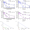
Figure 1. Cell proliferation of A2780 (A–B) and A2780/CP70 (C–D) cells on treatment with Dox and WFA, both alone or combination of WFA/DOX using MTT assays.
A2780 and A2780/CP cells were plated into 96 well plates. After 24 h of plating, cells were treated with various concentrations of Dox and WFA, both alone or in combination. After 48 h of treatment, cell viability was assayed using MTT assays. Values shown are mean ±SD of four independent experiments. *P<0.05 compared to control, #p<0.05 compared to Dox or WFA alone. (E) Isobologram analysis of A2780 cells (n = 4) and (F) A2780/CP70 (n = 4) using 7 doses of Dox and WFA maintained at a constant ratio and cell death was assayed by MTT assays. Results were analyzed with CalcuSyn software.
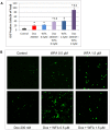
Figure 2. ROS generation in A2780 cells.
A2780 cells were plated into glass bottom dishes. After 24 h of plating, cells were treated with Dox and WFA, both alone or combination of WFA/DOX as described in Figure 1. After 24 h of treatment, medium was replaced with fresh medium containing H2DCFDA and incubated for 30 min. Cells were rinsed with PBS and examined under confocal microscope. (A) ROS positive cells (green color) were counted based on 3 low power fields. Mean ±SD. P values were determined by ANOVA analysis followed by Student-Newman-Keuls test for multiple comparisons. *P<0.05 from control, $P<0.05 compared to Dox, &P<0.05 compared to WFA. (B) Confocal microscopy analysis of cells indicating generation of ROS (green color).
Figure 3. Effect of non-enzymatic ROS antioxidant NAC on A2780 cell proliferation after 48 h of treatment.
A2780 cells were co-treated with NAC and Dox, WFA or combination of WFA/WFA for 48 h. Cell proliferation was determined using MTT assays. Values shown are mean ±SD of three independent experiments. P<0.05 compared to control, #P<0.05 compared to no NAC and NAC.
Figure 4. Effect of enzymatic antioxidant SOD (100 units/ml) on A2780 cell proliferation after 48 h of treatment. A2780 cells were co-treated with SOD and Dox, WFA or combination of WFA/Dox for 48 h.
Cell proliferation was determined using MTT assays. Values shown are mean ±SD of three independent experiments. *P<0.05 compared to control, #P<0.05 compared to no SOD and SOD.
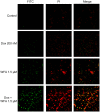
Figure 5. DNA damage (TUNEL) assay of A2780 cells after 24 h of treatment.
A2780 cells were treated with Dox, WFA both alone or combination of WFA/Dox. After 24 h of treatment, DNA damage was analyzed using TUNNEL assays. Images were obtained using confocal microscopy at 20X magnification.
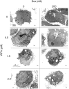
Figure 6. Analysis of autophagy using transmission electron microscope (TEM).
A2780 cells were treated with Dox and WFA, both alone or combination of WFA/Dox. After 24 h of treatment, cells were rinsed with PBS, fixed and processed for TEM analysis. Electron microscopic images at 5,600X magnification are shown.
Figure 7. Western blot analysis of autophagy pathway of A2780 cells treated for 24 hr. A2780 cells treated with Dox and WFA, both alone or combination of WFA/Dox.
After 24 h of treatment, cells were washed with PBS and lysed. Western blot analysis was performed for LC3B, Caspase 3 and GAPDH proteins.
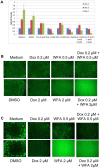
Figure 8. In vitro analysis of tumor growth using 3D tumor model.
(A) A2780 Cells were combined with Hubiogel® in a 1∶4 ratio and grown from 10 µL beads. Tumors were treated with Dox and WFA, both alone or combination of WFA/Dox. Tumors were treated twice/week by replacing the medium with medium containing fresh agent. Tumor growth was measured using MTT assays after day 3 or 7 of treatment. (B–C) Tumors after treatment were incubated with calcein AM for 30 min, and images were taken using fluorescence microscope after 3 days of treatment (B) or 7 days of treatment (C).
Figure 9. S.C. tumors were generated in nude mice and treated with PBS, Vehicle, Dox 1 mg/kg, Dox 9 mg/kg, WFA 2 mg/kg or Dox 1 mg/kg plus WFA 2 mg/kg.
(A) Tumors growth was measured from day 20–32 (post-cell injection) and treated every other day *P<0.05. (B) Tumor weight at day 32 collected immediately after sacrificing the animals.
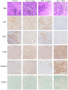
Figure 10. Immunohistochemistry compilation of tumor tissues developed with DAB (brown) and counterstained with hematoxylin to stain nuclei (blue).
Negative control samples were tissues without primary antibody. TUNEL assay was performed using ApopTag Plus Peroxidase Apoptosis Detection Kit.
Figure 11. Schematic summary of mechanisms of cell death induced by Dox/WFA combination treatment.
Similar articles
- Synergistic cytotoxic action of cisplatin and withaferin A on ovarian cancer cell lines.
Kakar SS, Jala VR, Fong MY. Kakar SS, et al. Biochem Biophys Res Commun. 2012 Jul 13;423(4):819-25. doi: 10.1016/j.bbrc.2012.06.047. Epub 2012 Jun 16. Biochem Biophys Res Commun. 2012. PMID: 22713472 Free PMC article. - Valproic Acid Induces Endocytosis-Mediated Doxorubicin Internalization and Shows Synergistic Cytotoxic Effects in Hepatocellular Carcinoma Cells.
Saha SK, Yin Y, Kim K, Yang GM, Dayem AA, Choi HY, Cho SG. Saha SK, et al. Int J Mol Sci. 2017 May 12;18(5):1048. doi: 10.3390/ijms18051048. Int J Mol Sci. 2017. PMID: 28498322 Free PMC article. - DOXIL when combined with Withaferin A (WFA) targets ALDH1 positive cancer stem cells in ovarian cancer.
Kakar SS, Worth CA, Wang Z, Carter K, Ratajczak M, Gunjal P. Kakar SS, et al. J Cancer Stem Cell Res. 2016;4:e1002. doi: 10.14343/JCSCR.2016.4e1002. Epub 2016 Apr 19. J Cancer Stem Cell Res. 2016. PMID: 27668267 Free PMC article. - Theanine and glutamate transporter inhibitors enhance the antitumor efficacy of chemotherapeutic agents.
Sugiyama T, Sadzuka Y. Sugiyama T, et al. Biochim Biophys Acta. 2003 Dec 5;1653(2):47-59. doi: 10.1016/s0304-419x(03)00031-3. Biochim Biophys Acta. 2003. PMID: 14643924 Review. - Doxorubicin-An Agent with Multiple Mechanisms of Anticancer Activity.
Kciuk M, Gielecińska A, Mujwar S, Kołat D, Kałuzińska-Kołat Ż, Celik I, Kontek R. Kciuk M, et al. Cells. 2023 Feb 19;12(4):659. doi: 10.3390/cells12040659. Cells. 2023. PMID: 36831326 Free PMC article. Review.
Cited by
- Withaferin A ameliorates ovarian cancer-induced cachexia and proinflammatory signaling.
Straughn AR, Kakar SS. Straughn AR, et al. J Ovarian Res. 2019 Nov 25;12(1):115. doi: 10.1186/s13048-019-0586-1. J Ovarian Res. 2019. PMID: 31767036 Free PMC article. - Protective effect of flavonoids from Rosa roxburghii Tratt on myocardial cells via autophagy.
Yuan H, Wang Y, Chen H, Cai X. Yuan H, et al. 3 Biotech. 2020 Feb;10(2):58. doi: 10.1007/s13205-019-2049-1. Epub 2020 Jan 22. 3 Biotech. 2020. PMID: 32015954 Free PMC article. - GREB1L overexpression is associated with good clinical outcomes in breast cancer.
Dong K, Geng C, Zhan X, Sun Z, Pu Q, Li P, Song H, Zhao G, Gao H. Dong K, et al. Eur J Med Res. 2023 Nov 14;28(1):510. doi: 10.1186/s40001-023-01483-y. Eur J Med Res. 2023. PMID: 37964281 Free PMC article. - Iron-Chelated Polydopamine Decorated Doxorubicin-Loaded Nanodevices for Reactive Oxygen Species Enhanced Cancer Combination Therapy.
Li XJ, Li WT, Li ZH, Zhang LP, Gai CC, Zhang WF, Ding DJ. Li XJ, et al. Front Pharmacol. 2019 Feb 6;10:75. doi: 10.3389/fphar.2019.00075. eCollection 2019. Front Pharmacol. 2019. PMID: 30787876 Free PMC article. - Withaferin A: From Ancient Remedy to Potential Drug Candidate.
Sultana T, Okla MK, Ahmed M, Akhtar N, Al-Hashimi A, Abdelgawad H, Haq IU. Sultana T, et al. Molecules. 2021 Dec 20;26(24):7696. doi: 10.3390/molecules26247696. Molecules. 2021. PMID: 34946778 Free PMC article. Review.
References
- Siegel R, Naishadham D, Jemal A (2012) Cancer statistics, 2012. CA Cancer J Clin 62: 10–29. - PubMed
- Pfisterer J, Ledermann JA (2006) Management of platinum-sensitive recurrent ovarian cancer. Semin Oncol 33: S12–16. - PubMed
- Singal PK, Li T, Kumar D, Danelisen I, Iliskovic N (2000) Adriamycin-induced heart failure: mechanism and modulation. Mol Cell Biochem 207: 77–86. - PubMed
- Carvalho C, Santos RX, Cardoso S, Correia S, Oliveira PJ, et al. (2009) Doxorubicin: the good, the bad and the ugly effect. Curr Med Chem 16: 3267–3285. - PubMed
Publication types
MeSH terms
Substances
LinkOut - more resources
Full Text Sources
Medical
Research Materials
