Characteristics of multi-organ lymphangiectasia resulting from temporal deletion of calcitonin receptor-like receptor in adult mice - PubMed (original) (raw)
Characteristics of multi-organ lymphangiectasia resulting from temporal deletion of calcitonin receptor-like receptor in adult mice
Samantha L Hoopes et al. PLoS One. 2012.
Abstract
Adrenomedullin (AM) and its receptor complexes, calcitonin receptor-like receptor (Calcrl) and receptor activity modifying protein 2/3, are highly expressed in lymphatic endothelial cells and are required for embryonic lymphatic development. To determine the role of Calcrl in adulthood, we used an inducible Cre-loxP system to temporally and ubiquitously delete Calcrl in adult mice. Following tamoxifen injection, Calcrl(fl/fl)/CAGGCre-ER™ mice rapidly developed corneal edema and inflammation that was preceded by and persistently associated with dilated corneoscleral lymphatics. Lacteals and submucosal lymphatic capillaries of the intestine were also dilated, while mesenteric collecting lymphatics failed to properly transport chyle after an acute Western Diet, culminating in chronic failure of Calcrl(fl/fl)/CAGGCre-ER™ mice to gain weight. Dermal lymphatic capillaries were also dilated and chronic edema challenge confirmed significant and prolonged dermal lymphatic insufficiency. In vivo and in vitro imaging of lymphatics with either genetic or pharmacologic inhibition of AM signaling revealed markedly disorganized lymphatic junctional proteins ZO-1 and VE-cadherin. The maintenance of AM signaling during adulthood is required for preserving normal lymphatic permeability and function. Collectively, these studies reveal a spectrum of lymphatic defects in adult Calcrl(fl/fl)/CAGGCre-ER™ mice that closely recapitulate the clinical symptoms of patients with corneal, intestinal and peripheral lymphangiectasia.
Conflict of interest statement
Competing Interests: The authors have declared that no competing interests exist.
Figures
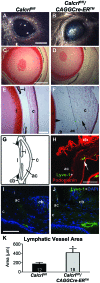
Figure 1. Acute-onset eye phenotype, eye inflammation, edema, and enlarged lymphatic vessels in Calcrlfl/fl/CAGGCre-ER™ mice. A,B,
Gross eye images indicating normal appearance of the control Calcrlfl/fl mice (A) and the distinct color change and disruption of the cornea of Calcrlfl/fl/CAGGCre-ER™ (B), (scale = 2 mm). C,D, Hematoxylin and eosin staining of mouse eyes indicating normal histology in Calcrlfl/fl (C) and disruption of the cornea in Calcrlfl/fl/CAGG-CreER™ mice (D), (4x objective,scale = 500 µm). E,F, Higher magnification of histological sections of eyes from Calcrlflf/fl mice (E) as compared to Calcrlfl/fl/CAGGCre-ER™ mice (F) exhibiting corneal edema (arrow) and inflammation (arrowhead) (10x objective, scale = 200 µm). Gross anatomy and histology images are representative from Calcrlflf/fl mice (n = 8) and Calcrlfl/fl/CAGGCre-ER™ mice (n = 9). G, Eye diagram indicating the location of components of the eye (l = lens, c = cornea, ac = anterior chamber, cb = ciliary body, i = iris). H, Lymphatic markers expressed in the eye shown by podoplanin(red) and Lyve-1(green) staining in a control mouse eye (20x objective, scale = 100 µm). I,J, Visualization of lymphatic vessels at the corneoscleral junction in the Calcrlflf/fl (I) and Calcrlfl/fl/CAGGCre-ER™ mice (J) indicating enlarged lymphatic vessels in Calcrlfl/fl/CAGGCre-ER™ mice (Lyve-1 = green; DAPI = blue; 20x objective, scale = 100 µm). K, Graph representing increased lymphatic vessel area at the corneoscleral junction in Calcrlfl/fl/CAGGCre-ER™ mice compared to control mice calculated using Image J software(*p<0.015). Mice used were 3–4 months of age.
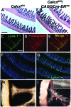
Figure 2. Dilated lacteals and submucosal lymphatics in Calcrlfl/fl/CAGGCre-ER™ mice and chyle-filled lymphatics after short-term Western diet. A,B,
Hematoxylin and eosin staining of mouse intestine showing normal histology in both Calcrlfl/fl(A) and Calcrlfl/fl/CAGGCre-ER™ mice(B) (6.3x objective, scale = 500 µm). C,D,E, Lymphatic marker expression in the lacteals and submucosal lymphatic vessels in wildtype mouse. Image was obtained from the jejunum of the intestine. Lyve-1 (C,green) and podoplanin(D,red) colocalize in the lymphatic vessels as seen in the merged image(E) (20x objective; scale = 100 µm). F,G Lyve-1(green) and DAPI(blue) staining in Calcrlfl/fl(F) and Calcrlfl/fl/CAGGCre-ER™ (G) mice indicating dilated lacteals and submucosal lymphatic vessels with temporal deletion of Calcrl in the jejunum of the intestine (4x objective, scale = 500 µm). Histology and immunofluorescent images are representative from Calcrlflf/fl mice (n = 7) and Calcrlfl/fl/CAGGCre-ER™ mice (n = 6). H,I, Chyle-filled mesenteric collecting lymphatic vessels in Calcrlfl/fl/CAGGCre-ER™ mice (I) relative to non-chyle filled vessels in control animals (H). Valves are distinctly visible in Calcrlfl/fl/CAGGCre-ER™ mice (arrows; inset refers to enlarged image of valve; scale = 3 mm; n = 4 per genotype). Mice used were 6–8 months of age.
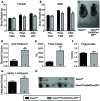
Figure 3. Calcrlfl/fl/CAGGCre-ER™ mice exhibit reduced body weight due to impaired lipid absorption. A,B,
Graphs of female(A) and male(B) body weights before injection of tamoxifen (Pre-TAM; 3–4 months of age), after injection of tamoxifen (Post-TAM; 3–4 months after), and after 1½ weeks on Western Diet (After WD). Both male and female Calcrlfl/fl/CAGGCre-ER™ mice were significantly smaller than Calcrlfl/fl mice after tamoxifen injection and after Western Diet. C, Image of Calcrlfl/fl and Calcrlfl/fl/CAGGCre-ER™ mice after 1½ weeks Western Diet. D, Acid steatocrit measurement in fecal samples from Calcrlfl/fl and Calcrlfl/fl/CAGGCre-ER™ mice after Western Diet for 1½ weeks indicating increased lipid excretion in the experimental mice. E, Lipase measurements in fecal samples from Calcrlfl/fl and Calcrlfl/fl/CAGGCre-ER™ mice on Western Diet for 1 ½ weeks indicating increased fecal lipase in experimental mice. F, Total triglyceride levels in Calcrlfl/fl and Calcrlfl/fl/CAGGCre-ER™ mice. G, Alpha-1 antitrypsin levels in fecal samples from Calcrlfl/fl and Calcrlfl/fl/CAGGCre-ER™ mice after 1½ weeks Western diet indicating lower levels in Calcrlfl/fl/CAGGCre-ER™ mice. (Integrated density values are scaled and should be multiplied by 105). H, Image of dot blot assay for alpha-1 antitrypsin in Calcrlfl/fl and Calcrlfl/fl/CAGGCre-ER™ mice fecal samples after Western diet (1∶2000 dilution of samples). (*p<0.03;**p<0.002).
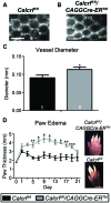
Figure 4. Dilated dermal lymphatic capillaries with exacerbated and prolonged edema. A,B,
Images of dermal lymphatic capillaries in the tail of Calcrlfl/fl(A) and Calcrlfl/fl/CAGGCre-ER™ (B) mice indicating increased diameter of these lymphatic vessels in Calcrlfl/fl/CAGGCre-ER™ mice (scale = 0.5 mm). C, Graphic representation of the increase in vessel diameter in the Calcrlfl/fl/CAGGCre-ER™ mice with respect to Calcrlfl/fl mice (*p≤0.05). D, Edema formation assay using hindpaw injections of CFA (4 µg/µl on Day 0). Assessment of paw thickness over 3 weeks (n = 5 for Calcrlfl/fl and n = 4 for Calcrlfl/fl/CAGGCre-ER™ mice) indicated enhanced and prolonged edema in Calcrlfl/fl/CAGGCre-ER™ mice relative to control mice (***p<0.05, **p<0.01, *p<0.001). Representative images of CFA-injected hindpaws at Day 11 for Calcrlfl/fl and Calcrlfl/fl/CAGGCre-ER™ mice (scale = 3 mm). Mice used were 6–8 months of age.
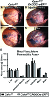
Figure 5. Increased lymphatic vascular permeability without change to blood vascular permeability in Calcrlfl/fl/CAGGCre-ER™ mice. A,B,C,D,
In vivo lymphatic permeability assay assessing the leakage of Evan’s blue dye from the dermal lymphatic vessels in the ear. Images represent Evan’s blue dye location directly after injection of the dye and 5 minutes post injection. There is an increase in leakage of the dye from the Calcrlfl/fl/CAGGCre-ER™ mice (B,D) relative to Calcrlfl/fl mice (A,C). Depicted are representative images from four independent experiments (mice 6–8 months of age). E, Blood vascular permeability assay indicating there is no difference in permeability between genotypes in the various tissues (n = 4 per genotype for each tissue).
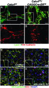
Figure 6. Inhibition of AM signaling disrupts lymphatic endothelial cell-cell junctions. A,B,C,D,
Confocal images of VE-Cadherin (red) and Lyve-1(green) expression in mesenteric lymphatic vessels of Calcrlfl/fl (A,C) and Calcrlfl/fl/CAGGCre-ER™ mice (B,D) (scale = 10 µm). Boxed region depicted in C and D. Junctional protein, VE-cadherin, is disorganized in Calcrlfl/fl/CAGGCre-ER™ mice relative to Calcrlfl/fl mice (representative images from n = 4 per genotype, age 6–8 months). E,F,G,H, Lymphatic endothelial cells stained with VE-Cadherin (green), ZO-1 (red) and DAPI (blue) after various treatments including a no treatment control (A), 10 nm AM (B), 1 µm AM22-52 (C), AM+AM22-52 (D) (arrow refers to inset region). Disorganization of cell-cell junctions occurs with inhibitor treatment (AM22-52) as compared to AM treatment. (VE-cadherin = red, Lyve-1 = green, DAPI = blue, 40x objective, scale = 100 µm; representative images from 3 independent experiments).
Similar articles
- Lymphatic deletion of calcitonin receptor-like receptor exacerbates intestinal inflammation.
Davis RB, Kechele DO, Blakeney ES, Pawlak JB, Caron KM. Davis RB, et al. JCI Insight. 2017 Mar 23;2(6):e92465. doi: 10.1172/jci.insight.92465. JCI Insight. 2017. PMID: 28352669 Free PMC article. - VE-Cadherin Is Required for Cardiac Lymphatic Maintenance and Signaling.
Harris NR, Nielsen NR, Pawlak JB, Aghajanian A, Rangarajan K, Serafin DS, Farber G, Dy DM, Nelson-Maney NP, Xu W, Ratra D, Hurr SH, Qian L, Scallan JP, Caron KM. Harris NR, et al. Circ Res. 2022 Jan 7;130(1):5-23. doi: 10.1161/CIRCRESAHA.121.318852. Epub 2021 Nov 18. Circ Res. 2022. PMID: 34789016 Free PMC article. - Adrenomedullin signaling is necessary for murine lymphatic vascular development.
Fritz-Six KL, Dunworth WP, Li M, Caron KM. Fritz-Six KL, et al. J Clin Invest. 2008 Jan;118(1):40-50. doi: 10.1172/JCI33302. J Clin Invest. 2008. PMID: 18097475 Free PMC article. - Shared and separate functions of the RAMP-based adrenomedullin receptors.
Kuwasako K, Kitamura K, Nagata S, Hikosaka T, Takei Y, Kato J. Kuwasako K, et al. Peptides. 2011 Jul;32(7):1540-50. doi: 10.1016/j.peptides.2011.05.022. Epub 2011 May 27. Peptides. 2011. PMID: 21645567 Review. - Regulation of cardiovascular development and homeostasis by the adrenomedullin-RAMP system.
Shindo T, Tanaka M, Kamiyoshi A, Ichikawa-Shindo Y, Kawate H, Yamauchi A, Sakurai T. Shindo T, et al. Peptides. 2019 Jan;111:55-61. doi: 10.1016/j.peptides.2018.04.004. Epub 2018 Apr 22. Peptides. 2019. PMID: 29689347 Review.
Cited by
- Adrenomedullin in lymphangiogenesis: from development to disease.
Klein KR, Caron KM. Klein KR, et al. Cell Mol Life Sci. 2015 Aug;72(16):3115-26. doi: 10.1007/s00018-015-1921-3. Epub 2015 May 8. Cell Mol Life Sci. 2015. PMID: 25953627 Free PMC article. Review. - Calcitonin-Receptor-Like Receptor Signaling Governs Intestinal Lymphatic Innervation and Lipid Uptake.
Davis RB, Ding S, Nielsen NR, Pawlak JB, Blakeney ES, Caron KM. Davis RB, et al. ACS Pharmacol Transl Sci. 2019 Jan 29;2(2):114-121. doi: 10.1021/acsptsci.8b00061. eCollection 2019 Apr 12. ACS Pharmacol Transl Sci. 2019. PMID: 32219216 Free PMC article. - Lymphatic deletion of calcitonin receptor-like receptor exacerbates intestinal inflammation.
Davis RB, Kechele DO, Blakeney ES, Pawlak JB, Caron KM. Davis RB, et al. JCI Insight. 2017 Mar 23;2(6):e92465. doi: 10.1172/jci.insight.92465. JCI Insight. 2017. PMID: 28352669 Free PMC article. - Mechanisms and functions of intestinal vascular specialization.
Bernier-Latmani J, González-Loyola A, Petrova TV. Bernier-Latmani J, et al. J Exp Med. 2024 Jan 1;221(1):e20222008. doi: 10.1084/jem.20222008. Epub 2023 Dec 5. J Exp Med. 2024. PMID: 38051275 Free PMC article. Review. - Standardizing protocols dealing with growth hormone receptor gene disruption in mice using the Cre-lox system.
Duran-Ortiz S, Bell S, Kopchick JJ. Duran-Ortiz S, et al. Growth Horm IGF Res. 2018 Oct-Dec;42-43:52-57. doi: 10.1016/j.ghir.2018.08.003. Epub 2018 Aug 29. Growth Horm IGF Res. 2018. PMID: 30195091 Free PMC article.
References
- Stacker SA, Farnsworth RH, Karnezis T, Shayan R, Smith DP, et al.. (2007) Molecular pathways for lymphangiogenesis and their role in human disease. Novartis Found Symp 281: 38–43; discussion 44–53, 208–209. - PubMed
- Stackert RA, Bursik K (2006) Ego development and the therapeutic goal-setting capacities of mentally ill adults. Am J Psychother 60: 357–374. - PubMed
- McColl BK, Paavonen K, Karnezis T, Harris NC, Davydova N, et al. (2007) Proprotein convertases promote processing of VEGF-D, a critical step for binding the angiogenic receptor VEGFR-2. FASEB J 21: 1088–1098. - PubMed
- Radhakrishnan K, Rockson SG (2008) The clinical spectrum of lymphatic disease. Ann N Y Acad Sci 1131: 155–184. - PubMed
- Rockson SG (2008) Diagnosis and management of lymphatic vascular disease. J Am Coll Cardiol 52: 799–806. - PubMed
Publication types
MeSH terms
Substances
LinkOut - more resources
Full Text Sources
Medical
Molecular Biology Databases
