Dynamic reorganization of metabolic enzymes into intracellular bodies - PubMed (original) (raw)
Review
Dynamic reorganization of metabolic enzymes into intracellular bodies
Jeremy D O'Connell et al. Annu Rev Cell Dev Biol. 2012.
Abstract
Both focused and large-scale cell biological and biochemical studies have revealed that hundreds of metabolic enzymes across diverse organisms form large intracellular bodies. These proteinaceous bodies range in form from fibers and intracellular foci--such as those formed by enzymes of nitrogen and carbon utilization and of nucleotide biosynthesis--to high-density packings inside bacterial microcompartments and eukaryotic microbodies. Although many enzymes clearly form functional mega-assemblies, it is not yet clear for many recently discovered cases whether they represent functional entities, storage bodies, or aggregates. In this article, we survey intracellular protein bodies formed by metabolic enzymes, asking when and why such bodies form and what their formation implies for the functionality--and dysfunctionality--of the enzymes that comprise them. The panoply of intracellular protein bodies also raises interesting questions regarding their evolution and maintenance within cells. We speculate on models for how such structures form in the first place and why they may be inevitable.
Figures
Figure 1
Bacterial microcompartments as exemplified by carboxysomes. (a) Transmission electron micrographs of Halothiobacillus neapolitanus cells showing carboxysomes (arrows) as polyhedral, protein-dense bodies. Adapted from Yeates et al. (2007). (b) The major shell protein (CsoS1ABC in H. neapolitanus) is a hexagonal subunit that oligomerizes into massive sheets that are bent into joined facets at the vertices by a second, pentagonal protein (CsoS4AB) to complete the shell. The interior of the shell is packed with RuBisCO and carbonic anhydrase to maximize CO2 capture. Adapted from Bonacci et al. (2011). (c) Fluorescence microscopy of carboxysomes (green) shows that their in vivo location within cyanobacteria is regulated such that they are centrally aligned and evenly spaced roughly 0.5 μm apart. Adapted from Savage et al. (2010).
Figure 2
Examples of microbodies visualized by thin-section transmission electron microscopy. (a) Peroxisomes (arrow) within sunflower cotyledon mesophyll cells. Crystalline inclusion bodies are formed from catalase, as shown by the immunogold nanoparticle localization (black dots). Adapted from Tenberge & Eising (1995) and Pavelka & Roth (2010). (b) Aspergillus nidulans showing a Woronin body filled with a HEX-1 protein crystal. Adapted from Yuan et al. (2003). Abbreviations: C, crystalline inclusion bodies; PM, peroxisomal matrix.
Figure 3
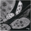
Green fluorescent protein C-terminal cytidine triphosphate (GFP-CTP) synthase fibers within Drosophila egg chamber cells. The fibers exhibit a diverse range of lengths. Adapted from Liu (2010).
Figure 4
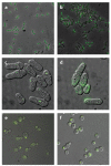
Hundreds of foci- and fiber-forming proteins have been discovered in systematic protein localization screens; most of these intracellular bodies are still largely uncharacterized. (a,b) Representative foci (ACCase β-subunit)- and fiber (UDP-N-acetylmuramate-alanine ligase)-forming green fluorescent protein (GFP) fusion proteins, respectively, from Caulobacter crescentus. Adapted from Werner et al. (2009). (c,d) Representative foci (Ade4)- and fiber (Pil1)-forming yellow fluorescent protein-fusion proteins, respectively, from Schizosaccharomyces pombe. Adapted from Matsuyama et al. (2006). (e,f) Representative foci (Gln1p)- and fiber (Asn2p)-forming GFP-fusion proteins, respectively, from Saccharomyces cerevisiae. Adapted from Narayanaswamy et al. (2009).
Figure 5
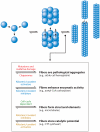
Proteins that assemble into symmetric quaternary structures should in principle have a higher propensity to form fibers, because effectors, whether allosteric, covalent, or mutational, that enhance binding between the oligomeric faces may be multiplied around the axis of symmetry, leading to enhanced fiber stability. The types of effectors and their contribution to fiber stability or destabilization can inform about an enzymatic fiber’s role within cells.
Figure 6
A sampling of metabolic enzymes that self-assemble into fibers. The quaternary structure of each enzyme is illustrated schematically (top row), following 90° rotation (middle row), and imaged in fiber form by electron microscopy (bottom row). (a) Acetyl-CoA carboxylase: crystal structure of Streptomyces coelicolor acetyl-CoA carboxylase β-subunit, PDB ID: 1XO6 (Diacovich et al. 2004) and electron micrograph of rat liver acetyl-CoA carboxylase (Nelson et al. 2008). (b) β-glucosidase: crystal structure of wheat β-glucosidase, PDB ID: 2DGA (Sue et al. 2006), and electron micrograph of oat β-glucosidase (Kim et al. 2005). (c) Glutamine synthetase: crystal structure of Escherichia coli glutamine synthetase, PDB ID: 1FY (Gill & Eisenberg 2001), and electron micrograph of E. coli glutamine synthetase (Frey et al. 1975). (d) Glutamate dehydrogenase: crystal structure of Clostridium symbiosum glutamate dehydrogenase, PDB ID: 1BGV (Stillman et al. 1993), and electron micrograph of cow liver glutamate dehydrogenase (Josephs & Borisy 1972). Scale not provided. (e) CTP synthase: human CTP synthase 2, PDB ID: 3IHL (M. Moche et al., unpublished data), and electron micrograph of Drosophila CTP synthase (Liu 2010). (f) Inosine monophosphate dehydrogenase: crystal structure of human type II inosine monophosphate dehydrogenase, PDB ID: 1NF7 (D. Risal, M.D. Strickler, B.M. Goldstein, unpublished data), and electron micrograph of human type II inosine monophosphate dehydrogenase (Ingerson-Mahar et al. 2010). (g) Human sickle-cell mutant hemoglobin: crystal structure, PDB ID: 2HBS (Harrington et al. 1997), and electron micrograph human sickle-cell mutant hemoglobin: (Ohtsuki et al. 1977). All crystal structure images created with Jmol.
Figure 7
An illustration of the analogous effects on cell morphology by cytidine triphosphate (CTP) synthase and sickle-cell mutant hemoglobin (HbS). (a) Bright field images of Caulobacter crescentus cells depleted of CTP synthase show severe bending, some to the point of circularization. (b) Cells overexpressing CTP synthase are straightened markedly. (c) Transmission electron micrograph of a C. crescentus cell with CTP synthase fibers (arrows) along the cell wall, altering cell morphology. Panels a_–_c adapted from Ingerson-Mahar et al. (2010). (d) Analogous images of red blood cells showing their normal, round morphology when oxygenated, and (e) their highly straightened morphology when the deoxygenated HbS forms into fibers. Adapted from Kaul et al. (1983). (f) Transmission electron micrograph showing HbS fibers (arrows) along the cell walls, altering cell morphology. Adapted from Döbler & Bertles (1968).
Similar articles
- Cellular stress leads to the formation of membraneless stress assemblies in eukaryotic cells.
van Leeuwen W, Rabouille C. van Leeuwen W, et al. Traffic. 2019 Sep;20(9):623-638. doi: 10.1111/tra.12669. Epub 2019 Jul 30. Traffic. 2019. PMID: 31152627 Free PMC article. Review. - Protein kinases are associated with multiple, distinct cytoplasmic granules in quiescent yeast cells.
Shah KH, Nostramo R, Zhang B, Varia SN, Klett BM, Herman PK. Shah KH, et al. Genetics. 2014 Dec;198(4):1495-512. doi: 10.1534/genetics.114.172031. Epub 2014 Oct 23. Genetics. 2014. PMID: 25342717 Free PMC article. - Inclusion body anatomy and functioning of chaperone-mediated in vivo inclusion body disassembly during high-level recombinant protein production in Escherichia coli.
Rinas U, Hoffmann F, Betiku E, Estapé D, Marten S. Rinas U, et al. J Biotechnol. 2007 Jan 1;127(2):244-57. doi: 10.1016/j.jbiotec.2006.07.004. Epub 2006 Jul 16. J Biotechnol. 2007. PMID: 16945443 - Yeast peroxisomes: How are they formed and how do they grow?
Akşit A, van der Klei IJ. Akşit A, et al. Int J Biochem Cell Biol. 2018 Dec;105:24-34. doi: 10.1016/j.biocel.2018.09.019. Epub 2018 Sep 27. Int J Biochem Cell Biol. 2018. PMID: 30268746 Review. - Patterns of indirect protein interactions suggest a spatial organization to metabolism.
Pérez-Bercoff Å, McLysaght A, Conant GC. Pérez-Bercoff Å, et al. Mol Biosyst. 2011 Nov;7(11):3056-64. doi: 10.1039/c1mb05168g. Epub 2011 Aug 31. Mol Biosyst. 2011. PMID: 21881679
Cited by
- Nanoreactor Design Based on Self-Assembling Protein Nanocages.
Ren H, Zhu S, Zheng G. Ren H, et al. Int J Mol Sci. 2019 Jan 30;20(3):592. doi: 10.3390/ijms20030592. Int J Mol Sci. 2019. PMID: 30704048 Free PMC article. Review. - Reversible, functional amyloids: towards an understanding of their regulation in yeast and humans.
Cereghetti G, Saad S, Dechant R, Peter M. Cereghetti G, et al. Cell Cycle. 2018;17(13):1545-1558. doi: 10.1080/15384101.2018.1480220. Epub 2018 Aug 2. Cell Cycle. 2018. PMID: 29963943 Free PMC article. Review. - Challenges in structural approaches to cell modeling.
Im W, Liang J, Olson A, Zhou HX, Vajda S, Vakser IA. Im W, et al. J Mol Biol. 2016 Jul 31;428(15):2943-64. doi: 10.1016/j.jmb.2016.05.024. Epub 2016 May 30. J Mol Biol. 2016. PMID: 27255863 Free PMC article. Review. - Quiescence Through the Prism of Evolution.
Daignan-Fornier B, Laporte D, Sagot I. Daignan-Fornier B, et al. Front Cell Dev Biol. 2021 Oct 29;9:745069. doi: 10.3389/fcell.2021.745069. eCollection 2021. Front Cell Dev Biol. 2021. PMID: 34778256 Free PMC article. - The structure of helical lipoprotein lipase reveals an unexpected twist in lipase storage.
Gunn KH, Roberts BS, Wang F, Strauss JD, Borgnia MJ, Egelman EH, Neher SB. Gunn KH, et al. Proc Natl Acad Sci U S A. 2020 May 12;117(19):10254-10264. doi: 10.1073/pnas.1916555117. Epub 2020 Apr 24. Proc Natl Acad Sci U S A. 2020. PMID: 32332168 Free PMC article.
References
- An S, Kumar R, Sheets ED, Benkovic SJ. Reversible compartmentalization of de novo purine biosynthetic complexes in living cells. Science. 2008;320(5872):103–6. - PubMed
Publication types
MeSH terms
Substances
LinkOut - more resources
Full Text Sources
Molecular Biology Databases


