Assembly of macromolecular complexes by satisfaction of spatial restraints from electron microscopy images - PubMed (original) (raw)
Assembly of macromolecular complexes by satisfaction of spatial restraints from electron microscopy images
Javier Velázquez-Muriel et al. Proc Natl Acad Sci U S A. 2012.
Abstract
To obtain a structural model of a macromolecular assembly by single-particle EM, a large number of particle images need to be collected, aligned, clustered, averaged, and finally assembled via reconstruction into a 3D density map. This process is limited by the number and quality of the particle images, the accuracy of the initial model, and the compositional and conformational heterogeneity. Here, we describe a structure determination method that avoids the reconstruction procedure. The atomic structures of the individual complex components are assembled by optimizing a match against 2D EM class-average images, an excluded volume criterion, geometric complementarity, and optional restraints from proteomics and chemical cross-linking experiments. The optimization relies on a simulated annealing Monte Carlo search and a divide-and-conquer message-passing algorithm. Using simulated and experimentally determined EM class averages for 12 and 4 protein assemblies, respectively, we show that a few class averages can indeed result in accurate models for complexes of as many as five subunits. Thus, integrative structural biology can now benefit from the relative ease with which the EM class averages are determined.
Conflict of interest statement
The authors declare no conflict of interest.
Figures
Fig. 1.
Flow chart of the scoring and sampling algorithms. (A) To calculate the em2D score, we project the model in evenly spaced directions on the hemisphere with positive _y-_axis values. The resulting projections and the input images are preprocessed and subsequently aligned in two dimensions to obtain an initial coarse alignment. The best alignments are refined using the Simplex algorithm to minimize the squared difference between the pixels of the image and the projection, providing the em2D score of the image. The total score for a model is the average of the individual image scores. (B) Inputs to the sampling protocol are the atomic structures of the assembly components and all the restraints. If chemical cross-linking data are available, we build a graph with nodes corresponding to assembly components and edges between cross-linked components; the edge weight is the number of cross-links. A pairwise rigid-body docking is performed between the elements connected by an edge in the maximum spanning tree of the graph. We use the docking solutions to constrain the possible moves of the components during the SA-MC sampling. The models coming from multiple SA-MC runs are improved using the DOMINO algorithm to obtain a set of models for the macromolecular assembly.
Fig. 2.
Clusters of the 100 best-scoring models for each structure in the benchmark. (A) Size of the largest cluster (asterisk on top of a bar indicates that the best-scoring model is part of the cluster). (B) Placement distance of the models in the cluster. (C) Placement angle of the models in the cluster. The error bars in B and C correspond to 1 SD. (D) Simulated density map for the native configuration of six structures in the benchmark set compared with the simulated density map generated by the 10 best-scoring solutions in the largest cluster. Each component of the assembly appears in a different color.
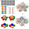
Fig. 3.
Model for the TfR–Tf complex. (A) Class averages used for modeling, corresponding to the 10 most populated class averages from the cryo-EM study. (B) Proximity restraints (colored ellipses) and cross-linking restraints (connected circles) used for modeling. Tf-1, first molecule of Tf (red); Tf-2, second molecule of Tf (green); TfR-A, first monomer of the receptor (gold); TfR-B, second monomer of the receptor (blue). (C) Comparison of the experimental cryo-EM density map (Left) with the simulated density map (8 Å) of the 10 best-scoring solutions in the largest cluster (Right). (D) Fitting of the best-scoring model into the experimental cryo-EM density map. The N- and C-terminal domains of Tf-1 are swapped with respect to the correct configuration (red arrow).
Similar articles
- Single particle electron microscopy reconstruction of the exosome complex using the random conical tilt method.
Liu X, Wang HW. Liu X, et al. J Vis Exp. 2011 Mar 28;(49):2574. doi: 10.3791/2574. J Vis Exp. 2011. PMID: 21490573 Free PMC article. - Optimod--an automated approach for constructing and optimizing initial models for single-particle electron microscopy.
Lyumkis D, Vinterbo S, Potter CS, Carragher B. Lyumkis D, et al. J Struct Biol. 2013 Dec;184(3):417-26. doi: 10.1016/j.jsb.2013.10.009. Epub 2013 Oct 24. J Struct Biol. 2013. PMID: 24161732 Free PMC article. - Multiple subunit fitting into a low-resolution density map of a macromolecular complex using a gaussian mixture model.
Kawabata T. Kawabata T. Biophys J. 2008 Nov 15;95(10):4643-58. doi: 10.1529/biophysj.108.137125. Epub 2008 Aug 15. Biophys J. 2008. PMID: 18708469 Free PMC article. - Visualizing molecular machines in action: Single-particle analysis with structural variability.
Scheres SH. Scheres SH. Adv Protein Chem Struct Biol. 2010;81:89-119. doi: 10.1016/B978-0-12-381357-2.00004-9. Adv Protein Chem Struct Biol. 2010. PMID: 21115174 Review. - The advent of near-atomic resolution in single-particle electron microscopy.
Cheng Y, Walz T. Cheng Y, et al. Annu Rev Biochem. 2009;78:723-42. doi: 10.1146/annurev.biochem.78.070507.140543. Annu Rev Biochem. 2009. PMID: 19489732 Review.
Cited by
- Archiving and disseminating integrative structure models.
Vallat B, Webb B, Westbrook J, Sali A, Berman HM. Vallat B, et al. J Biomol NMR. 2019 Jul;73(6-7):385-398. doi: 10.1007/s10858-019-00264-2. Epub 2019 Jul 5. J Biomol NMR. 2019. PMID: 31278630 Free PMC article. - The molecular architecture of the Dam1 kinetochore complex is defined by cross-linking based structural modelling.
Zelter A, Bonomi M, Kim JO, Umbreit NT, Hoopmann MR, Johnson R, Riffle M, Jaschob D, MacCoss MJ, Moritz RL, Davis TN. Zelter A, et al. Nat Commun. 2015 Nov 12;6:8673. doi: 10.1038/ncomms9673. Nat Commun. 2015. PMID: 26560693 Free PMC article. - Structural templates for modeling homodimers.
Kundrotas PJ, Vakser IA, Janin J. Kundrotas PJ, et al. Protein Sci. 2013 Nov;22(11):1655-63. doi: 10.1002/pro.2361. Epub 2013 Sep 20. Protein Sci. 2013. PMID: 23996787 Free PMC article. - Dissecting Fission Yeast Shelterin Interactions via MICro-MS Links Disruption of Shelterin Bridge to Tumorigenesis.
Liu J, Yu C, Hu X, Kim JK, Bierma JC, Jun HI, Rychnovsky SD, Huang L, Qiao F. Liu J, et al. Cell Rep. 2015 Sep 29;12(12):2169-80. doi: 10.1016/j.celrep.2015.08.043. Epub 2015 Sep 10. Cell Rep. 2015. PMID: 26365187 Free PMC article. - Reconstruction of 3D structures of MET antibodies from electron microscopy 2D class averages.
Chen Q, Vieth M, Timm DE, Humblet C, Schneidman-Duhovny D, Chemmama IE, Sali A, Zeng W, Lu J, Liu L. Chen Q, et al. PLoS One. 2017 Apr 13;12(4):e0175758. doi: 10.1371/journal.pone.0175758. eCollection 2017. PLoS One. 2017. PMID: 28406969 Free PMC article.
References
- Frank J. Three-Dimensional Electron Microscopy of Macromolecular Assemblies: Visualization of Biological Molecules in Their Native State. 2nd Ed. New York: Oxford Univ Press; 2006.
Publication types
MeSH terms
Substances
LinkOut - more resources
Full Text Sources
Other Literature Sources


