Cellular senescence induced by CD158d reprograms natural killer cells to promote vascular remodeling - PubMed (original) (raw)
Cellular senescence induced by CD158d reprograms natural killer cells to promote vascular remodeling
Sumati Rajagopalan et al. Proc Natl Acad Sci U S A. 2012.
Abstract
Natural killer (NK) cells, which have an essential role in immune defense, also contribute to reproductive success. NK cells are abundant at the maternal-fetal interface, where soluble HLA-G is produced by fetal trophoblast cells during early pregnancy. Soluble HLA-G induces a proinflammatory response in primary, resting NK cells on endocytosis into early endosomes where its receptor, CD158d, resides. CD158d initiates signaling through DNA-PKcs, Akt, and NF-κB for a proinflammatory and proangiogenic response. The physiological relevance of this endosomal signaling pathway, and how activation of CD158d through soluble ligands regulates NK cell fate and function is unknown. We show here that CD158d agonists trigger a DNA damage response signaling pathway involving cyclin-dependent kinase inhibitor p21 expression and heterochromatin protein HP1-γ phosphorylation. Sustained activation through CD158d induced morphological changes in NK cell shape and size, and survival in the absence of cell-cycle entry, all hallmarks of senescence, and a transcriptional signature of a senescence-associated secretory phenotype (SASP). SASP is a program that can be induced by oncogenes or DNA damage, and promotes growth arrest and tissue repair. The secretome of CD158d-stimulated senescent NK cells promoted vascular remodeling and angiogenesis as assessed by functional readouts of vascular permeability and endothelial cell tube formation. Retrospective analysis of the decidual NK cell transcriptome revealed a strong senescence signature. We propose that a positive function of senescence in healthy tissue is to favor reproduction through the sustained activation of NK cells to remodel maternal vasculature in early pregnancy.
Conflict of interest statement
The authors declare no conflict of interest.
Figures
Fig. 1.
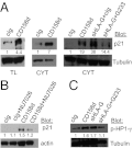
DDR signaling results in p21 induction and phosphorylation of HP1-γ. (A) Western blot analysis of total lysate (TL) or cytoplasmic (CYT) proteins from resting NK cells stimulated with control Ab (cIg), a mAb to CD158d or sHLA-G in the presence of cIg or anti-HLA-G (G233). Blots were probed with antibodies to p21 or tubulin (loading control). (B) Western blot analysis of p21 expression in resting NK cells stimulated with CD158d mAb in the presence or absence of DNA-PK inhibitor, NU7026. (C) Western blot analysis of phosphorylated HP1-γ in resting NK cells stimulated with CD158d mAb or sHLA-G in the presence of cIg or anti-HLA-G (G233). Normalized intensities of p21 and phosphorylated HP1-γ relative to the tubulin or actin loading controls are listed. Each experiment was performed using NK cells from at least three independent donors.
Fig. 2.
NK cells acquire features of senescence on CD158d-mediated activation. (A) Differential interference contrast images of resting NK cells stimulated with cIg or CD158d mAb for 16 h. The DIC image on the right (Magnification 2×). (B) NK cells treated as in (A) were stained with DRAQ5 (nuclei in red). (C) NK cells treated as in (A) and stained for SA-β−gal at 48 h. (D) Resting NK cells stimulated with cIg (circle), CD158d (red square), 2B4 (inverted triangle), and CD16 (triangle) were counted for live cells on a hemacytometer using Trypan Blue exclusion of dead cells (Scale bar, 10 μm). (E, F, and G) Resting NK cells stimulated with cIg (circle), CD158d mAb (red square), CD16 mAb (triangle), and IL-2 at 100 U/mL (diamond) were tested at the indicated time points for BrDU incorporation (E), G0/G1-positive NK cells (F), and Annexin V–positive NK cells (G). All data are representative of experiments using NK cells from at least three normal donors.
Fig. 3.
The transcriptional response to activation by CD158d reveals a senescence signature of up-regulated genes. (A) GSEA of microarray data of CD158d-induced gene expression compared with the molecular signature of oncogene ras-induced senescence. (B) Heat map showing relative expression of the top 115 up-regulated genes at 16 h compared with their expression at 4 h and 64 h. A high resolution image is shown in
Fig. S1
. (C) List of top 30 up-regulated genes at 16 h. Genes known to be involved in senescence are highlighted in blue. Function: S, senescence; RS, replicative senescence; I, inflammation; WH, wound healing; A, gremlin 2 is unique to angiogenesis. (D) The senescence signature after CD158d stimulation includes 40 genes at 16 h (
Table S4
). The schematic shows the overlap of genes involved in inflammation, wounding healing, and senescence responses. (E) Fold increase in gene expression values (CD158d/cIg stimulation) for the senescence signature at 16 h from resting NK cells of five different donors. Each panel has a different scale based on strength of gene expression. (F) Fold gene expression values for IL6, IL8, and IL1B from resting NK cells stimulated with sHLA-G in the presence of cIg or anti-HLA-G (G233).
Fig. 4.
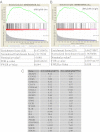
Up-regulation of senescence-associated genes in decidual NK cells. (A and B) GSEA of microarray data of decidual NK cells (30) compared with the molecular signature of oncogene ras-induced senescence (21). The transcriptional up-regulation of decidual NK cells versus peripheral blood CD56dim NK cells (A) and peripheral blood CD56bright NK cells (B) were analyzed separately. (C) List of senescence-associated genes up-regulated in decidual NK cells and in CD158d-stimulated resting NK cells, presented as fold change (F.C.) of expression in decidual NK (dNK) compared with peripheral blood NK (pNK) belonging to CD56dim or CD56bright subsets. ND, not detected.
Fig. 5.
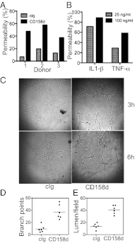
The senescence secretome induced by CD158d in NK cells increases vascular permeability and promotes angiogenesis. (A) Effect of CD158d-induced senescence secretome on vascular permeability. HUVEC were incubated with supernatants from resting NK cells stimulated with control or CD158d mAbs and permeability of the HUVEC monolayer is shown. (B) Effect of recombinant IL-1β and TNF-α on the permeability of HUVEC monolayers. (C) Representative images from the tube formation assay to test the effect of the CD158d-induced secretome. HUVEC seeded on a gel matrix were evaluated for their ability to form tubes in the presence of supernatants of NK cells stimulated for 16 h with either cIg or CD158d mAb. (D) Quantification of matrigel-tube formation assay in (C). The data are representative of experiments using NK cells from three independent donors.
Similar articles
- TNFR-associated factor 6 and TGF-β-activated kinase 1 control signals for a senescence response by an endosomal NK cell receptor.
Rajagopalan S, Lee EC, DuPrie ML, Long EO. Rajagopalan S, et al. J Immunol. 2014 Jan 15;192(2):714-21. doi: 10.4049/jimmunol.1302384. Epub 2013 Dec 11. J Immunol. 2014. PMID: 24337384 Free PMC article. - DNA-PKcs controls an endosomal signaling pathway for a proinflammatory response by natural killer cells.
Rajagopalan S, Moyle MW, Joosten I, Long EO. Rajagopalan S, et al. Sci Signal. 2010 Feb 23;3(110):ra14. doi: 10.1126/scisignal.2000467. Sci Signal. 2010. PMID: 20179272 Free PMC article. - HLA-G-mediated NK cell senescence promotes vascular remodeling: implications for reproduction.
Rajagopalan S. Rajagopalan S. Cell Mol Immunol. 2014 Sep;11(5):460-6. doi: 10.1038/cmi.2014.53. Epub 2014 Jul 7. Cell Mol Immunol. 2014. PMID: 24998350 Free PMC article. Review. - Endosomal signaling and a novel pathway defined by the natural killer receptor KIR2DL4 (CD158d).
Rajagopalan S. Rajagopalan S. Traffic. 2010 Nov;11(11):1381-90. doi: 10.1111/j.1600-0854.2010.01112.x. Epub 2010 Sep 20. Traffic. 2010. PMID: 20854369 Review. - KIR2DL4 (CD158d): An activation receptor for HLA-G.
Rajagopalan S, Long EO. Rajagopalan S, et al. Front Immunol. 2012 Aug 20;3:258. doi: 10.3389/fimmu.2012.00258. eCollection 2012. Front Immunol. 2012. PMID: 22934097 Free PMC article.
Cited by
- Aging, Cellular Senescence, and Progressive Multiple Sclerosis.
Papadopoulos D, Magliozzi R, Mitsikostas DD, Gorgoulis VG, Nicholas RS. Papadopoulos D, et al. Front Cell Neurosci. 2020 Jun 30;14:178. doi: 10.3389/fncel.2020.00178. eCollection 2020. Front Cell Neurosci. 2020. PMID: 32694983 Free PMC article. Review. - The structure of the atypical killer cell immunoglobulin-like receptor, KIR2DL4.
Moradi S, Berry R, Pymm P, Hitchen C, Beckham SA, Wilce MC, Walpole NG, Clements CS, Reid HH, Perugini MA, Brooks AG, Rossjohn J, Vivian JP. Moradi S, et al. J Biol Chem. 2015 Apr 17;290(16):10460-71. doi: 10.1074/jbc.M114.612291. Epub 2015 Mar 10. J Biol Chem. 2015. PMID: 25759384 Free PMC article. - HLA-G Orchestrates the Early Interaction of Human Trophoblasts with the Maternal Niche.
Gregori S, Amodio G, Quattrone F, Panina-Bordignon P. Gregori S, et al. Front Immunol. 2015 Mar 30;6:128. doi: 10.3389/fimmu.2015.00128. eCollection 2015. Front Immunol. 2015. PMID: 25870595 Free PMC article. Review. - Cellular senescence: from growth arrest to immunogenic conversion.
Burton DG, Faragher RG. Burton DG, et al. Age (Dordr). 2015;37(2):27. doi: 10.1007/s11357-015-9764-2. Epub 2015 Mar 20. Age (Dordr). 2015. PMID: 25787341 Free PMC article. Review. - Modelling the impact of decidual senescence on embryo implantation in human endometrial assembloids.
Rawlings TM, Makwana K, Taylor DM, Molè MA, Fishwick KJ, Tryfonos M, Odendaal J, Hawkes A, Zernicka-Goetz M, Hartshorne GM, Brosens JJ, Lucas ES. Rawlings TM, et al. Elife. 2021 Sep 6;10:e69603. doi: 10.7554/eLife.69603. Elife. 2021. PMID: 34487490 Free PMC article.
References
- Moffett-King A. Natural killer cells and pregnancy. Nat Rev Immunol. 2002;2(9):656–663. - PubMed
- Bilinski MJ, et al. Uterine NK cells in murine pregnancy. Reprod Biomed Online. 2008;16(2):218–226. - PubMed
- Hanna J, et al. Decidual NK cells regulate key developmental processes at the human fetal-maternal interface. Nat Med. 2006;12(9):1065–1074. - PubMed
Publication types
MeSH terms
Substances
LinkOut - more resources
Full Text Sources
Molecular Biology Databases
Research Materials
Miscellaneous


