Functional characterization of nonmetastatic paraganglioma and pheochromocytoma by (18) F-FDOPA PET: focus on missed lesions - PubMed (original) (raw)
Multicenter Study
Functional characterization of nonmetastatic paraganglioma and pheochromocytoma by (18) F-FDOPA PET: focus on missed lesions
Sophie Gabriel et al. Clin Endocrinol (Oxf). 2013 Aug.
Abstract
Aims and methods: To evaluate the clinical value of (18) F-fluorodihydroxyphenylalanine ((18) F-FDOPA) PET in relation to tumour localization and the patient's genetic status in a large series of pheochromocytoma/paraganglioma (PHEO/PGL) patients and to discuss in detail false-negative results. A retrospective study of PGL patients who were investigated with (18) F-FDOPA PET or PET/CT imaging in two academic endocrine tumour centres was conducted (La Timone University Hospital, Marseilles, France and National Institutes of Health (NIH), Bethesda, MD, USA).
Results: One hundred sixteen patients (39·7% harbouring germline mutations in known disease susceptibility genes) were evaluated for a total of 195 PHEO/PGL foci. (18) F-FDOPA PET correctly detected 179 lesions (91·8%) in 107 patients (92·2%). Lesion-based sensitivities for parasympathetic PGLs (head, neck, or anterior/middle thoracic ones), PHEOs, and extra-adrenal sympathetic (abdominal or posterior thoracic) PGLs were 98·2% [96·5% for Timone and 100% for NIH], 93·9% [93·8 and 93·9%] and 70·3% [47·1 and 90%] respectively (P < 0·001). Sympathetic (adrenal and extra-adrenal) SDHx-related PGLs were at a higher risk for negative (18) F-FDOPA PET than non-SDHx-related PGLs (14/24 vs 0/62, respectively, P < 0·001). In contrast, the risk of negative (18) F-FDOPA PET was lower for parasympathetic PGLs regardless of the genetic background (1/90 in SDHx vs 1/19 in non-SDHx tumours, P = 0·32). (18) F-FDOPA PET failed to detect two head and neck PGLs (HNPGL), likely due to their small size, whereas most missed sympathetic PGL were larger and may have exhibited a specific (18) F-FDOPA-negative imaging phenotype. (18) F-FDG PET detected all the missed sympathetic lesions.
Conclusions: (18) F-FDOPA PET appears to be a very sensitive functional imaging tool for HNPGL regardless of the genetic status of the tumours. Patients with false-negative tumours on (18) F-FDOPA PET should be tested for SDHx mutations.
© 2012 John Wiley & Sons Ltd.
Figures
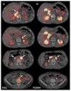
Figure 1
Multicentric SDHD-related PGL syndrome (2 HNPGL, 2 PHEO, and 4 extra-adrenal PGL). A. Fused axial 18F-FDG PET/CT images centered over the tumours (6 positive tumours, arrows). B. Matching axial 18F-FDOPA PET/CT images show 2 positive tumours (arrows). Missed tumours on 18F-FDOPA PET/CT were located as follows: 9 mm left PHEO, 26 mm interaortocaval, 17 mm lateral caval, 6 mm iliac bifurcation.
Figure 2
Multicentric SDHD-related PGL syndrome (5 HNPGL, 1 thoracic and 1 extra-adrenal PGL). A. 18F-FDG PET (maximal intensity projection (MIP)). B. 18F-FDOPA PET (MIP). C. Axial contrast-enhanced CT showing a 5 mm cervical PGL of the vagus nerve missed by 18F-FDOPA PET/CT (arrow). D. Axial contrast-enhanced CT image centered on a 14 mm abdominal extra-adrenal PGL located lateral to the celiac trunk (arrow). E. Axial 18F-FDG PET image centered over the positive tumour (arrow). F. Axial 18F-FDOPA PET image centered over the false-negative abdominal extra-adrenal PGL.
Figure 3
Multicentric SDHB-related PGL (2 extra-adrenal PGL, 1 adrenal PHEO). A. Enhanced coronal and axial CT slices at the level of the tumours (reconstruction in the lower left image). B. Fused axial 18F-FDG PET/CT images centered over the tumours (2 positive extra adrenal tumours 28 and 13 mm in diameter, arrows). C. Fused axial 18F-FDOPA PET/CT images centered over the tumours. 18F-FDOPA-negative tumour sites were lateral aortic (at the level of the superior mesenteric artery) and preaortic (PGL derived from the organ of Zuckerkandl). False-positive uptake was seen in an enlarged left adrenal gland (top row). The adrenal gland weighed 8 g (normal 5 to 6 g) and showed cortical hyperplasia, but the adrenal medulla was normal.
Figure 4
Multicentric SDHD-related PGL syndrome (1 HNPGL, 1 cardiac, 2 adrenal PHEO). A. 18F-FDG PET (MIP image) showing positive cardiac PGL and bilateral PHEO (arrows). Non-specific uptake in the mediastinum corresponds to brown fat. The HNPGL is not visible on this projection. B. Coronal fused 18F-FDG PET/CT image centered over the PHEO (2 positive tumours, arrows). C. 18F-FDOPA PET (MIP image) showing positive HNPGL and cardiac PGL, negative bilateral PHEO.
Figure 5
Multicentric SDHD-related PGL syndrome (multiple HNPGL, 1 parasympathetic thoracic PGL, 1 sympathetic thoracic (cardiac) PGL, and 2 sympathetic retroperitoneal PGL). A. 18F-FDG PET/CT (MIP image) shows multiple foci, including uptake in a paravertebral sympathetic legion (arrow). B. Axial CT and fused 18F-FDG PET/CT images centered over the sympathetic thoracic PGL (arrows). C. 18F-FDOPA PET (MIP image) showing positive parasympathetic and extra-adrenal sympathetic PGL, but negative sympathetic thoracic PGL.
Similar articles
- Prospective comparison of (68)Ga-DOTATATE and (18)F-FDOPA PET/CT in patients with various pheochromocytomas and paragangliomas with emphasis on sporadic cases.
Archier A, Varoquaux A, Garrigue P, Montava M, Guerin C, Gabriel S, Beschmout E, Morange I, Fakhry N, Castinetti F, Sebag F, Barlier A, Loundou A, Guillet B, Pacak K, Taïeb D. Archier A, et al. Eur J Nucl Med Mol Imaging. 2016 Jul;43(7):1248-57. doi: 10.1007/s00259-015-3268-2. Epub 2015 Dec 5. Eur J Nucl Med Mol Imaging. 2016. PMID: 26637204 Clinical Trial. - 18F-fluorodihydroxyphenylalanine PET/CT in pheochromocytoma and paraganglioma: relation to genotype and amino acid transport system L.
Feral CC, Tissot FS, Tosello L, Fakhry N, Sebag F, Pacak K, Taïeb D. Feral CC, et al. Eur J Nucl Med Mol Imaging. 2017 May;44(5):812-821. doi: 10.1007/s00259-016-3586-z. Epub 2016 Nov 29. Eur J Nucl Med Mol Imaging. 2017. PMID: 27900521 Free PMC article. - 6-18F-fluoro-L-dihydroxyphenylalanine positron emission tomography is superior to 123I-metaiodobenzyl-guanidine scintigraphy in the detection of extraadrenal and hereditary pheochromocytomas and paragangliomas: correlation with vesicular monoamine transporter expression.
Fottner C, Helisch A, Anlauf M, Rossmann H, Musholt TJ, Kreft A, Schadmand-Fischer S, Bartenstein P, Lackner KJ, Klöppel G, Schreckenberger M, Weber MM. Fottner C, et al. J Clin Endocrinol Metab. 2010 Jun;95(6):2800-10. doi: 10.1210/jc.2009-2352. Epub 2010 Apr 6. J Clin Endocrinol Metab. 2010. PMID: 20371665 - Clinical aspects of SDHx-related pheochromocytoma and paraganglioma.
Timmers HJ, Gimenez-Roqueplo AP, Mannelli M, Pacak K. Timmers HJ, et al. Endocr Relat Cancer. 2009 Jun;16(2):391-400. doi: 10.1677/ERC-08-0284. Epub 2009 Feb 3. Endocr Relat Cancer. 2009. PMID: 19190077 Free PMC article. Review. - Role of (18) F-FDOPA PET/CT imaging in endocrinology.
Santhanam P, Taïeb D. Santhanam P, et al. Clin Endocrinol (Oxf). 2014 Dec;81(6):789-98. doi: 10.1111/cen.12566. Epub 2014 Sep 1. Clin Endocrinol (Oxf). 2014. PMID: 25056984 Review.
Cited by
- Overexpression of L-Type Amino Acid Transporter 1 (LAT1) and 2 (LAT2): Novel Markers of Neuroendocrine Tumors.
Barollo S, Bertazza L, Watutantrige-Fernando S, Censi S, Cavedon E, Galuppini F, Pennelli G, Fassina A, Citton M, Rubin B, Pezzani R, Benna C, Opocher G, Iacobone M, Mian C. Barollo S, et al. PLoS One. 2016 May 25;11(5):e0156044. doi: 10.1371/journal.pone.0156044. eCollection 2016. PLoS One. 2016. PMID: 27224648 Free PMC article. Clinical Trial. - Current and future trends in the anatomical and functional imaging of head and neck paragangliomas.
Taïeb D, Varoquaux A, Chen CC, Pacak K. Taïeb D, et al. Semin Nucl Med. 2013 Nov;43(6):462-73. doi: 10.1053/j.semnuclmed.2013.06.005. Semin Nucl Med. 2013. PMID: 24094713 Free PMC article. Review. - Exploring the link between tumour metabolism and succinate dehydrogenase deficiency: A 18 F-FDOPA PET/CT study in head and neck paragangliomas.
Reichert T, Fakhry N, Lavieille JP, Amodru V, Sebag F, Romanet P, Loundou A, Castinetti F, Pacak K, Montava M, Taïeb D. Reichert T, et al. Clin Endocrinol (Oxf). 2019 Dec;91(6):879-884. doi: 10.1111/cen.14086. Epub 2019 Oct 1. Clin Endocrinol (Oxf). 2019. PMID: 31479526 Free PMC article. - A Clinical Roadmap to Investigate the Genetic Basis of Pediatric Pheochromocytoma: Which Genes Should Physicians Think About?
Dias Pereira B, Nunes da Silva T, Bernardo AT, César R, Vara Luiz H, Pacak K, Mota-Vieira L. Dias Pereira B, et al. Int J Endocrinol. 2018 Mar 20;2018:8470642. doi: 10.1155/2018/8470642. eCollection 2018. Int J Endocrinol. 2018. PMID: 29755524 Free PMC article. Review. - 18F-FLT PET/CT in the Evaluation of Pheochromocytomas and Paragangliomas: A Pilot Study.
Blanchet EM, Taieb D, Millo C, Martucci V, Chen CC, Merino M, Herscovitch P, Pacak K. Blanchet EM, et al. J Nucl Med. 2015 Dec;56(12):1849-54. doi: 10.2967/jnumed.115.159061. Epub 2015 Sep 10. J Nucl Med. 2015. PMID: 26359261 Free PMC article.
References
- Taieb D, Neumann H, Rubello D, Al-Nahhas A, Guillet B, Hindie E. Modern nuclear imaging for paragangliomas: beyond SPECT. Journal of nuclear medicine : official publication, Society of Nuclear Medicine. 2012;53:264–274. - PubMed
- Gimenez-Roqueplo AP, Favier J, Rustin P, Rieubland C, Crespin M, Nau V, Khau Van Kien P, Corvol P, Plouin PF, Jeunemaitre X. Mutations in the SDHB gene are associated with extra-adrenal and/or malignant phaeochromocytomas. Cancer research. 2003;63:5615–5621. - PubMed
- Amar L, Baudin E, Burnichon N, Peyrard S, Silvera S, Bertherat J, Bertagna X, Schlumberger M, Jeunemaitre X, Gimenez-Roqueplo AP, Plouin PF. Succinate dehydrogenase B gene mutations predict survival in patients with malignant pheochromocytomas or paragangliomas. The Journal of clinical endocrinology and metabolism. 2007;92:3822–3828. - PubMed
Publication types
MeSH terms
Substances
LinkOut - more resources
Full Text Sources
Other Literature Sources
Medical
Miscellaneous


