Selective excision of 5-carboxylcytosine by a thymine DNA glycosylase mutant - PubMed (original) (raw)
Selective excision of 5-carboxylcytosine by a thymine DNA glycosylase mutant
Hideharu Hashimoto et al. J Mol Biol. 2013.
Abstract
The mammalian thymine DNA glycosylase (TDG) excises the mismatched base, uracil, thymine or 5-hydroxymethyluracil (5hmU), as well as removes 5-formylcytosine (5fC) and 5-carboxylcytosine (5caC) when paired with a guanine. In the previously solved structure of TDG in complex with DNA containing 5caC, the side chain of asparagine 157 (N157) contacts the 5-carboxyl moiety of 5caC via a weak hydrogen bond. We examined the role of N157 in recognition of 5caC by mutagenesis. The asparagine-to-alanine (N157A) mutant has no detectable base excision activity for a G:T mismatch, and its excision activity is reduced for other substrates including G:5caC. Unexpectedly, the asparagine-to-aspartate (N157D) mutant has a comparable base excision rate for G:5caC substrate to that of wild type, but it only has residual activity for G:U and no detectable activity for other substrates. We further show that the N157D mutant has higher activity for 5caC at a lower pH (6.0), suggesting that increased protonation of the carboxylate of 5caC and the aspartate facilitates base excision. The N157D mutant remains highly specific for 5caC even in the presence of large excess of genomic DNA, a property that can potentially be used for mapping the very low amount of 5caC in genomes.
Copyright © 2013 Elsevier Ltd. All rights reserved.
Figures
Figure 1. N157 of human TDG is involved in DNA binding
(a) Structure of TDG in complex with an A:5caC mismatch (PDB 3UO7) . The side chain amino group of Asn157 forms a weak hydrogen bond with the 5-carboxyl moiety of 5caC, which is flipped out from the double stranded DNA. (b) Structure of TDG in complex with a uracil analog (2’-deoxy-2’-fluoroarabinouridine) (PDB 3UFJ) . (c) A post-reactive complex structure of TDG containing a G:5hmU mismatch with the cleaved 5hmU base remaining in the active site pocket (PDB 4FNC) . The side chain of Asn157 interacts with the 5’ phosphate of the target nucleotide. (d-f) The activities of TDG wild type (WT) (panel d), mutants N157A (panel e) and N157D (panel f) on G:X under the single turnover condition ([SDNA]=0.25 µM and [ETDG]=2.5 µM) after 10 min reaction at 30°C in 0.1% BSA, 1 mM EDTA, 100 mM NaCl and 10 mM BisTris HCl, pH 6.0. Various 32 bp oligonucleotides labeled with 6-carboxy-fluorescein (FAM) were used as substrates: (FAM)-5’-TCG GAT GTT GTG GGT CAG
X
GC ATG ATA GTG TA-3’ (where
X
=C, 5mC, 5hmC, 5fC, 5caC, U, T and 5hmU) and 5’-TAC ACT ATC ATG CGC TGA CCC ACA ACA TCC GA-3’. Human TDG residues 111–308 (pXC1056) and its mutants N157A (pXC1155) and N157D (pXC1138) were prepared as described . The excision reaction was monitored, as described , by denaturing gel electrophoresis following NaOH hydrolysis of the abasic site.
Figure 2. N157D mutant selectively cleaves 5caC
(a) The time course (0–60 min) of N157D mutant reaction on seven oligonucleotides with various modifications under the single turnover condition at 30°C, pH 6.0 and 100 mM NaCl. For G:5caC substrate, more time points between 0 to 10 min were also taken. (b) N157D has enhanced activity on G:5caC at lower pH. Four different pH values between 6.0 and 7.2 with 0.4 increments were used at 10 mM BisTris HCl, 100 mM NaCl and 30°C. The intensities of the FAM labeled DNA were determined by Typhoon Trio+, and quantified by the image-processing program ImageJ. The data were fitted to non-linear regression using software GraphPad PRISM 5.0d (GraphPad Software Inc.): [Product] = _P_max(1-e−_k_t), where _P_max is the product plateau level, k(min−1) is the observed rate constant, and t is the reaction time. (c) The activities of N157D on G:5caC at 30°C were measured at pH 6.0 with varying salt concentration between 0–250 mM NaCl with 25 or 50 mM increments. The _K_obs values (min−1) were calculated for reactions between 0–10 min. Plot of activity as a function of NaCl concentration after 2.5 min reactions. (d) The log plots of activities of WT TDG (lines 1, 2 and 3) and N157D mutant (line 4) as a function of pH. For lines 1 and 2, the data were measured from a mixture of three buffers , whereas lines 3 and 4 were measured from 10 mM BisTrisHCl as indicated in the legend of panel b.
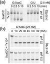
Figure 3. Detection of 5caC in the presence of large excess of genomic DNA
(a) Specificity of N157D (5caC vs. U) in the presence of large access of genomic DNA. The reaction (200 µl) contains 25 µM of N157D, 2.5 nM of 32-bp FAM-labeled G:X (X=5caC or U) oligonucleotides and 100 µg (or 0.5 mg/ml) of salmon sperm genome DNA (Rockland Inc.). The G+C contents of salmon DNA are approximately 44.4% , and thus the salmon DNA used in the reaction contains 757 µM of base pair and 360 µM G:C pairs. The 5caC/C ratio (2.5 nM/360 µM) was estimated to be 7×10⁻⁶. (b) Excision reactions (20 µl) contain 25 µM (top panel) or 2.5 µM (bottom panel) of N157D, 25 nM of 32-bp FAM-labeled G:5caC oligonucleotides and 10 µg of salmon sperm genome DNA.
Similar articles
- Activity and crystal structure of human thymine DNA glycosylase mutant N140A with 5-carboxylcytosine DNA at low pH.
Hashimoto H, Zhang X, Cheng X. Hashimoto H, et al. DNA Repair (Amst). 2013 Jul;12(7):535-40. doi: 10.1016/j.dnarep.2013.04.003. Epub 2013 May 13. DNA Repair (Amst). 2013. PMID: 23680598 Free PMC article. - Excision of 5-hydroxymethyluracil and 5-carboxylcytosine by the thymine DNA glycosylase domain: its structural basis and implications for active DNA demethylation.
Hashimoto H, Hong S, Bhagwat AS, Zhang X, Cheng X. Hashimoto H, et al. Nucleic Acids Res. 2012 Nov 1;40(20):10203-14. doi: 10.1093/nar/gks845. Epub 2012 Sep 8. Nucleic Acids Res. 2012. PMID: 22962365 Free PMC article. - Nei-like 1 (NEIL1) excises 5-carboxylcytosine directly and stimulates TDG-mediated 5-formyl and 5-carboxylcytosine excision.
Slyvka A, Mierzejewska K, Bochtler M. Slyvka A, et al. Sci Rep. 2017 Aug 21;7(1):9001. doi: 10.1038/s41598-017-07458-4. Sci Rep. 2017. PMID: 28827588 Free PMC article. - Structural and mutation studies of two DNA demethylation related glycosylases: MBD4 and TDG.
Hashimoto H. Hashimoto H. Biophysics (Nagoya-shi). 2014 Oct 18;10:63-8. doi: 10.2142/biophysics.10.63. eCollection 2014. Biophysics (Nagoya-shi). 2014. PMID: 27493500 Free PMC article. Review. - Epigenetic modifications in DNA could mimic oxidative DNA damage: A double-edged sword.
Ito S, Kuraoka I. Ito S, et al. DNA Repair (Amst). 2015 Aug;32:52-57. doi: 10.1016/j.dnarep.2015.04.013. Epub 2015 May 1. DNA Repair (Amst). 2015. PMID: 25956859 Review.
Cited by
- The carboxy-terminal domain of ROS1 is essential for 5-methylcytosine DNA glycosylase activity.
Hong S, Hashimoto H, Kow YW, Zhang X, Cheng X. Hong S, et al. J Mol Biol. 2014 Nov 11;426(22):3703-3712. doi: 10.1016/j.jmb.2014.09.010. Epub 2014 Sep 21. J Mol Biol. 2014. PMID: 25240767 Free PMC article. - Differential stabilities and sequence-dependent base pair opening dynamics of Watson-Crick base pairs with 5-hydroxymethylcytosine, 5-formylcytosine, or 5-carboxylcytosine.
Szulik MW, Pallan PS, Nocek B, Voehler M, Banerjee S, Brooks S, Joachimiak A, Egli M, Eichman BF, Stone MP. Szulik MW, et al. Biochemistry. 2015 Feb 10;54(5):1294-305. doi: 10.1021/bi501534x. Epub 2015 Jan 29. Biochemistry. 2015. PMID: 25632825 Free PMC article. - Connections between TET proteins and aberrant DNA modification in cancer.
Huang Y, Rao A. Huang Y, et al. Trends Genet. 2014 Oct;30(10):464-74. doi: 10.1016/j.tig.2014.07.005. Epub 2014 Aug 14. Trends Genet. 2014. PMID: 25132561 Free PMC article. Review. - Activity and crystal structure of human thymine DNA glycosylase mutant N140A with 5-carboxylcytosine DNA at low pH.
Hashimoto H, Zhang X, Cheng X. Hashimoto H, et al. DNA Repair (Amst). 2013 Jul;12(7):535-40. doi: 10.1016/j.dnarep.2013.04.003. Epub 2013 May 13. DNA Repair (Amst). 2013. PMID: 23680598 Free PMC article. - Role of Base Excision "Repair" Enzymes in Erasing Epigenetic Marks from DNA.
Drohat AC, Coey CT. Drohat AC, et al. Chem Rev. 2016 Oct 26;116(20):12711-12729. doi: 10.1021/acs.chemrev.6b00191. Epub 2016 Aug 8. Chem Rev. 2016. PMID: 27501078 Free PMC article. Review.
References
- Booth MJ, Branco MR, Ficz G, Oxley D, Krueger F, Reik W, Balasubramanian S. Quantitative sequencing of 5-methylcytosine and 5- hydroxymethylcytosine at single-base resolution. Science. 2012;336:934–937. - PubMed
Publication types
MeSH terms
Substances
LinkOut - more resources
Full Text Sources
Other Literature Sources


